
Gabriele Masselli1 , Maria Chiara Colaiacomo1, Giuseppe Rossi1 and Gianfranco Gualdi1
(1)
Department of Radiology, Umberto I Hospital, Sapienza University, Rome, Italy
Gabriele Masselli
Email: g.masselli@policlinicoumberto1.it
Keywords
DeliveryMRIMusculoskeletalPregnancy
24.1 Introduction
Pregnancy, childbirth, and breastfeeding may result in various musculoskeletal (MSK) disorders [1–9]. Many changes occurring during pregnancy can be related to MSK disorders, such as increased body weight, fluid retention, increased calcium need, and altered glucose/insulin ratio thus leading to stress on skeletal bones and joints, in particular overload on the lumbar spine and pelvic girdle. Indeed, pregnancy induces a major gain of body weight over a short period which dramatically increases the stress on the lower skeleton, and most of disorders occur late in the course of pregnancy [1]. Most common symptom is pain due to joint involvement and neurologic compression, but also hematogenous infection can occur.
24.2 Pregnancy-Related Low Back Pain and Pelvic Girdle Pain
Pregnancy-related low back pain and pelvic girdle pain are very common with around 45 % of women during pregnancy and 25 % in postpartum suffering from these conditions [2], which are mostly temporary and self-limiting, recovering in a few weeks or months after delivery, even if 8–10 % have persistent pain for 1–2 years [2, 6, 10]. However, 25 % of all those pregnant women have severe pain and 8 % have severe disability [2]. There have been reported an increasing number of patients who are requesting induction of labor or cesarean section in order to relief symptoms, with potential risk for the mother and the baby and resource implications [4, 11, 12].
These complaints start usually around the 18th week of pregnancy but can also start earlier or be delayed as late as 3 weeks portpartum [2, 4, 13, 14].
Risk factors for low back pain and pelvic girdle are strenuous work, previous low back pain, and previous history of pregnancy-related low back pain or pelvic girdle syndrome, while factors that don’t affect the risk are contraceptive pills, time interval since last pregnancy, height, weight, smoking, and age [15]; in particular there is a high risk for young women, or the fist pregnancy, and for much older women or much later pregnancy which has a higher risk of preceding tissue damage.
The mechanisms underlying these conditions are probably a combination of biomechanical, traumatic, hormonal, metabolic, and degenerative [16, 17].
During pregnancy, increased levels of relaxin, a polypeptide hormone produced by the corpus luteum and decidua, lead to greater ligamentous laxity particularly in the joints of the pelvis, resulting in widening of the symphysis pubis and sacroiliac joints [16, 17]. This leads to increased motion of the pelvic joints, inflammation, and reduced stability of the pelvis with an altered efficiency of load transmission from the trunk to legs and increased shear forces across the joints [4, 18]. In normal condition in fact the stability of the pelvis is gained by the integrity of the sacroiliac joints, symphysis pubis, the sacrum, and all the muscles, fascia, and ligaments that attach to the pelvis [19, 20].
Diagnosis is mostly clinical; however, imaging is required for differential diagnosis [21, 22].
Ultrasound has been used to measure the interpubic gap between the two pubic bones at the symphysis pubis, but there is no correlation between the severity of symptoms and the degree of separation [17]. X-ray imaging techniques such as computed tomography scans are not ideal in pregnancy [4]. Therefore, currently, the use of MRI (Fig. 24.1) should be reserved for the investigation of pain in pregnancy where there is a strong suspicion of abnormality such as the presence of neurological signs and in case of focal inflammatory signs suggesting osteomyelitis (Fig. 24.2) or pathologic bone infiltration (Fig. 24.3) or neurologic symptoms and cauda equina syndrome, to rule out lumbar disk herniation or other compressive conditions. Osteomyelitis and septic arthritis of the sacroiliac or pubic symphysis are rare conditions due to hematogenous spread and microbic contamination of the joint. It occurs more frequently in the early postpartum period, but can also be observed during pregnancy, due to simple injury of the urinary tract or endometritis, and staphylococcus aureus is the most prevalent organism encountered [23–26]. In case of suspicion of osteomyelitis or septic arthritis, MRI should be performed immediately. Findings include involvement of the subchondral bone and surrounding soft tissue, with possible fluid collections or abscesses.

Fig. 24.1
Pubic osteitis in a 32-year-old woman at 20 weeks of gestational age, with recurrent pubic pain. Coronal (a) and axial STIR (b) and coronal (c) and axial (d) T1-weighted MRI images show sclerosis of the subchondral bone at the level of the pubic symphysis (arrows) with small subchondral erosions, finding typical of pubic instability

Fig. 24.2
Pubic osteomyelitis in a 35-year-old woman presents with pubic pain, fever, and elevated inflammatory markers. Sagittal (a), coronal (b), and axial (c) T2-weighted HASTE images, and axial STIR images (d) show fluid collection (arrows) at the level of the symphysis pubic bone with hyperintensity of the pubic bone (short arrows). DWI image (e) shows the presence of restriction of diffusion within the fluid collection, which is indicative of an abscess (arrows). These findings are in agreement with osteomyelitis

Fig. 24.3
Pubic chondrosarcoma in a 30-year-old woman at 34 weeks of gestational age, with recurrent pubic pain. (a) Coronal STIR image demonstrates soft tissue mass (arrow) with erosion of the right pubic bone and pericortical extension. Note lobular growth and deep endosteal scalloping. (b) Coronal T1-weighted sequence shows bone destruction and cortical penetration (arrow). A chondrosarcoma was found at biopsy and confirmed at pathology after the delivery
24.2.1 Low Back Pain, Disk Disease, and Sciatica
Low back pain is one of the most common complain during pregnancy and postpartum period, affecting more than 50 % of woman during pregnancy [27, 28]. It is characterized by lumbar region pain, exacerbated in forward flexion, with a restriction of spine movement and painful palpation of the erector spinae muscle [4, 19, 29].
Several factors contribute to pain during pregnancy, including lumbar lordosis, direct pressure from the gravid uterus, and ligamentous laxity due to relaxin hormone.
Overload on the spine is due to increased body weight, anterior displacement of the body’s center of gravity, and loss of the normal abdominal muscle tone that normally acts as a spine stabilizer [19, 20].
True sciatica is rare affecting less than 1 % of women [28]. Clinical diagnosis is easy and MRI is to be reserved in case of uncontrolled symptoms, especially if related to spinal canal stenosis or strength and motor deficiency [1].
Disk herniation causing radiculopathy occurs in 1/10,000 pregnancies; in these cases L5 or S1 root is commonly affected by lateral compression, while large central herniation can cause cauda equina syndrome [27, 28]. Disk herniation is the most common cause of sciatica, but a rare cause could be compression of lumbar nerves from epidural varices [28, 30, 31]. Conditions such as congestive heart failure, hepatic failure, obesity, pregnancy, abdominal compression, and arteriovenous malformation can produce dilatation of the epidural veins, which are part of the valveless venous network communicating with the inferior vena cava, the azygos system, and the pelvic veins [15, 27, 28, 32]. Epidural veins have been reported to cause radiculopathy, urinary retention, myelopathy, and low back pain [30–33].
24.2.2 Pelvic Girdle Syndrome
Pelvic girdle pain is common during pregnancy and childbearing period [2–4]. It is due to overload on the hips, sacroiliac joints, and pubic symphysis and to increased levels of the hormone relaxin that contributes to ligamentous laxity [16].
Pain is usually located in the lower spine, between the posterior iliac crest and the gluteal folds, centered on sacroiliac joints, and radiates posteriorly to the thighs without a true radicular distribution [15]. The pain is usually intermittent and can be precipitated by prolonged sustained postures and simple activities such as walking, sitting, or standing [34]. This condition is usually well assessed clinically and imaging is required only in case of severe uncontrolled symptoms [4, 27].
Stress on the symphysis pubis during pregnancy may cause acute changes, especially in the postpartum period. Even in asymptomatic pregnant women, MRI can depict higher water content in the cartilage and edema in the parasymphyseal bone [35].
Rarely increased load can lead to disruption of the pubic symphysis, with widening of the pubic space and a gap larger than 10 mm as measured by ultrasound or fluid collection in the symphysis [36].
Another rare condition is septic arthritis. It can involve the sacroiliac joints or the symphysis. It is generally due to hematogenous contamination of the joints from a lesion to urinary or genital tract during delivery (Fig. 24.2). However, it can rarely occur also during pregnancy [23–26].
24.2.3 Coccygodynia
Childbirth is a common cause of coccygodynia which consists in pain in the coccyx area in the sitting position [37]. Coccygodynia appears usually the day after delivery, when the patient assumes a sitting position. This condition can be evaluated with radiographies that can show a rigid (fused with the sacrum), dislocated, or hypermobile (>35° on dynamic radiography in flexion or extension) coccyx [38]. However, if coccygodynia appears during pregnancy, as in cases of short perineum [37], MRI is the best imaging modality to assess this condition [39]. Intraosseous edema and changes to the soft tissue surrounding the sacrococcygeal joint are well assessed by MRI fat suppression images and can be considered the source of pain (Fig. 24.4).
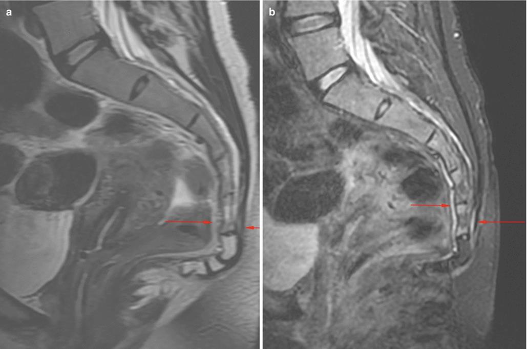
Fig. 24.4
Coccygeal instability with anteverted coccyx in a 29-year-old woman with coccydynia 8 weeks after childbirth. Sagittal FSE T2-weighted (a) and STIR (b) images show hypersignal areas at the level of pre-coccygeal and post-coccygeal spaces indicative of edema (arrows)
24.2.4 Osteitis Condensans Ilii
Osteitis condensans ilii consists in bone sclerosis in the inferior aspect of the iliac bone adjacent to sacroiliac joint [6, 40]. This condition has a high prevalence in women with previous pregnancy but it is not unique of this population [41]. Clinically, osteitis condensans manifests with low back pain and tenderness in the sacroiliac joints. Imaging is required if a differential diagnosis with chronic sacroiliitis is needed (Figs. 24.5, 24.6, and 24.7).

Fig. 24.5
Osteitis condensans ilii in a 38-year-old woman. Patient reported progressive left sacral pain occurring at 8 months of pregnancy. Axial oblique T1-weighted (a) and STIR (b) images obtained during early postpartum period show severe subchondral bone sclerosis (arrows) particularly evident on the iliac side. There is also mild marrow bone edema, visible on the axial oblique STIR (arrows in b). Note the absence of bone erosions and reactive synovitis, finding that allows the differential diagnosis with chronic sacroiliitis

Fig. 24.6
Chronic osteitis condensans ilii in a 35-year-old woman at 23 weeks of gestational age. The findings of hypointense signal intensity (arrows) on axial T1-weighted (a) and STIR (b) images at the level of the iliac side are indicative of subchondral bone sclerosis, confirmed by CT (c), performed after delivery

Fig. 24.7
Osteitis condensans ilii, in a 38-year-old woman. Patient reported progressive left sacral pain occurring at 7 months of pregnancy. Coronal T1-weighted (a) and STIR (b) images obtained during early postpartum period show mild marrow subchondral edema (arrow) of the left sacrum iliac joint. Same patients also suffered from lateral hip rotators overload: axial T1-weighted (c) and STIR (d) images show partial tear of the gluteus medius at the trochanteric insertion (arrows) associated with reactive trochanteric bursitis
CT well depicts the presence of triangular-shaped subchondral sclerosis along the iliac side of the sacroiliac joints bilaterally [42]. MRI demonstrates vertical T1 and T2 hypointense linear foci along the sacral alae representing fracture lines and T1 hypointense and T2 hyperintense signal in the surrounding bone marrow representing associated edema [1, 6].
The clinical differential diagnoses include sacroiliitis with inflammatory or infectious causes, osteitis condensans ilii, lumbosacral degenerative spondylosis, and transient regional osteoporosis of the sacrum [6]. The diagnosis of osteitis condensans ilii is more consistent in the presence of only subchondral sclerosis along the iliac side of the sacroiliac joint and absent joint effusion or erosions in conjunction with the clinical history of recent pregnancy and labor [1]. Treatment is usually conservative including bed rest and analgesics until resolution of the pain [40, 41].
24.3 Bone Mineral Density
Major changes occur in maternal calcium homeostasis during pregnancy and breastfeeding [7]. There are discrepant data with some studies reporting that BMD changes in response to the transfer of calcium to the fetus in combination with decreased renal calcium reabsorption, while others have reported unchanged or even increased BMD due to high levels of estradiol and increased intestinal calcium reabsorption due to 1,25-dihidroxyvitamin D levels which protects against bone loss [7, 43–52].
However, there is a large accordance that pregnancy could be a risk factor for developing a new onset or worsening of a preexisting osteoporosis [43–57]. Bone mineral density decreases gradually during pregnancy and breastfeeding, but the occurrence of true osteoporosis is rare [1]. Pregnancy and breastfeeding cause a reversible bone loss, which is in early phases limited to the trabecular bone, but it involves cortical bone during prolonged breastfeeding [46, 58–61].
A controlled cohort study [7] demonstrated that compared to controls BMD decreased in pregnant women of 1.8 % at the lumbar spine, of 3.2 % at the total hip, of 2.4 % at the whole body, and of 4.2 % at the ultradistal forearm. Breastfeeding has an additional effect on BMD decreased in postpartum period. At 9 months postpartum, women who had breastfed for less than 9 months had a BMD similar to that of controls, while those who were still breastfeeding had a further decrease in BMB at the lumbar spine and hip.
During prolonged breastfeeding, BMD started to be regained at sites of most trabecular bone, but it decreases further at sites rich of cortical bone [7]. Similarly amenorrhea length has an impact on BMD changes, being correlated to breastfeeding length [7]. At 19 months postpartum, BMD returns at prepregnancy level, independently of breastfeeding length. Calcium and vitamin D intake is not associated to BMD changes [7].
Body composition also changes during pregnancy with an increase of fat and lean tissue mass. In postpartum, fat mass tends to have a slower decline in women who continue breastfeeding.
BMD changes in pregnancy and breastfeeding may predispose to osteoporosis and increased risk of fractures.
24.4 Fractures
Pregnancy-related fractures can have different origin. Fractures can be caused by an overload on a normal bone, fatigue or stress fractures, or to a normal load on a weakened osteoporotic bone, insufficiency fractures [1]. The vertebras, sacrum, iliac bone, and femoral head can be affected. MRI detects bone fractures as a line of hypointensity in all sequences lining deeply the articular surface, surrounded by bone edema [1].
Risk factors for fatigue sacral stress related to pregnancy and postpartum include vaginal delivery of a high birth weight infant, increased lumbar lordosis, excessive weight gain, and rapid vaginal delivery [1]. Sacral fatigue fractures can occur also in healthy young woman other than in relation to pregnancy, particularly in long-distance runners or athletes [1]. Imaging features of pregnancy-related fractures are similar to those related to involutional osteoporosis. Radiographs may be normal or demonstrate unilateral or bilateral areas of sclerosis along the sacral wings. Sacral stress fracture lines run parallel to the sacroiliac bone with a typical T1 and T2 hypointense vertical line surrounded by T2 hyperintensity representing edema [6, 62, 63]. Differential diagnosis includes sacroiliitis, with inflammatory or infectious cause, osteitis condensans ilii, lumbosacral degenerative spondylosis, and transient regional osteoporosis of the sacrum.
Femoral head stress fractures are usually located on an area of maximum load, the anterior and superior aspect of the femoral head [62]. Subchondral fractures are seen as curvilinear lines or bands paralleling the subchondral bone and surrounded by edema [1, 5, 62, 63]. This finding distinguishes fractures from osteonecrosis where a serpiginous line of demarcation with double signal intensity extends to the subchondral bone on both sides and delineates the zone of infarction [1, 5, 62, 63].
24.5 Hip Disorders
Despite a reported 38 % of hip pain during pregnancy, true hip disease is very rare during pregnancy and early postpartum (with reported data of 3/4900) [5]. However, they cause a significant clinical impairment and must be considered by the clinician. Hip disorders during pregnancy usually involve the femoral head, and the most frequent are transient osteoporosis and stress fractures, while osteonecrosis occurs less frequently [5, 64].
Transient osteoporosis is related to pregnancy, which is a predisposing factor for this condition. It is associated to the third trimester of pregnancy [5, 65–67]. Clinical examination usually doesn’t show a limitation of movements while the pain is so intense to prevent gait [67]. MRI shows bone marrow edema in the femoral head and neck (Fig. 24.8), joint effusion, and capsular thickening (66–74), while x-rays can show regional osteoporosis [67] and osteopenia at DEXA examination [5]. Transient osteoporosis can complicate with secondary spontaneous fractures, subchondral fractures, seen as a line of hypointensity lining deeply of the subchondral bone. Pathogenesis of transient osteoporosis is related to altered calcium homeostasis during pregnancy even if other less convincing hypotheses have been advocated, such as algodystrophy, impaired venous flow in the femoral head, or nerve compression [5, 66–74].

Fig. 24.8
Bilateral transient osteoporosis of the hip in a 35-year-old woman; pain initially occurred on the right lip, after 6 months of pregnancy, followed by contralateral side 1 month later. Coronal T1-weighted (a) and STIR (b) and axial T1-weighted (c) and STIR (d) images obtained during early postpartum period show bilateral bone marrow edema (arrows) of femoral heads and joint effusions. Coronal STIR image (e) after 5 months of treatment detects edema resolution
In stress fractures symptoms occur very late, at 32nd–41st gestational week with severe pain during walking relieved by rest, and resolved spontaneously 2–5 months after onset [1]. This condition has been reported to be the second cause of pregnancy-related hip disease [5]. An explanation could be bone fragility due to transient osteoporosis. They could be secondary fracture, such as complication of osteoporosis, or true stress fractures due to preexisting bone fragility associated to overload or a combination of both [5]. The hypothesis of true stress fracture is supported by the fact that they appear early during pregnancy and visible at MRI if performed soon, as conventional x-ray examinations are usually negative [5, 62]. In later phases x-rays, to be better performed after delivery, can show a subchondral linear radiolucency representing a fracture line or a focal area of sclerosis and sequently a subtle focal depression on femoral head contour [62]. MRI shows fracture as a thin hypointense line in all sequences (Fig. 24.9) occurring in the femoral head and the upper part of the neck or both or a linear band parallel to the subchondral bone [62, 63]. Other findings are marked bone marrow edema, contour abnormalities of the femoral head, and joint effusion, despite lower compared to transient osteoporosis [5, 62]. The finding of a linear hypointense band surrounded by edema is quite specific for subchondral fractures, and it distinguishes them from the well-defined zonal pattern of osteonecrosis [62, 63]. However, in case of advance femoral head collapse, this finding may not be evident so a distinction between the two conditions is more challenging [62].

Fig. 24.9
Stress fracture of the right femoral neck in a 40-year-old woman at 22 weeks of gestational age. Coronal T1-weitghted (a) and STIR (b) sequences show hypointense signal of the fracture line (arrow), surrounded by edema. Coronal reformatted CT (c) performed after delivery shows a thin hyperattenuating line
MRI is the modality of choice respect to conventional x-rays, owing to the lack of ionizing radiation and because it can show signs of very early stage (bone edema and joint effusion).
Transient osteoporosis and stress fractures are reversible and self-limiting conditions, with a clinical and radiographic resolution after conservative treatment, and they need to be distinguished from osteonecrosis of the femoral head [1].
Osteonecrosis (ON) of the femoral head occurs rarely. Pregnancy-related ON represents about 2 % of all cases [5, 64, 75–78]. Imaging features do not differ from idiopathic ON; however, a predilection for unilateral involvement, especially the left one, has been reported; moreover women with pregnancy-related ON were also more likely to undergo their first pregnancy at an older age (5). The cause of pregnancy-related ON is not clear; some mechanisms have been postulated such as pelvic venous congestion, lipid disturbance, higher serum cortisol, and state of hypercoagulability [5].
MRI is highly sensitive for the diagnosis of ON of the femoral head [79–84] (Fig. 24.10). A normal fat intensity area with a surrounding low-intensity band on T1-w sequences and a double line sign on T2-w, which represent the boundary of the necrotic lesion, are frequently seen in pre-radiological (stage 1) and radiologically non-collapsed stage (stage 2) of ON, according to ARCO (Association Research Circulation Osseous) classification. In stage 3 (radiological collapsed) and stage 4 diseases, necrotic tissue and reparative tissue coexist with homogeneous and inhomogeneous hypointense areas on T1, although T2 can sometimes differentiate between necrotic tissue (fat like or low intensity) and reactional tissue (intermediate intensity). Hypointense bands should represent necrosis although necrotic cells can still be hyperintense on T2 until the area of necrosis becomes more extensive [63]. A peripheral rim of enhancement after i.v. injection of gadolinium at the boundary of the altered signal intensity area is consistent with reparative fibrous tissue and repaired marrow; the peripheral rim of enhancement corresponds to necrotic trabecular bone [63]. However, the use of gadolinium should be reserved only in case of doubt as potential adverse effects on the fetus are not known. Extralesional reactive changes consist of dilated vessels and bone marrow edema [63]. The presence of edema represents an inflammatory change in the reparative process both in case of collapsed head and in non-collapsed head; in this latter change, it may indicate that the femoral head has begun to collapse microscopically [63].
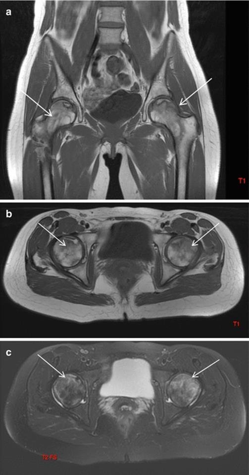
Fig. 24.10
Bilateral avascular necrosis of femoral head in a 25-year-old woman who accused severe hip pain during late pregnancy, treated with conservative treatment. Coronal and axial T1-weighted (a, b) and axial STIR (c) images performed after delivery show areas of avascular necrosis in the head of femurs, with typical map-like morphology (arrows)
24.6 Neural Disorders
24.6.1 Carpal Tunnel Syndrome
Carpal tunnel syndrome is the most frequent compression syndrome during pregnancy, especially in the third trimester, and it is often bilateral [9].
Pregnant women with CTS are more likely to be nulliparous, over 30 years of age, in the last trimester of pregnancy and have pregnancies complicated by hand or generalized edema [9].
This condition is related to fluid retention associated to hormonal changes with alteration of normal content-to-container ratio in the carpal tunnel and compression of the tendon sheaths on the median nerve [9–88].
MRI and ultrasound have been used to investigate carpal tunnel syndrome. MRI with diffusion tensor imaging and tractography can add useful information to grade the severity of nerve involvement [89]. However, imaging is usually not performed as symptoms generally resolve after delivery and do not require surgical intervention [9].
24.6.2 Meralgia Paresthetica and Other Neuropathies
Stretching and compression of superficial nerves passing in the abdominal wall due to mass effect of pregnant uterus can cause a pain syndrome called meralgia paresthetica [8, 90]. It is a pure sensitive neuropathy caused by compression of the lateral cutaneous nerve under or within the inguinal ligament [90]. Increased abdominal girth and lumbar lordosis are predisposing factors [91, 92]. Abnormalities of the lateral cutaneous nerve of the thigh can be seen on ultrasound [93]. Other nerves involved are lower intercostal nerve and iliohypogastric nerve [94, 95]. Treatment is only supportive as spontaneous recovery after delivery is the norm.
Other neuropathies during pregnancy are femoral and obturator neuropathy [92]; proposed mechanisms are compression of the fetal head on the nerves during pregnancy or delivery. Postpartum foot drop is due to injury to lumbar root, plexus, sciatic nerve, or the common peroneal nerve during labor and delivery [96] .
24.7 Conclusion
Pregnancy, delivery, and breastfeeding are associated to peculiar MSK disorders. Even if imaging features of these conditions are shared by those occurring in general population, radiologists should be aware of such disorders, as they require most often imaging than in the past.
References
1.
Proisy M, Rouili A, Raoult H, Rozel C, Guggenbuhl P, Jacob D, Guilin R (2014) Imaging of musculoskeletal disorders related to pregnancy. AJR Am J Roentgenol 202:828–838CrossRefPubMed
2.
Wu WH, Meijer OG, Uegaki K, Mens JMA, van Dieen JH, Wuisman PIJM, Ostgaard NC (2004) Pregnancy-related pelvic girdle pain (PPP), I: terminology, clinical presentation, and prevalence. Eur Spine J 13:575–589PubMedCentralCrossRefPubMed
3.
Bergstrom C, Persson M, Mogren I (2014) Pregnancy-related low back pain and pelvic girdle pain approximately 14 months after pregnancy – pain status, self-rated health and family situation. BMC Pregnancy childbirth 14:48PubMedCentralCrossRefPubMed
4.
Vermani E, Mittal R, Weeks A (2010) Pelvic girdle and low back pain in pregnancy: a review. Pain Pract 10(1):60–71CrossRefPubMed
5.
Steib-Furno S, Mathieu L, Pham T, Armingeat T, Porcu G, Gamerre M, Chagnaud C, Lafforgue P (2007) Pregnancy-related hip diseases: incidence and diagnosis. Joint Bone Spine 74:373–378CrossRefPubMed
6.
Beltran LS, Bencardino JT (2011) Lower back pain after recently living birth: postpartum sacral stress fractures. Skeletal Radiol 40:481–482CrossRef
7.
Moller UK, Vio Streym S, Mosekilde L, Rejnmark L (2012) Changes in bone mineral density and body composition during pregnancy and postpartum. A controller cohort study. Osteoporos Int 23:1213–1223CrossRefPubMed
8.
William Sax T, Rosenbaum RB (2006) Neuromuscular disorders in pregnancy. Muscle Nerve 34:559–571CrossRef
9.
Padua L, Di Pasquale A, Pazzaglia C, Liotta GA, Librante A, Mondelli M (2010) Systematic review of pregnancy-related carpal tunnel syndrome. Muscle Nerve 42:697–702CrossRefPubMed
10.
Waynberger S, Potin J, Chevillot M, Perrotin F (2005) Physiologie de l’appareil locomoteur au cours de la grossesse. Rev Rhum Ed Fr 72:681–685CrossRef
11.
National Institute for Clinica Excellence. Cesarean section: clinical guideline 13 (2004) Avalable at: http://www.nice.org/CG013NICEguideline Accessed 20 Apr 2009
12.
Deneux-Tharaux C, Carmona E, Bouvier-Colle MH, Beart G (2006) Postpartum maternal mortality and cesarean delivery. Obstet Gynecol 108(Pt 1):541–548CrossRefPubMed
13.
Gutke A, Ostgaard HC, Oberg B (2006) Pelvic girdle pain and lumbar pain in pregnancy: a cohort study of the consequence in terms of health and functioning. Spine 31:E149–E155CrossRefPubMed
14.
Sihvonen T, Huttunen M, Makkonen M, Airaksinen O (1998) Functional changes in back muscle activity correlate with pain intensity and prediction of low back pain during pregnancy. Arch Phys Med Rehabil 79:1,210–1,212CrossRef
15.
Vleeming A, Albert HB, Ostgaard HC, Sturesson B, Stuge B (2008) European guidelines for the diagnosis and treatment of pelvic girdle pain. Eur Spine J 17:794–819PubMedCentralCrossRefPubMed
16.
MacLennan AH, Nicolson R, Green RC, Bath M (1986) Serum relaxin and pelvic pain of pregnancy. Lancet 2:243–245CrossRefPubMed
17.
Bjorklund K, Naessen T, Nordstrom ML, Bergstrom S (1999) Pregnancy related back and pelvic pain and changes in bone density. Acta Obstet Gynecol Scand 78:681–685CrossRefPubMed
18.
Rungee JL (1993) Low back pain during pregnancy. Orthopedics 16:1339–1344PubMed
19.
Ostgaard HC, Zetherstrom G, Roos-Hansson E, Svanberg B (1994) Reduction of back and posterior pelvic pain during pregnancy. Spine 19:894–900CrossRefPubMed
20.
Hansen A, Jensen DV, Wormslev M et al (1999) Symptom-giving pelvic girdle relaxation in pregnancy II. Symptoms and clinical signs. Acta Obstet Gynecol Scand 78:111–115CrossRefPubMed
21.
Wang PI, Chong ST, Kielar AZ, Kelly AM, Knoepp UD, Mazza MB, Goodsitt MM (2012) Imaging of pregnant and lactating patients: part 1, evidence-based review and recommendations. AJR Am J Roentgenol 198:778–784CrossRefPubMed
22.
Wang PI, Chong ST, Kielar AZ, Kelly AM, Knoepp UD, Mazza MB, Goodsitt MM (2012) Imaging of pregnant and lactating patients: part 1, evidence-based review and recommendations. AJR Am J Roentgenol 198:785–792CrossRefPubMed
23.
Gamble K, Dardarian TS, Finstein J, Fox E, Sehdev H, Randall TC (2006) Osteomyelitis of the pubic symphysis in pregnancy. Obstet Gynecol 107(2 Pt 2):477–481CrossRefPubMed
24.
Almoujahed MO, Khatib R, Baran J (2003) Pregnancy associated pyogenic sacroiliitis: case report and review. Infect Dis Obstet Gynecol 11:53–57PubMedCentralCrossRefPubMed
25.
Ducrotoy V, Fournet P, Vittecoq O, Daragon A (1998) Postpartum septic arthritis: two case reports. J Gynecol Obstet Biol Reprod (Paris) 27:449–454
26.
Dunk RA, Langhoff-Roos J (2010) Osteomyelitis of the pubic symphysis after spontaneous vaginal delivery. BMJ Case Rep 2010:ii
27.
Weinreb JC, Wolbarsht LB, Cohen JM, Brown CE, MAravilla KR (1998) Prevalence of lumbosacral intervertebral disk abnormalities on MR images in pregnant and asymptomatic nonpregnant women. Radiology 170:125–128CrossRef
28.
Ostgaard HC, Andersson GB, Karlsson K (1991) Prevalence of back pain in pregnancy. Spine 16:549–552CrossRefPubMed
29.
Lile J, Perkins J, Hammer RL, Loubert PV (2003) Diagnostic and management strategies for pregnant women with back pain. JAAPA 16:31–44PubMed
30.
Wong C, Thng PLK, Thoo F, Low C (2003) Symptomatic spinal epidural varices presenting with nerve impingement. Spine 28:E347–E350CrossRefPubMed
31.
Genevay S, Palazzo E, Huten D, Fossati P, Meyer O (2002) Lumboradiculopathy due to epidural varices: two cases reports and a review of the literature. Join Bone Spine 69:214–217CrossRef
32.
Bastiaanssen JM, de Bie RA, Bastiaenen CH, Essed CG, van den Brandt PA (2005) A historical perspective on pregnancy related low back and/or pelvic girdle pain. Eur J Obstet Gynecol Reprod Biol 120:3–14CrossRefPubMed
33.
Paksoy Y, Gormus N (2004) Epidural venous plexus enlargement presenting with radiculopathy and back pain in patients with inferior vena cava obstruction and occlusion. Spine 29:2419–2424CrossRefPubMed
34.
Roost CC, Jaqueline J, Kaiser A, Verhagen AP, Koes BW (2004) Pelvic pain during pregnancy: a descriptive study of signs and symptoms of 870 patients in primary care. Spine 29:2567–2572CrossRef
35.
Kurzel RB, Au AH, Rooholamini SA, Smith W (1996) Magnetic resonance imaging of peripartum rupture of the symphysis pubis. Obstet Gynecol 87:826–829PubMed
36.
Bjorklund K, Nordstrom ML, Bergstrom S (1999) Sonographic assessment of symphyseal joint distension during pregnancy and post partum with special reference to pelvic pain. Acta Obstet Gynecol Scand 78:125–130CrossRefPubMed
37.
Maigne JY, Rusakiewicz F, Diouf M (2012) Postpartum coccydynia: a case series study of 57 women. Eur J Phys Rehabil Med 48:387–392PubMed
38.
Maigne JY, Doursounian L, Chatellier G (2000) Causes and mechanisms of common coccydynia: role of body mass index and coccigea trauma. Spine 25:3072–3079CrossRefPubMed
39.
Maigne JY, Pigeau I, Roger B (2012) Magnetic resonance imaging findings in the painful adult coccyx. Eur Spine J 21:2097–2104PubMedCentralCrossRefPubMed
40.
Jenks K, Meikle G, Gray A, Stebbings S (2009) Osteitis condensans ilii: a significant association with sacroiliac joint tenderness in women. Int J Rheum Dis 12:39–43CrossRefPubMed
41.
Mitra R (2010) Osteitis condensans ilii. Rheumatol Int 30:293–296CrossRefPubMed
42.
Gemmel F, de Coningh A, Collins J, Rijk P (2011) SPECT/CT of osteitis condensans ilii: one-stop shop imaging. Clin Nucl Med 36(1):59–61CrossRefPubMed
43.
Black AJ, Topping J, Durham B, Farquharson RG, Fraser WD (2000) A detailed assessment of alterations in bone turnover, calcium homeostasis, and bone density in normal pregnancy. J Bone Miner Res 15:557–563CrossRefPubMed
44.
Cross NA, Hillman LS, Allen SH, Krause GF, Vieira NE (1995) Calcium homeostasis and bone metabolism during pregnancy, lactation, and postweaning: a longitudinal study. Am J Clin Nutr 61:514–523PubMed
45.
Ritchie LD, Fung EB, Halloran BP, Turnlund JR, Van Loan MD, Cann CE, King JC (1998) A longitudinal study of calcium homeostasis during human pregnancy and lactation and after resumption of menses. Am J Clin Nutr 67:693–701PubMed
46.
Gallacher SJ, Fraser WD, Owens OJ, Dryburgh FJ, Logue FC, Jenkins A, Kennedy J, Boyle IT (1994) Changes in calciotrophic hormones and biochemical markers of bone turnover in normal human pregnancy. Eur J Endocrinol 131:369–374CrossRefPubMed
47.
Kent GN, Price RI, Gutteridge DH, Allen JR, Rosman KJ, Smith M, Bhagat CI, Wilson SG, Retallack RW (1993) Effect of pregnancy and lactation on maternal bone mass and calcium metabolism. Osteoporos Int 3(Suppl 1):44–47CrossRefPubMed
48.
Gertner JM, Coustan DR, Kliger AS, Mallette LE, Ravin N, Broadus AE (1986) Pregnancy as state of physiologic absorptive hypercalciuria. Am J Med 81:451–456CrossRefPubMed
49.
Lopez JM, Gonzalez G, Reyes V, Campino C, Diaz S (1996) Bone turnover and density in healthy women during breastfeeding and after weaning. Osteoporos Int 6:153–159CrossRefPubMed
50.
Drinkwater BL, Chesnut CH III (1991) Bone density changes during pregnancy and lactation in active women: a longitudinal study. Bone Miner 14:153–160CrossRefPubMed
51.
Karlsson C, Obrant KJ, Karlsson M (2001) Pregnancy and lactation confer reversible bone loss in humans. Osteoporos Int 12:828–834CrossRefPubMed
52.
Laskey MA, Prentice A (1999) Bone mineral changes during and after lactation. Obstet Gynecol 94:608–615CrossRefPubMed
53.
Ulrich U, Miller PB, Eyre DR, Chesnut CH III, Schlebusch H, Soules MR (2003) Bone remodeling and bone mineral density during pregnancy. Arch Gynecol Obstet 268:309–316CrossRefPubMed
54.
Pearson D, Kaur M, San P, Lawson N, Baker P, Hosking D (2004) Recovery of pregnancy mediated bone loss during lactation. Bone 34:570–578CrossRefPubMed
55.
Naylor KE, Iqbal P, Fledelius C, Fraser RB, Eastell R (2000) The effect of pregnancy on bone density and bone turnover. J Bone Miner Res 15:129–137CrossRefPubMed
56.
Fiore CE, Pennisi P, DiStefano A, Riccobene S, Caschetto S (2003) Pregnancy-associated changes in bone density and bone turnover in the physiological state: prospective data on sixteen women. Horm Metab Res 35:313–318CrossRefPubMed
57.
More C, Bettembuk P, Bhattoa HP, Balogh A (2001) The effects of pregnancy and lactation on bone mineral density. Osteoporos Int 12:732–737CrossRefPubMed
58.
Kolthoff N, Eiken P, Kristensen B, Nielsen SP (1998) Bone mineral changes during pregnancy and lactation: a longitudinal cohort study. Clin Sci (Lond) 94:405–412CrossRef
59.
Affinito P, Tommaselli GA, di Carlo C, Guida F, Nappi C (1996) Changes in bone mineral density and calcium metabolism in breastfeeding women: a one year follow-up study. J Clin Endocrinol Metab 81:2314–2318PubMed
60.
Krebs NF, Reidinger CJ, Robertson AD, Brenner M (1997) Bone mineral density changes during lactation: maternal, dietary, and biochemical correlates. Am J Clin Nutr 65:1738–1746PubMed
61.
Hopkinson JM, Butte NF, Ellis K, Smith EO (2000) Lactation delays postpartum bone mineral accretion and temporarily alters its regional distribution in women. J Nutr 130:777–783PubMed
62.
Davies M, Cassar-Pullicino VN, Darby AJ (2004) Subchondral insufficiency fractures of the femoral head. Eur Radiol 14:201–207CrossRefPubMed
63.
Sakai T, Sugano N, Nishii T, Haraguchi K, Ochi T, Ohzono K (2000) MR findings of necrotic lesions and the extralesional area of osteonecrosis of the femoral head. Skeletal Radiol 29:133–141CrossRefPubMed
64.
Montella BJ, Nunley JA, Urbaniak JR (1999) Osteonecrosis of the femoral head associated with pregnancy: a preliminary report. J Bone Joint Surg Am 81:790–798CrossRefPubMed
65.
Lafforgue P, Furno-Steib S (2005) Coxopathies induites par la grossesse. Rev Rhum Ed Fr 72:733–738CrossRef
66.
Arayssi TK, Tawbi HA, Usta IM, Hourani MH (2003) Calcitonin in the treatment of transient osteoporosis of the hip. Semin Arthritis Rheum 32:388–39CrossRefPubMed
67.
Brodell JD, Burns JE Jr, Heiple KG (1989) Transient osteoporosis of the hip of pregnancy: two cases complicated by pathological fracture. J Bone Joint Surg Am 71:1252–1257PubMed
68.
Holzer G, von Skrbensky G, Holzer LA, Pichl W (2009) Hip fractures and the contribution of cortical versus trabecular bone to femoral neck strength. J Bone Miner Res 24:468–474CrossRefPubMed
69.
Scapinelli C, Candiotto S, Scapinelli A (1997) Reflex decalcifying algodystrophy of the hip in pregnancy (so-called transient osteoporosis). Review of the literature and four cases report. J Obstet Gynaecol 17:134e8
70.
Schapira D (1992) Transient osteoporosis of the hip. Semin Arthiritis Rheum 22:98e105
71.
Hauzeur JP, Hanquinet S, Gevenois PA, Appelboom T (1991) Study of magnetic resonance imaging in transient osteoporosis of the hip. J Rheumatol 18:1211e7
72.
Vande Berg BE, Malghem JJ, Labaisse MA, Noel HM, Maldague BE (1993) MR imaging of avascular necrosis and transient marrow edema of the femoral head. Radiographics 13:501e20CrossRef
73.
Hayes CH, Conway WF, Daniel WW (1993) MR imaging of bone marrow edema pattern: transient osteoporosis, transient bone marrow edema syndrome, or osteonecrosis. Radiographics 13:1001e11CrossRef
74.
Watson RM, Roach NA, Dalinka MK (2004) Avascular necrosis and bone marrow edema syndrome. Radiol Clin North Am 42:207e19CrossRef
75.
Hasegawa Y, Iwase T, Iwasada S, Kitamura S, Iwata H (1999) Osteonecrosis of the femoral head associated with pregnancy. Arch Orthop Trauma Surg 119:112e4
76.
Spencer C, Smith P, Rafla N, Weatherell R (1999) Corticosteroids in pregnancy and osteonecrosis of the femoral head. Obstet Gynecol 94:848CrossRefPubMed
77.
Gribble RK, Emanuel Berres L (2001) Idiopathic osteonecrosis of the hip during pregnancy: outcome in a subsequent gestation. Obstet Gynecol 98:911e3CrossRef
78.
Vandenbussche E, Madhar M, Nich C, Zribi W, Abdallah T, Augereau B (2005) Bilateral osteonecrosis of the femoral head after pregnancy. Arch Orthop Trauma Surg 125:201e3CrossRef
79.
Mitchell DG, Joseph PM, Fallon M et al (1987) Chemical-shift MR imaging of the femoral head: an in vitro study of normal hips and hips with avascular necrosis. AJR Am J Roentgenol 148:1159–1164CrossRefPubMed
80.
Bassett LW, Mirra JM, Cracchiolo A, Gold RH (1987) Ischemic necrosis of the femoral head: correlation of magnetic resonance imaging and histologic sections. Clin Orthop 223:181–187PubMed
81.
Jergesen HE, Lang P, Moseley M, Genant HK (1990) Histologic correlation in magnetic resonance imaging of femoral head osteonecrosis. Clin Orthop 253:150–163PubMed
82.
Lang P, Jergesen HE, Moseley ME, Block JE, Chafetz NI, Genant HK (1988) Avascular necrosis of the femoral head: high-field-strength MR imaging with histologic correlation. Radiology 169:517–524CrossRefPubMed
83.
Takatori Y, Kamogawa M, Kokubo T et al (1987) Magnetic resonance imaging and histopathology in femoral head necrosis. Acta Orthop Scand 58:499–503CrossRefPubMed
84.
Vande Berg B, Malghem J, Labaisse MA, Noel H, Maldague B (1992) Avascular necrosis of the hip: comparison of contrast-enhanced and nonenhanced MR imaging with histologic correlation. Radiology 182:445–450CrossRefPubMed
85.
Wand JS (1990) Carpal tunnel syndrome in pregnancy and lactation. J Hand Surg Br 15:93–95CrossRefPubMed
86.
Voitk AJ, Mueller JC, Farlinger DE, Johnston RU (1983) Carpal tunnel syndrome in pregnancy. Can Med Assoc J 128:277–281PubMedCentralPubMed
87.
Shaafi S, Naimian S, Itomlou H, Sayyah Melli M (2006) Prevalence and severity of carpal tunnel syndrome (CTS) during pregnancy based on electrophysiologic studies. Shiraz E-Med J 3:1–6, Available at: http://semj.sums.ac.ir/vol7/jul2006/cts.htm
88.
Turgut F, Cetinsahinahim M, Turgut M, Bolukbasi O (2001) The management of carpal tunnel syndrome in pregnancy. J Clin Neurosci 8:332–334CrossRefPubMed
89.
Brienza M, Pujia F, Colaiacomo MC, Anastasio MG, Pierelli F, Di Biasi C, Andreoli C, Gualdi G, Valente GO (2014) 3T diffusion tensor imaging and electroneurography of peripheral nerve: a morphofunctional analysis in carpal tunnel syndrome. J Neuroradiol 41(2):124–130CrossRefPubMed
90.
Ritchie JR (2003) Orthopedic considerations during pregnancy. Clin Obstet Gynecol 46:456–466CrossRefPubMed
91.
Tsen LC (2002) Neurologic complications of labor analgesia and anesthesia. Int Anesthesiol Clin 40:67–88CrossRefPubMed
92.
Wong CA, Scavone BM, Dugan S, Smith JC, Prather H, Ganchiff JN et al (2003) Incidence of postpartum lumbosacral spine and lower extremity nerve injuries. Obstet Gynecol 101:279–282CrossRefPubMed
93.
Tagliafico A, Serafini G, Lacelli F, Perrone N, Valsania V, Martinoli C (2011) Ultrasound-guided treatment of meralgia paresthetica (lateral femoral cutaneous neuropathy): technical description and results of treatment in 20 consecutive patients. J Ultrasound Med 30:1341–1346PubMed
94.
Peleg R, Gohar J, Koretz M, Peleg A (1997) Abdominal wall pain in pregnant women caused by thoracic lateral cutaneous nerve entrapment. Eur J Obstet Gynecol Reprod Biol 74:169–171CrossRefPubMed
95.
Carter BL, Racz GB (1994) Iliohypogastric nerve entrapment in pregnancy: diagnosis and treatment. Anesth Analg 79:1193–1194CrossRefPubMed
96.
Babayev M, Bodack MP, Creatura C (1998) Common peroneal neuropathy secondary to squatting during childbirth. Obstet Gynecol 91:830–832CrossRefPubMed