
Bradley S. Hurst1 , Andrea Tinelli2, 3, 4, 5 , Antonio Malvasi6, 7 and William H. Parker8, 9
(1)
Reproductive Endocrinology and Infertility, Carolinas HealthCare System, 1025 Morehead Medical Drive, Suite 500, Charlotte, NC 28204, USA
(2)
Department of Obstetrics and Gynecology, Vito Fazzi Hospital, Lecce, Italy
(3)
Laboratory of Human Physiology, The International Translational Medicine and Biomodelling Research Group, Department of Informatics and Applied Mathematics, Moscow Institute of Physics and Technology (State University), Dolgoprudny, Moscow Region, Russia
(4)
Institute of Physics and Technology (State University), Moscow, Russia
(5)
Division of Experimental Endoscopic Surgery, Imaging, Technology and Minimally Invasive Therapy, Department of Obstetrics & Gynecology, Vito Fazzi Hospital, Lecce, Italy
(6)
Department of Obstetrics and Gynecology, Santa Maria Hospital, G.V.M. Care and Research, Bari, Italy
(7)
International Translational Medicine and Biomodelling Research Group, Department of Applied Mathematics, Moscow Institute of Physics and Technology (State University), Moscow Region, Russia
(8)
Minimally Invasive Gynecologic Surgery, UCLA Medical Center, Santa Monica, CA 90401, USA
(9)
Department of Obstetrics and Gynecology, UCLA School of Medicine, Los Angeles, CA 90401-2831, USA
Bradley S. Hurst (Corresponding author)
Email: bhurst@carolinas.org
Andrea Tinelli
Email: andreatinelli@gmail.com
Antonio Malvasi
Email: antoniomalvasi@gmail.com
7.1 Introduction
Uterine fibroids can be identified by first-trimester ultrasound in approximately 8–20 % of women (Fig. 7.1). Most women with uterine fibroids can expect to have a normal pregnancy and delivery. However, complications from fibroids may occur during pregnancy depending on the size, number, and location of fibroids, and preconception treatment of fibroids may also pose risks to the pregnant woman and her fetus.
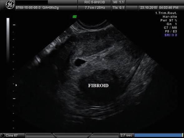
Fig. 7.1
A sagittal section of a pregnant uterus at 6 weeks with a posterior intramural fibroid of 3 cm in diameter
Very rarely does the presence of a fibroid during pregnancy lead to an unfavorable outcome. Submucous fibroids can prevent implantation and cause infertility and can increase the risk of first- and second-trimester miscarriage. During delivery, fibroids may rarely obstruct the normal passageway for delivery (Fig. 7.2a, b) and, when cesarean section is needed, can increase the complexity and risks associated with the procedure (Fig. 7.3). Uterine fibroids can increase postpartum bleeding, sometimes requiring transfusion and occasionally leading to life-threatening bleeding. Finally, in the postpartum interval, uterine fibroids may limit contraceptive options or reduce the efficacy of contraception.
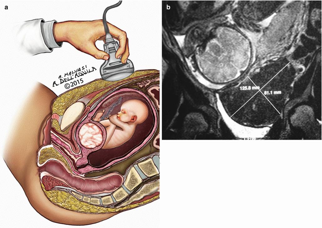
Fig. 7.2
(a) A draft of ultrasonographic scan of a cervical anterior fibroid, at the beginning of pregnancy. (b) MRI image of a larger cervical fibroid obstructing the normal passageway for delivery (Courtesy of Prof. Dr. Josè Palacios de Jaraquemada)

Fig. 7.3
A large anterior fibroid enucleated during cesarean section, after delivery of the newborn (Courtesy of Prof. Dr. Josè Palacios de Jaraquemada)
Treatment of fibroids before conception is also associated with risks to the pregnant patient and her fetus, beyond the risks of surgery, anesthesia, adhesions, and possible decreased fertility and the discomfort and time away from work and family. For women who have undergone myomectomy, there may be an increased risk of dehiscence of the myomectomy incisions during pregnancy or delivery (Fig. 7.4), and hysteroscopic or laparoscopic myomectomy may increase the incidence of placental abnormalities such as placenta accreta. Pregnancy is usually not recommended after uterine artery embolization, magnetic resonance-guided focused ultrasound-directed surgery, or myolysis due to the uterine abnormalities that persist after these procedures, possibly associated with myometrial abnormalities.
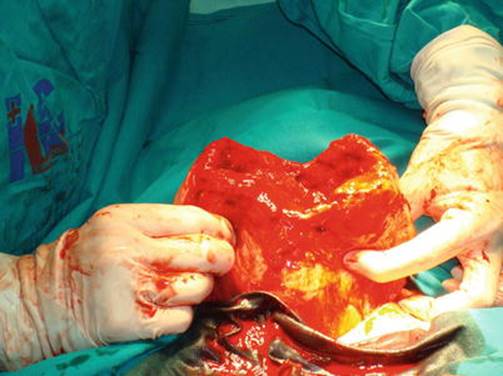
Fig. 7.4
A dehiscence of uterine scar after myomectomy during pregnancy, at 25 weeks (Courtesy of Dr. Radmila Sparic)
In this chapter, we will review the pathophysiology of uterine fibroids and discuss the consequences and management of uterine fibroids during pregnancy.
7.2 Background
7.2.1 Epidemiology and Pathophysiology
Uterine fibroids can be identified in approximately 70–80 % of women (Fig. 7.5) by the time they reach menopause, although most are asymptomatic [1]. Most of these benign tumors develop independently. Growth is stimulated by estradiol, progesterone, and local growth factors and promoted by angiogenesis. Because of stimulation by estrogen and progesterone, the peak prevalence of fibroids occurs during the 40s. Many fibroids have chromosomal abnormalities including translocations, aneuploidy, gene mutations, deletions, inactivation, or overexpression [2]. Vitamin D deficiency is likely to play a role in the development of uterine fibroids, as vitamin D3 reduces fibroid cell proliferation in vitro and fibroid tumor growth in animal models [3]. It has been suggested that vitamin D deficiency may be a factor to explain why women of African descent have a higher incidence and a greater number of uterine fibroids than other ethnic groups. There is also familial tendency to develop these tumors between first-degree relatives and twins [4].

Fig. 7.5
A transvaginal sagittal uterine scanning with multiple fibroids
About 40 % of fibroids grow during pregnancy (Fig. 7.6), and most of the growth takes place during the first trimester (Fig. 7.7). During pregnancy, uterine fibroids are exposed to high levels of estrogen and progesterone. Estrogen produced by the ovary and the placenta, as well as additional growth factors, stimulates the growth of fibroids. Additionally, local conversion of androgens to estrogens by aromatase occurs within the tumors [5]. The primary action of estrogen and the estrogen receptor α (ERα) appears to be mediated by induction of progesterone receptors (PRs), which makes the tumor responsive to progesterone. Progesterone stimulates the growth of the fibroid through genes that regulate both apoptosis and cell proliferation.

Fig. 7.6
An ultrasonographic transvaginal uterine section showing an anterior isthmic fibroid of 3.5 cm in diameter in a pregnant at 7 weeks
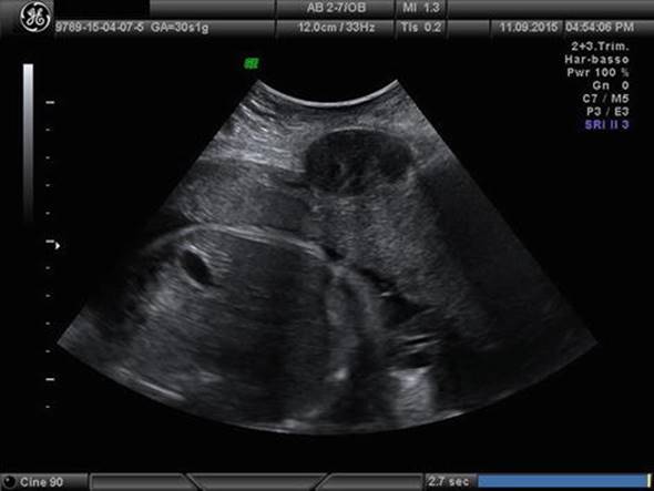
Fig. 7.7
A transabdominal uterine scanning of a patient at 29 weeks with an anterior subserosal fibroid of 4.5 cm in diameter
Since fibroid growth is stimulated by estrogen and progesterone and both of these hormones are elevated during pregnancy, it is reasonable to expect growth of these masses during pregnancy. Although the growth or degeneration of a fibroid is not linear throughout the course of pregnancy, there is remarkable growth during the early pregnancy. This was shown in a prospective case-controlled study of women with fibroids undergoing IVF, in which fibroids were serially measured by ultrasound in 25 women who became pregnant and in 25 who failed to conceive [6]. A significant 34 % increase in the mean diameter of fibroids was found in early pregnancy, compared to a 2 % increase in those who failed to conceive. There was no correlation between ovarian responsiveness and fibroid growth; therefore the changes were attributed solely to pregnancy-associated factors. The observation that fibroids grow in diameter by approximately 30–35 % during the early pregnancy is concerning, as it is possible that an asymptomatic or seemingly “innocent” fibroid near the endometrium could enlarge and lead to unexpected problems during pregnancy. However, it is important to consider that there is no evidence that “prophylactic surgical treatment” is beneficial.
The fibroid is surrounded by a “pseudocapsule” (Fig. 7.8), and recent studies have demonstrated the importance of the pseudocapsule. The pseudocapsule is a fibro-neurovascular structure surrounding a fibroid (Fig. 7.9), separating it from normal peripheral myometrium (Fig. 7.10). The fibroid pseudocapsule is composed of a neurovascular network rich in neurofibers and contains neurotensin, neuropeptide tyrosine, and protein gene product 9.5, as well as substance P and vasoactive intestinal polypeptide [6]. There is a significant increase in endoglin expression level in the pseudocapsule compared to the myometrium or the uterine fibroid, indicating that an active neoangiogenesis is present in pseudocapsule, whereas angiogenic factors including von Willebrand factor (vWF) and vascular endothelial growth factor A (VEGF-A) seem to have little influence on the pseudocapsule angiogenesis. Endoglin is preferentially expressed in proliferating endothelial cells, whereas the vWF and VEGF-A are preferentially expressed in preexisting endothelial cells [7]. Combined, these important findings suggest healing after myomectomy is likely promoted by preservation of the pseudocapsule.

Fig. 7.8
A macroscopic image of a uterine fibroid and its surrounding pseudocapsule in the red ring
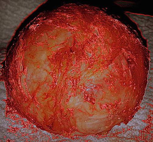
Fig. 7.9
A white fibroid surrounded by its branches of pseudocapsule, enhanced in red

Fig. 7.10
A laparotomic image showing the fibroid pseudocapsule detached from fibroid by surgical scissor
7.2.2 Clinical Presentation in Nonpregnant Women
Most women with uterine fibroids are asymptomatic. In these women, fibroids may be diagnosed during a pelvic examination or when a pelvic ultrasound is performed (Fig. 7.11). When symptomatic, uterine fibroids cause morbidity corresponding to the size, number, and location of the masses (Fig. 7.12). Common symptoms in the nonpregnant woman related to a submucous fibroid include heavy menstrual bleeding or intermenstrual bleeding, pain, pressure, and infertility. Bulk symptoms may be present, based on size and location of the fibroids, typically occurring with large subserosal or intramural fibroids (Fig. 7.13). These tumors may also cause urinary frequency if they compress the bladder, dyspareunia if the mass distorts the posterior cul-de-sac or cervical position, or difficulty with defecation for a posterior fibroid (Fig. 7.14).
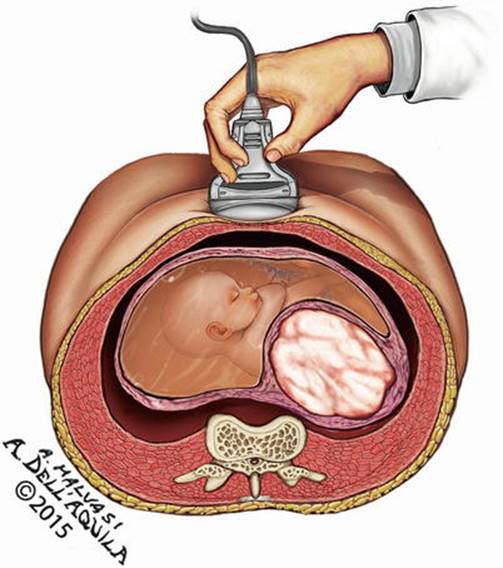
Fig. 7.11
A transabdominal ultrasonographic scanning showing a posterior uterine fibroid

Fig. 7.12
A giant uterus with multiple fibroids causing compressive symptoms and massive bleeding during menses

Fig. 7.13
A large subserosal/intramural fibroid, whose upper limit reaches the liver
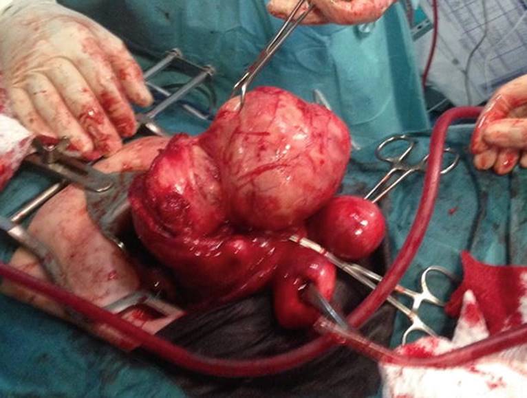
Fig. 7.14
A uterus with visceral anatomy subverted by multiple fibroids of different diameters (from 4 to 18 cm), aspect (intramural, subserosal, and pedunculated), and location (anterior, posterior, fundal) contemporary causing urinary frequency, dyspareunia, and difficulty with defecation for a posterior fibroid
While the most common reasons women seek treatment for uterine fibroids are bleeding and pressure, fertility and obstetric issues play a role as well. There is a clear cause and effect for submucosal tumors affecting fertility (Fig. 7.15). Pregnancy outcomes improve after removal of submucous fibroids. There is a general consensus that subserosal fibroids do not cause infertility (Fig. 7.16). There is more controversy regarding intramural fibroids on fertility (Fig. 7.17). However, in some women with otherwise unexplained infertility, myomectomy may improve pregnancy outcomes [8].

Fig. 7.15
A left submucous fibroid of 3 cm in diameter largely occupying uterine cavity
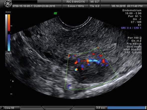
Fig. 7.16
An ultrasonographic sagittal scan of a retroverted uterus of a patient wishing pregnancy with a posterior subserosal fibroid

Fig. 7.17
A 30-year-old patient with an intramural fibroid in the posterior uterine body of 7 cm in diameter
7.2.3 Treatment of Fibroids in Nonpregnant Women
Treatment of fibroids in a nonpregnant woman is important since all treatments can have important implications during pregnancy and in the peripartum interval. Management options include medical therapy; primarily gonadotropin-releasing hormone agonists (GnRHas) and progesterone antagonists; surgery including hysteroscopic myomectomy (Fig. 7.18), laparoscopic myomectomy (Fig. 7.19), or abdominal myomectomy (Fig. 7.20); radiologic interventions including uterine artery embolization (UAE) and magnetic resonance-guided focused ultrasound surgery (MRgFUS); and myolysis procedures.

Fig. 7.18
A hysteroscopic myomectomy of anterior G2 fibroid
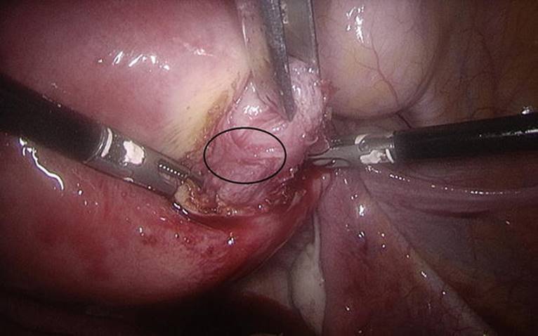
Fig. 7.19
A laparoscopic myomectomy; in the black ring the myoma pseudocapsule is highlighted
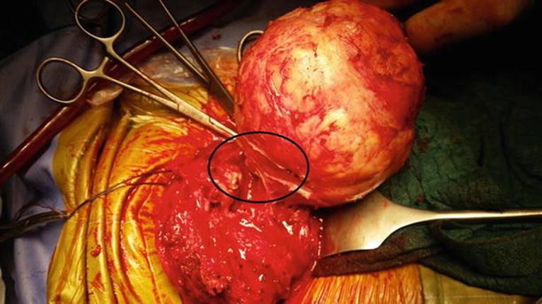
Fig. 7.20
A laparotomic myomectomy; in the black ring the myoma pseudocapsule is highlighted
Medical treatments such as combined oral contraceptive pills or continuous progestin pills have limited evidence for efficacy, especially in women with distortion of the uterine cavity from fibroids, and are primarily considered as temporizing measures [9]. GnRHa may be used to decrease menorrhagia, especially in preparation for surgery, to allow for recovery of anemia, thin the endometrial lining, and facilitate hysteroscopic resection of a submucous fibroid. GnRHa reduces the diameter and volume of fibroids while the patient is hypoestrogenic, but rapid regrowth of fibroids occurs when the medication is discontinued. GnRHa should generally be avoided prior to a myomectomy as it can make the procedure more difficult, and administration before myomectomy increases the likelihood of persistent or recurrent fibroids [8]. Cost and side effects of these medications limit their long-term use and many women go on to other forms of treatment.
Ulipristal acetate (Fig. 7.21) is a selective progesterone receptor modulator that is approved for treatment of symptomatic uterine fibroids in Europe and Canada. Approximately 62 and 73 % of women become amenorrheic during treatment, and bleeding is controlled in over 80 % of women with abnormal bleeding due to fibroids [10]. Menstruation resumes after each treatment course and is diminished compared with the baseline. When a second treatment course is administered, fibroid volume is reduced approximately 55 % compared to the baseline, and pain and quality-of-life measures improve. Since repeated courses may reduce the need for surgery for some women with fibroids, conception without further intervention may occur.

Fig. 7.21
Ulipristal acetate, a selective progesterone receptor modulator
Pregnancy outcome data is limited after treatment with ulipristal. One study reported that 15 of 21 women who attempted to conceive were successful after participating in one of the ulipristal clinical trials [11]. Among the 18 pregnancies in these women, 12 resulted in births of healthy babies and six ended in early miscarriage. No further growth of fibroids was found during pregnancy. While this early observation is encouraging, it is too soon to determine if this medication will be a good option for women with symptomatic fibroids who wish to avoid surgery.
7.2.4 Surgery
Surgical management of uterine fibroids is appropriate in the following situations (1): abnormal uterine bleeding not responding to conservative treatments (2), high level of suspicion of pelvic malignancy (3), growth after menopause (4), infertility when there is distortion of the endometrial cavity or tubal obstruction (5), recurrent pregnancy loss (with distortion of the endometrial cavity) (6), pain or pressure symptoms (that interfere with quality of life) (7), urinary tract symptoms (frequency and/or obstruction), and (8) iron-deficiency anemia secondary to chronic blood loss [12].
Myomectomy refers to the excision of one or more uterine fibroids. It is considered the best option for young women with symptomatic fibroids who desire preservation of fertility. The benefit of myomectomy in infertile women is difficult to assess since the incidence of fibroids increases with age as does the incidence of infertility. The route of myomectomy, including hysteroscopy, laparoscopy, or open myomectomy, is chosen based on patient symptoms and the size, number, and location of the fibroids.
7.2.5 Hysteroscopic Myomectomy
Transcervical hysteroscopic myomectomy (Fig. 7.22) is appropriate for women with a symptomatic submucous fibroid and a desire for childbearing or uterine preservation. Following hysteroscopic myomectomy, the risk of early miscarriage is significantly reduced, and there is a corresponding increase in viable term deliveries [13]. When needed, hysteroscopic myomectomy can be performed concurrently with abdominal or laparoscopic myomectomy to reduce bulk symptoms. During hysteroscopy, the uterine cavity is filled with distention media, and the fibroid is resected with scissors, with monopolar or bipolar cautery, or with a hysteroscopic morcellator [14]. Typically hysteroscopic resection of fibroids is appropriate for tumors 6 cm or less, while larger masses may require a two-step surgery [15]. The surgical technique used during hysteroscopic myomectomy may be important for women as it relates to rates of conception, spontaneous abortion, and pregnancy outcomes. Some methods may be more likely than others to damage the endometrium, some procedures and techniques may allow for a greater percentage of complete removal of the submucous fibroid, and some approaches could damage the myometrium. Data to recommend one procedure over another, however, is lacking at present.

Fig. 7.22
Transcervical posterior hysteroscopic myomectomy, by a bipolar resector
Pregnancy outcomes are favorable after hysteroscopic myomectomy. In one study, 86 % of women conceived after hysteroscopic myomectomy, and there was no difference in outcomes for fibroids that were completely, mostly, or partially intracavitary [16]. Pregnancy and birth rates were not related to the number, localization, or diameter of the fibroids. Miscarriage was more likely to occur when the fibroid was resected in the anterior uterine wall, and preterm delivery was more common when a fundal fibroid was removed. It is reasonable to monitor women carefully for miscarriage and preterm labor after undergoing hysteroscopic myomectomy.
7.2.6 Abdominal Myomectomy
In 1845, Washington Atlee reported his experience performing a successful abdominal myomectomy in the American Journal of the Medical Sciences [17]. Mortality associated with this procedure was high until Victor Bonney mastered the techniques of myomectomy, inspired by his nulligravida wife’s emotionally devastating hysterectomy for a submucous fibroid in 1908. Bonney introduced many surgical techniques still used today, including compression of the uterine arteries with a clamp, now referred to as a “Bonney clamp,” to reduce bleeding and elevate the uterus to improve exposure during myomectomy. He recognized the problem caused by dead space from the myoma bed and described a technique to obliterate the cavity by under-sewing the deeper layers. Bonney eventually performed over 700 myomectomies, and his mortality rate was only 1.1 %, remarkable in an era before blood transfusion and antibiotics were available. By the 1930s, Bonney advocated abdominal myomectomy for any woman with fibroids wishing to have children under the age of 41.
Recently, Tinelli and colleagues have emphasized the importance of surgical technique to preserve the fibroid pseudocapsule during myomectomy (Fig. 7.23a, b), to reduce intraoperative bleeding, promote postoperative healing, and preserve function of the uterus [18]. Since a “classical” myomectomy technique was not used for controls, the findings should be interpreted with caution. However, myomectomy with preservation of the pseudocapsule resulted in a reduction of the uterine healing area from 78 % of the previous fibroid size on the first day to <4 % on the 45th day after surgery [19].

Fig. 7.23
Two laparotomic myomectomies (a, b) by intracapsular technique showing the surgical scissors cutting the pseudocapsule branches attaching myoma to the pseudocapsule
Long-term outcomes following abdominal myomectomy are typically good and patient satisfaction is high, but adhesions and recurrent fibroids may compromise the results in some individuals. Myomectomy often reduces bleeding and improves fertility in women with excessive bleeding or infertility primarily caused by endometrial distortion from submucous myomas or large intramural myomas. Slightly more than 50 % of women conceive after open myomectomy [20]. Adhesions form in more than 90 % of abdominal myomectomies, with the incidence highest (94 %) with posterior incisions and lower (56 %) with fundal or anterior uterine incisions [21]. When severe, adhesions can result in bowel obstruction and require additional intervention. Adhesions can also increase the complexity of cesarean delivery. For this reason, adhesion barriers are recommended to minimize the extent of adhesions. Seprafilm and Interceed (Fig. 7.24) both have been shown to reduce the extent and severity of post-myomectomy adhesions [22, 23]. Growth of new fibroids is not uncommon after myomectomy, but additional surgery is required in a minority of patients.

Fig. 7.24
An Interceed adhesion barrier applied on the uterine sutured surface after cesarean section, to minimize the extent of adhesions (Courtesy of Prof. Dr. Josè Palacios de Jaraquemada)
7.2.7 Laparoscopic Myomectomy
Laparoscopic myomectomy was first described in the 1970s. Advances in instruments and surgical knowledge have progressed so that laparoscopic myomectomy is considered preferable to abdominal myomectomy when feasible. Laparoscopic myomectomy provides some obvious advantages compared to abdominal myomectomy. Decreased postoperative pain, shorter recovery time, reduced febrile morbidity, decreased blood loss, and decreased adhesion formation are the clear advantages seen in the minimally invasive approach. The risk of recurrence of fibroids and pregnancy outcomes are comparable after resection of fibroids from abdominal versus laparoscopic route [8]. In a study looking at intracapsular subserous and intramural myomectomy preserving the fibroid pseudocapsule (Fig. 7.25a, b), women who underwent the procedure for infertility (74 %) eventually conceived [24]. To optimize laparoscopic myomectomy, it is important to properly select patients, place trocars to optimize visualization and prevent instrument collision, use the principles of traction and countertraction to the best advantage, and utilize appropriate instruments and suture material to better facilitate the case. The fibroid is enucleated from the surrounding pseudocapsule (Fig. 7.26) and the uterus repaired in single layer for subserosal fibroids (Fig. 7.27) and double layer for those extending into the myometrium (Fig. 7.28). Appropriate candidates for a laparoscopic myomectomy traditionally have less than three fibroids and size less than 8–10 cm. However, depending on individual surgical expertise, fibroids larger than this can be attempted, though typically with longer operating time and more anesthetic complications [25].

Fig. 7.25
Two laparoscopic myomectomies (a, b) by intracapsular technique showing, in the black (a) and white (b) ring, the pseudocapsule branches attaching myoma to the pseudocapsule

Fig. 7.26
The final result of laparoscopic intracapsular myomectomy, with intramural fibroid enucleated by uterus-sparing myoma pseudocapsule, with few bleeding from hysterotomy, before suturing

Fig. 7.27
A laparoscopic hysterorrhaphy by a single layer suturing after a subserosal fibroid enucleating
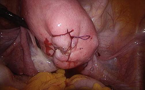
Fig. 7.28
The final result of a laparoscopic hysterorrhaphy by a double layer suturing after an intramural fibroid enucleating: the suture is always introflexing as a “basketball suture”
While there is a steep learning curve associated with the advanced laparoscopic techniques such as intracorporeal suturing, robotic-assisted laparoscopy allows for laparoscopic myomectomy in a wider scope of practice. Advantages include improved dexterity and three-dimensional view. While operative times tend to be longer in robotic-assisted cases than traditional laparoscopic, robotic-assisted laparoscopy has a shorter learning curve and does not add morbidity to the procedure. Pitfalls include loss of tactile sensation during surgery and increased cost [26]. Advanced laparoscopic technique with the rise of single-port laparoscopic surgery to myomectomy presents a steep learning curve but has proposed benefits such as improved cosmetic outcome [27].
Surgical technique for any myomectomy is important in considering subsequent obstetric outcomes. Regardless of technique, it is important to recognize that a prior myomectomy increases the risk of uterine rupture. While this risk is less than 1 % when performed by a skilled surgeon [8], uterine rupture is important as rupture is an obstetrical emergency and can have catastrophic consequences for the mother and fetus (Fig. 7.29) [28]. The risk of uterine rupture is not known when the surgery is performed in most communities. The risk is likely higher when multiple large and deep uterine incisions are required to complete the myomectomy or suboptimal surgical techniques are used or if the myomectomy incision becomes infected postoperatively [29]. Although the risk of uterine dehiscence during pregnancy is low with proper myometrial closure, data are insufficient to determine if cesarean section should be routinely advised for delivery after resecting large, deep, or multiple fibroids (Fig. 7.30a, b).

Fig. 7.29
Uterine rupture is an obstetrical emergency for massive bleeding and painful–hemorrhagic shock, with possible catastrophic consequences for the mother and fetus

Fig. 7.30
Images of urgent longitudinal laparotomy at 23 weeks, in a patient with a previous laparotomic anterior isthmic myomectomy: (a) after the opening of abdominal cavity, the placenta appears completely free floating on the abdominal cavity; (b) after fetus and placental removal, surgeons show the uterine rupture on the previous uterine scar
7.2.8 Endometrial Ablation
Endometrial ablation can be performed after a hysteroscopic myomectomy for women with submucous fibroids who do not desire future fertility. However, ablation of the uterine cavity is not intended as a method of contraception, and some women have conceived, often with serious complications, after undergoing this procedure.
Since the endometrium is severely and irreversibly damaged by any endometrial ablation technique, it is not surprising that serious complications have been reported in women who died of a uterine rupture and massive internal bleeding at 24 weeks of gestation and a woman who required an emergency hysterectomy after a pregnancy termination was complicated by placenta increta [30]. Another report described a nonfatal uterine rupture that occurred at 26 5/7 weeks in a woman who conceived following ablation [31]. Because of the high morbidity associated with pregnancy after endometrial ablation, reliable contraception should be used. When pregnancy occurs, close monitoring for placental abnormalities, premature rupture of membranes, pregnancy distorted by uterine synechiae, or uterine abruption and the need for emergency hysterectomy may occur [32]. Even pregnancy termination should be considered to be a potentially high-risk procedure associated with complications including septic abortion and may necessitate hysterectomy [33]. Any pregnancy after endometrial ablation should be considered to be potentially dangerous, and the patient should be informed that hysterectomy may be needed if complications arise.
Several techniques and methods for endometrial ablation have been described since the procedure was introduced in the 1980s. Initially, ablation was performed using a rollerball, a resectoscope loop, or a contact laser. The technique requires technical expertise with operative hysteroscopy, and potential complications include uterine perforation and fluid overload. In the late 1990s and early twenty-first century, several ablation devices were introduced for endometrial ablation including a balloon system that heats the endometrium, a cryoablation system under ultrasound guidance, and a system that delivers heated saline under hysteroscopic guidance. A microwave mesh system, NovaSure (Novacept, Palo Alto, CA), is performed by placing a triangular electrode in the uterus, and negative pressure pulls the endometrium into contact with the electrode. The NovaSure system is the only FDA-approved device to treat small (<2 cn) polyps, and it can be used to treat a fibroid uterus, as long as there is no distortion of the shape of the cavity. However, none of these devices is intended for endometrial distortion from uterine fibroids, so the utility for ablation with submucous fibroids is limited [34].
7.2.9 Uterine Artery Embolization
If future fertility is desired, myomectomy is preferred over embolization, as pregnancy outcomes are uncertain. Uterine artery embolization (UAE) is primarily performed by interventional radiologists by a catheterization of the femoral artery and bilateral occlusion of uterine arteries with substances such as polyvinyl foam particles. The selective perfusion of these vessels causes infarction and the fibroids decrease in volume by approximately 50 %. UAE is a reasonable alternative to hysterectomy and abdominal myomectomy with favorable short-term outcomes including improved bleeding, pain, and quality of life and similar long-term outcomes [35]. Uterine artery embolization is most appropriate for women with symptomatic fibroids who are past the childbearing years and poor surgical candidates or those who wish to avoid hysterectomy.
In one study, 14 of 23 women who desired pregnancy conceived, resulting in 13 uncomplicated term deliveries and two miscarriages at 12 and 16 weeks [36]. In another study, approximately 28 % of women who tried to conceive were successful after UAE, and only about 14 % had a live birth [37]. In another study, only 1 in 31 women who desired fertility eventually conceived, and she experienced a first-trimester miscarriage [38].
Fibroids may degenerate but do not disappear after UAE; therefore the remaining fibroids may still compromise the gravid uterus. Furthermore, during embolization, the particles used to occlude the vascular flow of the fibroid also may occlude normal vessels of the myometrium and endometrium. Although the uterus receives flow from the cervical and ovarian arteries and blood flow is maintained to the uterus, the particles used for UAE are trapped within the small arterial branches, although they have not been shown to cause long-term effects. Therefore, it is not surprising that UAE results in infertility and more pregnancy complications such as spontaneous abortion, preterm delivery, malpresentation, abnormal placentation, and postpartum hemorrhage compared to myomectomy [39]. Reported pregnancy outcomes in women who have undergone UAE include first-trimester miscarriage [38], uterine rupture, and placenta increta [40]. Although the Society of Interventional Radiology Standards of Practice Committee states UAE may be preferred for women with prior myomectomy and that patient’s preference for UAE as primary therapy for fibroids should be respected [41], currently, we do not recommend UAE for women who desire future fertility, and pregnancies that occur should be considered high risk for uterine or placental abnormalities.
7.2.10 MRgFUS
Magnetic resonance-guided focused ultrasound surgery (MRgFUS) is a noninvasive approach to treat fibroids that utilizes magnetic resonance imaging (MRI) to visualize anatomy and monitor tissue temperature and thermal dose during delivery of focused ultrasound to the target tissue. The ExAblate system (InSightec, Haifa, Israel) was approved by the FDA in 2004 after initial clinical studies showed significant improvement in quality-of-life scores [42]. Fibroid volume is decreased approximately 29 % 6 months after treatment [43]. Reintervention rate after MRgFUS is approximately 13 %, after a mean follow-up of approximately 19 months [44].
Although the procedure is not recommended for women who desire future fertility, by 2010 there were 54 pregnancies reported in 51 women after MRgFUS treatment of fibroids, resulting in a 41 % live birth rate, a 20 % ongoing pregnancy rate beyond 20 weeks, a 28 % spontaneous abortion rate, and an 11 % pregnancy termination rate [45]. Although favorable pregnancy outcomes following MRgFUS have been reported, until more safety data is available, MRgFUS should not be chosen for women who desire future fertility, and pregnancies that occur should be monitored for uterine or placental abnormalities.
Appropriate candidates for MRgFUS include premenopausal women who have completed childbearing and have accessible MR-enhancing symptomatic uterine fibroids (i.e., not shielded by the bowel or bone) less than 24 weeks in size. Ineligible women include those who are pregnant and lactating, have active pelvic inflammatory disease or any active infection, and have chronic leg or lower back pain, severe claustrophobia precluding the use of MRI, weight that exceeds MR capability (approximately 113 kg), implanted materials or devices that are contraindicated for MR, extensive abdominal scarring in the ultrasound beam path that cannot be avoided, dermoid cyst in the beam path, intrauterine device, pedunculated fibroids, known or suspected hyperplasia or malignancy, and undiagnosed uterine bleeding. Overall, less than half of women are eligible for MRgFUS, with the most common exclusions being fibroid size, high cost, and desired fertility [46].
MRgFUS-related complications include skin and sciatic nerve injury, and there is a small risk of bowel and bladder injury. A study prospectively comparing MRgFUS and hysterectomy found fewer complications and faster recovery after MRgFUS but better quality-of-life scores 6 months after hysterectomy [47].
7.2.11 Myolysis
Electrical, thermal, and ultrasound energy sources have been used to coagulate and devascularize symptomatic myomas. Current methods of myolysis have achieved success in relieving symptoms relating to myoma volume, but little is known about its safety for women wishing pregnancy.
Myolysis involves placement of the ablation device using either ultrasound or directly inserted into the uterus during laparoscopy. Many types of ablation devices have been described, including monopolar electrical or radiofrequency devices, thermal devices, or laser probes. Older devices placed during laparoscopy often caused uterine serosal injury and induced dense pelvic adhesions [48]. Bipolar electrode probe myolysis reduced myoma volume by 89 % and had a 97 % patient satisfaction rate at 6 months from surgery [49]. In another case series, the addition of bipolar electrode probe myolysis to endometrial ablation reduced the need for repeat surgery by 66 % and increased the postsurgical amenorrhea rate from 37 to 57 % [50]. However, bipolar myolysis also caused a high rate of dense adhesions. Fibroid volume reduction following laparoscopic cryomyolysis was only about 10 % in one study, and postoperative adhesion formation was high [51]. A different laparoscopic cryomyolysis study of 20 patients, however, produced fibroid volume reduction of 80 % and improved symptoms [52].
A newer form of myolysis, the Acessa procedure, has provided renewed interest in myolysis. The approach uses radiofrequency energy to a fibroid through a needle array during laparoscopy. A laparoscopic ultrasound determines the size and location of fibroids. The introducer is placed through the serosa into the fibroid, and the electrode array is placed with laparoscopic and ultrasound visualization [53]. In a study of 135 women undergoing this type of myolysis, menstrual blood loss was reduced by approximately 40 %, and fibroid volume decreased by 45 % by 12 months. By 36 months, the cumulative repeat intervention rate was 11 % [54]. A post-marketing study is under way to report pregnancy outcomes for women who conceive after treatment [55].
As is the case after UAE and MRgFUS, the fibroid size is reduced but still remains; therefore pregnancy outcomes could be compromised, at least compared to a woman without fibroids. It is possible that some techniques damage the myometrium and could increase the risk of uterine rupture during pregnancy and delivery. Furthermore, dense adhesions induced by laparoscopic techniques could increase the difficulty and complexity of cesarean delivery.
So far, there is limited data that describes pregnancy outcomes after myolysis. One report identified two women who delivered 15 and 18 months after undergoing ultrasound-directed transvaginal myolysis, and no complications arose during or after delivery [56]. However, another report identified three women who conceived soon after undergoing laparoscopic myolysis, and two experienced uterine rupture at 32 and 39 weeks, with death of the 32 week fetus [57]. The third delivered at the term. While it is possible that these pregnancy complications were related to expensive damage to the myometrium due to imprecise placement of the myolysis device during laparoscopy, pregnancy should be discourage for the woman who has undergone myolysis until more data are available.
7.3 Fibroids and Pregnancy
7.3.1 Incidence of Fibroids During Pregnancy
The prevalence of fibroids in pregnancy is 18 % in African-American, 8 % in white, and 10 % in Hispanic women, based on first-trimester sonography [58].
The mean size of the fibroids is approximately 2.5 cm. However, when ultrasound is delayed after first visit (Fig. 7.31), at 7–10 weeks, they may be more difficult to identify, as the fibroids are detected in only 3.2 % of pregnancies in the second trimester (Fig. 7.32) [59]. Clinical exam detects 42 % of fibroids larger than 5 cm during pregnancy, but only 12.5 % of those smaller than 5 cm [60].
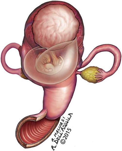
Fig. 7.31
An ultrasonographic scan of a fetus at 8 weeks with an overlying fundal fibroid

Fig. 7.32
An ultrasonographic scan of a fetus at 14 weeks with an overlying fundal fibroid
7.4 Effect of Pregnancy on Fibroids
Although fibroids grow during the first trimester, pregnancy has a variable and unpredictable effect on fibroid growth after the early pregnancy. One study found that fibroid size remains unchanged after it is first discovered by ultrasound during the first trimester in 69 % of women with a single fibroid (Fig. 7.33) [61]. In those who had enlargement of the fibroids, the greatest increase in size occurred before 10 weeks of gestation (Fig. 7.34). Fibroid growth was independent of the initial fibroid volume. After delivery, a reduction in fibroid size was noted.

Fig. 7.33
A single fundal fibroid discovered during the first trimester of pregnancy

Fig. 7.34
A single fundal fibroid increasing in size before 10 weeks of gestation
7.5 Fibroid Degeneration During Pregnancy
Degeneration occurs in approximately 9 % of women with uterine fibroids during pregnancy, based on clinical symptoms and instrumental evidence (Fig. 7.35) [62]. Infarction of a fibroid may occur if the vascular supply is insufficient to keep up with fibroid growth. It may also occur if there is a sudden occlusion of the vascular flow to the fibroid, such as with torsion of a pedunculated tumor (Fig. 7.36), or sudden change in estradiol and progesterone levels, which may occur during miscarriage. Among 113 women followed during pregnancy with serial sonography, ten (9 %) developed anechoic spaces or coarse heterogeneous patterns consistent with fibroid degeneration. Seven of ten women also had severe abdominal pain requiring hospitalization consistent with infarction and degeneration of the fibroids. No sonographic changes were observed in the other 103 women, of whom only 12 % had similar pain. A small study found that ibuprofen shortened the hospital stay and decreased readmission [63].
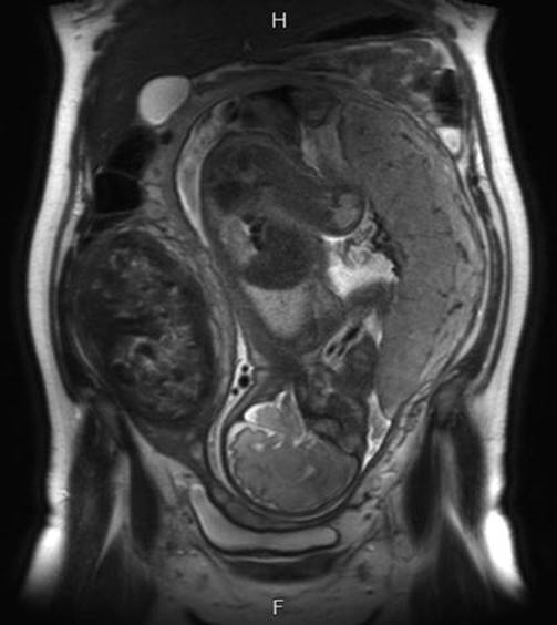
Fig. 7.35
MRI scan showing a fetus in longitudinal position at 34 weeks in a uterus with a fibroid of 12 cm in diameter in degeneration (Courtesy of Prof. Dr. Josè Palacios de Jaraquemada)

Fig. 7.36
A torsion of a pedunculated fibroid, with its degeneration
On occasion, myomectomy has been required in cases of torsion of a pedunculated fibroid associated with intense pain (Fig. 7.37). In one case, a woman who presented with an acute abdomen at 11 weeks was found to have a normal pelvis with the exception of an 8 cm pedunculated fibroid [64]. Another case described a successful pregnancy after laparoscopic myomectomy for a woman who presented with an acute abdomen at 10 weeks [64]. In both cases, laparoscopic myomectomy resulted in rapid pain relief and term delivery of a healthy baby.

Fig. 7.37
A sagittal scan by MRI of a large pedunculated fibroid in an early pregnant women, with initial fibroid torsion on its axis (Courtesy of Prof. Dr. Josè Palacios de Jaraquemada)
Uterine torsion, rotation of the uterus more than 45° along the long axis, can occur when the fibroid uterus is markedly enlarged. In one case, a 27-year-old woman presented at 15 week 3 day pregnancy in shock with an acute abdomen [65]. Laparotomy demonstrated complete axial torsion of the uterus due to a large fundal myoma and a massive abruption. When acute uterine torsion occurs near term, an emergency cesarean section can be performed, but may necessitate what is actually an anatomically posterior uterine incision [66]. A high index of suspicion is needed to rapidly diagnose and manage women with fibroids who present with an acute abdomen, and fibroid-related causes should be included in the differential diagnosis.
7.6 Influence of Fibroids on Pregnancy
Most women with uterine fibroids can expect to have a pregnancy without fibroid-related complications (Fig. 7.38). However, the incidence of complications has varied in different study groups.

Fig. 7.38
A pregnant with a posterior uterine fibroid; she can even expect to have a pregnancy without fibroid-related complications
In one study of 12,600 pregnant women, 167 were found to have uterine fibroids, and there was no difference in the incidence of preterm delivery, premature rupture of membranes, fetal growth restriction, placenta previa, placental abruption, postpartum hemorrhage, or retained placenta [67]. Only cesarean deliveries were more common among women with fibroids (23 % vs. 12 %).
In contrast to this relatively reassuring study, other investigators have found a higher incidence of fibroid-related complications during pregnancy (Fig. 7.39). In a retrospective study of 15,104 pregnancies that included 401 women diagnosed with fibroids by second-trimester ultrasound, there was no increase in premature rupture of membranes, operative vaginal delivery, chorioamnionitis, or endomyometritis [68]. However, compared to women without fibroids, there was a higher incidence of preterm delivery (19 % vs. 13 %), placenta previa (3.5 % vs. 1.8 %, a difference of 1.7 %), postpartum hemorrhage (8.3 % vs. 2.9 %), and cesarean delivery (49 % vs. 21 %). Other studies have found that women with untreated fibroids have an increased risk of poor obstetric outcomes including increased incidence of growth restriction, intrauterine fetal demise, placental abruption, placenta previa, preterm birth, breech presentation, premature rupture of membranes, blood transfusion, as well as an increased cesarean delivery rate [69]. Stout and colleagues found placenta previa in 1.4 % with fibroids compared with 0.5 % with no fibroids, a 0.9 % difference; placental abruption in 1.4 % with fibroids compared with 0.7 % with no fibroids, a 0.7 % difference; and preterm rupture of membranes in 3.3 % of fibroids compared to 2.4 % with no fibroids, a 1.1 % difference [59]. The preterm birth rate at 34–37 weeks was 15.1 % with fibroids compared with 10.5 % without, a 4.6 % difference, but the clinical significance of this is small since these babies do very well. The preterm birth rate less than 34 weeks was 3.9 % compared with 2.8 % without, a 1.1 % difference that was statistically significant.

Fig. 7.39
A longitudinal laparotomy in a patient at 44 years old with a massive hemorrhage at 9 weeks from a giant uterus with multiple fibroids; such woman gives the consent to urgently remove the uterus as a life-treating therapy
The size of uterine fibroids is an important factor in determining prognosis and pregnancy-related complications [70]. In one study that compared women with no fibroids or small fibroids, women with fibroids larger than 5 cm (Fig. 7.40) delivered at a significantly earlier gestational age (38.6 vs. 38.4 vs. 36.5 weeks). The rates of preterm premature rupture of membranes and preterm delivery were significantly higher with large fibroids, and the prognosis was also related with the number of fibroids >5 cm in diameter (Fig. 7.41). Blood loss at delivery was significantly higher in the large fibroid group, and 12 % of women with large fibroids needed a postpartum blood transfusion.
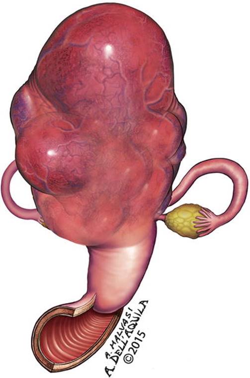
Fig. 7.40
A fundal fibroid of 6 cm in diameter in a uterus with multiple fibroids in pregnancy

Fig. 7.41
A laparotomic image showing a large fibroid causing a premature rupture of membranes and preterm delivery by urgent cesarean section; the fibroid was enucleated after fetal delivery (Courtesy of Prof. Dr. Josè Palacios de Jaraquemada)
The location of fibroids is another factor that contributes to pregnancy-related complications. Posterior fibroids that were 3 cm or larger (Fig. 7.42a, b) are associated with significantly more pelvic pain (p = 0.001) and a significantly higher miscarriage rate compared to those with anterior fibroids of comparable size [71]. However, no difference was observed between those with anterior or posterior fibroids related to the rates of preterm delivery, bleeding in early pregnancy, infants with small for gestational age, and hospitalization period during pregnancy. Women with posterior located myomas had significantly higher miscarriage rates.

Fig. 7.42
An ultrasonographic scan showing a fetus at 18 weeks in a uterus with an anterior placenta and a posterior fibroid of 8 cm in diameter (a); the draft shows a fetus at 15 weeks in a uterus with anterior placenta and posterior uterine fibroid
Fetal injury attributed to mechanical compression by fibroids has been reported to occur very infrequently. A search of the PubMed database from 1980 to 2010 revealed one case of fetal head anomalies with fetal growth restriction [72], one case of a postural deformity [73], one case of a limb reduction [74], and one case of fetal head deformation with torticollis [75].
It is clear that the incidence of cesarean delivery is increased in women with uterine fibroids, especially with fibroid 3 cm or larger [76]. In some cases, a fibroid located at or near the site of the preferred cesarean incision site (Fig. 7.43) can increase the complexity and complications of the procedure. In some cases, myomectomy during cesarean section (Fig. 7.44) may be appropriate, and in other cases it may be in the patient’s interest to undergo myomectomy for large fibroids during cesarean delivery. However, when myomectomy is performed during cesarean section, it is important to observe meticulous surgical techniques (Fig. 7.45).
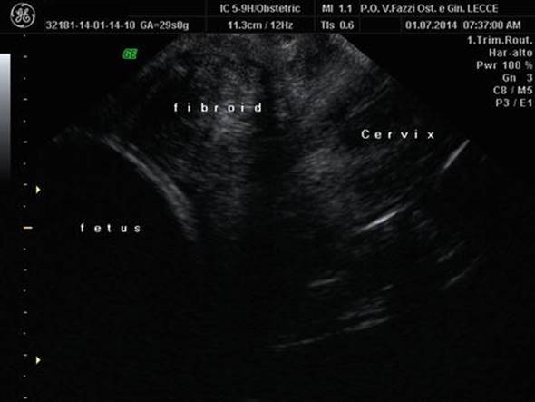
Fig. 7.43
An ultrasonographic scan showing an anterior fibroid located at or near the site of the preferred cesarean incision site
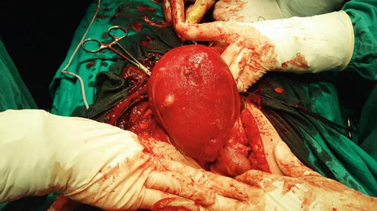
Fig. 7.44
A hysterorrhaphy after anterior myomectomy during cesarean section in a patient with multiple adhesions after a previous myomectomy

Fig. 7.45
The image shows a low uterine segment myomectomy performed during cesarean section; it is important to observe meticulous surgical techniques during fetal extraction and fibroid enucleating
Tinelli and colleagues described a prospective case–control study of 68 women who underwent (Fig. 7.46), compared to a control group of 72 women who underwent cesarean delivery without myomectomy [77]. Most of the fibroids were subserosal or intramural, and 54 % were fundal, 32 % were in the body of the uterus, and 19 % were in the lower uterine segment. Surgical techniques including focal gentle hemostasis, sharp pseudocapsule dissection, careful approximation of the myometrial edges, and closure of dead space was performed with meticulous attention to prevent hematoma formation. Comparing the two groups, there was no difference in the duration of hospital stay or postoperative anemia. Another study compared myomectomy at cesarean section in 76 women with fibroids and 60 women who underwent cesarean delivery without myomectomy and demonstrated that myomectomy could be a safe option for some women with fibroids [78]. These studies demonstrated that myomectomy can safely be performed by experienced surgeons during cesarean delivery.

Fig. 7.46
An intracapsular cesarean myomectomy
7.7 Conclusion
Because of the high incidence of uterine fibroids during the reproductive years, prepregnancy treatment for uterine fibroids and pregnancy with uterine fibroids is a common occurrence (Fig. 7.47). Sometimes fibroids in pregnancy lead to a complicated pregnancy (Fig. 7.48). Most women with asymptomatic fibroids experience a normal pregnancy and birth. When pregnancy is desired by a woman who has symptomatic fibroids, hysteroscopic myomectomy and laparoscopic or abdominal intracapsular myomectomy provide better outcomes than procedures that reduce the size of fibroids, including uterine artery embolization, magnetic resonance-guided focused ultrasound surgery, and myolysis. However, pregnancy-related complications can occur with all of these approaches. In women who conceive with uterine fibroids, the incidence of cesarean delivery is increased, also for contemporary cesarean myomectomy (Fig. 7.49). Maternal and fetal complication related to the size and location of fibroids may include growth restriction, placental abruption, placenta previa, preterm birth, breech presentation, premature rupture of membranes, and blood transfusion, in addition to an increased cesarean delivery rate and possibility of cesarean hysterectomy (Fig. 7.50). Because of the potential for these complications, all women who are pregnant and have been treated for fibroids or have fibroids identified before or during pregnancy should be followed closely to improve the likelihood of delivering a healthy baby at term.
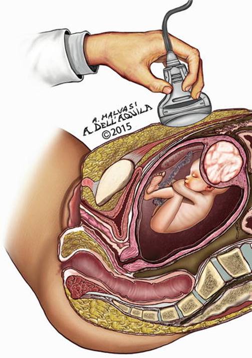
Fig. 7.47
A pregnant with a fetus at 15 weeks and a fundal uterine fibroid

Fig. 7.48
A sagittal scan by MRI of a large symptomatic cervical fibroid in a pregnant woman at 19 weeks (Courtesy of Prof. Dr. Josè Palacios de Jaraquemada)
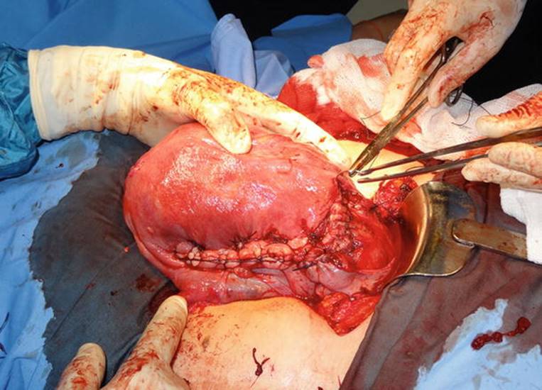
Fig. 7.49
A cesarean myomectomy result: after fetal delivery, the incision of low uterine segment had been prolonged on the right upper lateral anterior part of the uterine body, to remove large fibroid
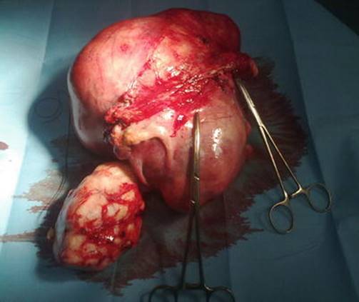
Fig. 7.50
A cesarean hysterectomy: the pregnant uterus, deformed by multiple large fibroids, was removed after the delivery of newborn
References
1.
Baird DD, Dunson DB, Hill MC, Cousins D, Schectman JM (2003) High cumulative incidence of uterine leiomyoma in black and white women: ultrasound evidence. Am J Obstet Gynecol 188:100–107CrossRefPubMed
2.
Mehine M, Mäkinen N, Heinonen HR, Aaltonen LA, Vahteristo P (2014) Genomics of uterine fibroids: insights from high-throughput sequencing. Fertil Steril 102:621–629CrossRefPubMed
3.
Brakta S, Diamond JS, Al-Hendy A, Diamond MP, Halder SK (2015) Role of vitamin D in uterine fibroid biology. Fertil Steril 104:698–706CrossRefPubMedPubMedCentral
4.
Commandeur AE, Styer AK, Teixeira JM (2015) Epidemiological and genetic clues for molecular mechanisms involved in uterine fibroid development and growth. Hum Reprod Update 21:593–615CrossRefPubMedPubMedCentral
5.
Moravek MB, Yin P, Ono M, 5th Coon JS, Dyson MT, Navarro A, Marsh EE, Chakravarti D, Kim JJ Wei JJ, Bulun SE (2015) Ovarian steroids, stem cells and uterine fibroid: therapeutic implications. Hum Reprod Update 21:1–12CrossRefPubMed
6.
Benaglia L, Cardellicchio L, Filippi F, Paffoni A, Vercellini P, Somigliana E, Fedele L (2014) The rapid growth of fibroids during early pregnancy. PLoS One 9:e85933CrossRefPubMedPubMedCentral
7.
Di Tommaso S, Massari S, Malvasi A, Bozzetti MP, Tinelli A (2013) Gene expression analysis reveals an angiogenic profile in uterine fibroid pseudocapsule. Mol Hum Reprod 19:380–387CrossRefPubMed
8.
Hurst BS, Matthews ML, Marshburn PB (2005) Laparoscopic myomectomy for symptomatic uterine myomas. Fertil Steril 83:1–23CrossRefPubMed
9.
Matteson KA, Anderson BL, Pinto SB, Lopes V, Schulkin J, Clark MA (2011) Practice patterns and attitudes about treating abnormal uterine bleeding: a national survey of obstetricians and gynecologists. Am J Obstet Gynecol 205:321.e1–8CrossRef
10.
Donnez J, Hudecek R, Donnez O, Matule D, Arhendt HJ, Zatik J, Kasilovskiene Z, Dumitrascu MC, Fernandez H, Barlow DH, Bouchard P, Fauser BC, Bestel E, Terrill P, Osterloh I, Loumaye E (2015) Efficacy and safety of repeated use of ulipristal acetate in uterine fibroids. Fertil Steril 103:519–27.e3CrossRefPubMed
11.
Luyckx M, Squifflet JL, Jadoul P, Votino R, Dolmans MM, Donnez J (2014) First series of 18 pregnancies after ulipristal acetate treatment for uterine fibroids. Fertil Steril 102:1404–1409CrossRefPubMed
12.
Wallach EE, Vlahos NF (2004) Uterine myomas: an overview of development, clinical features, and management. Obstet Gynecol 104:393–406CrossRefPubMed
13.
Roy KK, Singla S, Baruah J, Sharma JB, Kumar S, Singh N (2010) Reproductive outcome following hysteroscopic myomectomy in patients with infertility and recurrent abortions. Arch Gynecol Obstet 282:553–560CrossRefPubMed
14.
Hamerlynck TW, Dietz V, Schoot BC (2011) Clinical implementation of the hysteroscopic morcellator for removal of intrauterine myomas and polyps. A retrospective descriptive study. Gynecol Surg 8:193–196CrossRefPubMed
15.
Camanni M, Bonino L, Delpiano EM, Ferrero B, Migliaretti G, Deltetto F (2010) Hysteroscopic management of large symptomatic submucous uterine myomas. J Minim Invasive Gynecol 17:59–65CrossRefPubMed
16.
Litta P, Conte L, De Marchi F, Saccardi C, Angioni S (2014) Pregnancy outcome after hysteroscopic myomectomy. Gynecol Endocrinol 30:149–152CrossRefPubMed
17.
Chamberlain G (2003) The master of myomectomy. J R Soc Med 96:302–304CrossRefPubMedPubMedCentral
18.
Tinelli A, Mettler L, Malvasi A, Hurst B, Catherino W, Mynbaev OA, Guido M, Alkatout I, Schollmeyer T (2014) Impact of surgical approach on blood loss during intracapsular myomectomy. Minim Invasive Ther Allied Technol 23:87–95CrossRefPubMed
19.
Tinelli A, Hurst BS, Mettler L, Tsin DA, Pellegrino M, Nicolardi G, Dell'Edera D, Malvasi A (2012) Ultrasound evaluation of uterine healing after laparoscopic intracapsular myomectomy: an observational study. Hum Reprod 27:2664–2670CrossRefPubMed
20.
Malzoni M, Tinelli R, Cosentino F, Iuzzolino D, Surico D, Reich H (2010) Laparoscopy versus minilaparotomy in women with symptomatic uterine myomas: short-term and fertility results. Fertil Steril 93:2368–2373CrossRefPubMed
21.
The Myomectomy Adhesion Multicenter Study Group (1995) An expanded polytetrafluoroethylene barrier (Gore-Tex Surgical Membrane) reduces post-myomectomy adhesion formation. Fertil Steril 63:491–493CrossRef
22.
Diamond MP (1996) Reduction of adhesions after uterine myomectomy by Seprafilm membrane (HAL-F): a blinded, prospective, randomized, multicenter clinical study. Seprafilm Adhesion Study Group. Fertil Steril 66:904–910CrossRefPubMed
23.
Ahmad G, O’Flynn H, Hindocha A, Watson A (2015) Barrier agents for adhesion prevention after gynaecological surgery. Cochrane Database Syst Rev (4):CD000475
24.
Tinelli A, Hurst BS, Hudelist G, Tsin DA, Stark M, Mettler L, Guido M, Malvasi A (2012) Laparoscopic myomectomy focusing on the myoma pseudocapsule: technical and outcome reports. Hum Reprod 27:427–435CrossRefPubMed
25.
Sizzi O, Rossetti A, Malzoni M, Minelli L, La Grotta F, Soranna L, Panunzi S, Spagnolo R, Imperato F, Landi S, Fiaccamento A, Stola E (2007) Italian multicenter study on complications of laparoscopic myomectomy. J Minim Invasive Gynecol 14:453–462CrossRefPubMed
26.
Tinelli A, Malvasi A, Gustapane S, Buscarini M, Gill IS, Stark M, Nezhat FR, Mettler L (2011) Robotic assisted surgery in gynecology: current insights and future perspectives. Recent Pat Biotechnol 5:12–24CrossRefPubMed
27.
Lee HJ, Kim JY, Kim SK, Lee JR, Suh CS, Kim SH (2015) Learning curve analysis and surgical outcomes of single-port laparoscopic myomectomy. J Minim Invasive Gynecol 22:607–611CrossRefPubMed
28.
Landon MB, Lynch CD (2011) Optimal timing and mode of delivery after cesarean with previous classical incision or myomectomy: a review of the data. Semin Perinatol 35:257–261CrossRefPubMed
29.
Parker WH, Einarsson J, Istre O, Dubuisson JB (2010) Risk factors for uterine rupture after laparoscopic myomectomy. J Minim Invasive Gynecol 17:551–554CrossRefPubMed
30.
Laberge PY (2008) Serious and deadly complications from pregnancy after endometrial ablation: two case reports and review of the literature. J Gynecol Obstet Biol Reprod (Paris) 37:609–613CrossRef
31.
Bowling MR, Ramsey PS (2010) Spontaneous uterine rupture in pregnancy after endometrial ablation. Obstet Gynecol 115:405–406CrossRefPubMed
32.
Hamar BD, Wolff EF, Kodaman PH, Marcovici I (2006) Premature rupture of membranes, placenta increta, and hysterectomy in a pregnancy following endometrial ablation. J Perinatol 26:135–137CrossRefPubMed
33.
Gill LA, Baldwin E, Lessard-Anderson C, White W (2015) Septic abortion with placenta accreta in pregnancy after endometrial ablation. Obstet Gynecol 125:822–824CrossRefPubMed
34.
Bren L (2001) Alternatives to hysterectomy. New technologies, more options. FDA Consum 35:23–28PubMed
35.
van der Kooij SM, Hehenkamp WJ, Birnie E, Ankum WM, Mol BW, Scherjon S, Reekers JA (2013) The effect of treatment preference and treatment allocation on patients' health-related quality of life in the randomized EMMY trial. Eur J Obstet Gynecol Reprod Biol 169:69–74CrossRefPubMed
36.
Firouznia K, Ghanaati H, Sanaati M, Jalali AH, Shakiba M (2009) Pregnancy after uterine artery embolization for symptomatic fibroids: a series of 15 pregnancies. AJR Am J Roentgenol 192:1588–1592CrossRefPubMed
37.
Hamoda H, Pepas L, Tasker F, Reidy J, Khalaf Y (2015) Intermediate and long-term outcomes following uterine artery fibroid embolization. Eur J Obstet Gynecol Reprod Biol 191:33–38CrossRefPubMed
38.
Torre A, Paillusson B, Fain V, Labauge P, Pelage JP, Fauconnier A (2014) Uterine artery embolization for severe symptomatic fibroids: effects on fertility and symptoms. Hum Reprod 29:490–501CrossRefPubMed
39.
Usadi RS, Marshburn PB (2007) The impact of uterine artery embolization on fertility and pregnancy outcome. Curr Opin Obstet Gynecol 19:279–283CrossRefPubMed
40.
Yeaton-Massey A, Loring M, Chetty S, Druzin M (2014) Uterine rupture after uterine artery embolization for symptomatic leiomyomas. Obstet Gynecol 123:418–420CrossRefPubMed
41.
Dariushnia SR, Society of Interventional Radiology Standards of Practice Committee, Nikolic B, Stokes LS, Spies JB (2014) Quality improvement guidelines for uterine artery embolization for symptomatic leiomyomata. J Vasc Interv Radiol 25:1737–1747CrossRefPubMed
42.
Rabinovici J, Inbar Y, Revel A, Zalel Y, Gomori JM, Itzchak Y, Schiff E, Yagel S (2007) Clinical improvement and shrinkage of uterine fibroids after thermal ablation by magnetic resonance-guided focused ultrasound surgery. Ultrasound Obstet Gynecol 30:771–777CrossRefPubMed
43.
Ikink ME, Voogt MJ, Verkooijen HM, Lohle PN, Schweitzer KJ, Franx A, Mali WP, Bartels LW, van den Bosch MA (2013) Mid-term clinical efficacy of a volumetric magnetic resonance-guided high-intensity focused ultrasound technique for treatment of symptomatic uterine fibroids. Eur Radiol 23:3054–3061CrossRefPubMed
44.
Mindjuk I, Trumm CG, Herzog P, Stahl R, Matzko M (2015) MRI predictors of clinical success in MR-guided focused ultrasound (MRgFUS) treatments of uterine fibroids: results from a single centre. Eur Radiol 25:1317–1328CrossRefPubMed
45.
Rabinovici J, David M, Fukunishi H, Morita Y, Gostout BS, Stewart EA, MRgFUS Study Group (2010) Pregnancy outcome after magnetic resonance-guided focused ultrasound surgery (MRgFUS) for conservative treatment of uterine fibroids. Fertil Steril 93:199–209CrossRefPubMed
46.
Behera MA, Leong M, Johnson L, Brown H (2010) Eligibility and accessibility of magnetic resonance-guided focused ultrasound (MRgFUS) for the treatment of uterine leiomyomas. Fertil Steril 94:1864–1868CrossRefPubMed
47.
Taran FA, Tempany CM, Regan L, Inbar Y, Revel A, Stewart EA, MRgFUS Group (2009) Magnetic resonance-guided focused ultrasound (MRgFUS) compared with abdominal hysterectomy for treatment of uterine leiomyomas. Ultrasound Obstet Gynecol 34:572–578CrossRefPubMed
48.
Nisolle M, Smets M, Malvaux V (1993) Laparoscopic myolysis with Nd:YAG laser. J Gynecol Surg 9:95–99CrossRefPubMed
49.
Phillips DR (1995) Laparoscopic fibroid coagulation (myolysis). Gynaecol Endosc 4:5–9
50.
Goldfarb HA (2000) Myoma coagulation (Myolysis). Obstet Gynecol Clin N Am 27:421–430CrossRef
51.
Zreik TG, Rutherford TJ, Palter SF, Troiano RN, Williams E, Brown JM, Olive DL (1998) Cryomyolysis, a new procedure for the conservative treatment of uterine fibroids. J Am Assoc Gynecol Laparosc 5:33–38CrossRefPubMed
52.
Zupi E, Piredda A, Marconi D, Townsend D, Exacoustos C, Arduini D, Szaboles B (2004) Directed laparoscopic cryomyolysis: a possible alternative to myomectomy and/or hysterectomy for symptomatic fibroids. Am J Obstet Gynecol 190:639–643CrossRefPubMed
53.
Chudnoff SG, Berman JM, Levine DJ, Harris M, Guido RS, Banks E (2013) Outpatient procedure for the treatment and relief of symptomatic uterine myomas. Obstet Gynecol 121:1075–1082CrossRefPubMed
54.
Berman JM, Guido RS, Garza Leal JG, Pemueller RR, Whaley FS, Chudnoff SG, Halt Study Group (2014) Three-year outcome of the Halt trial: a prospective analysis of radiofrequency volumetric thermal ablation of myomas. J Minim Invasive Gynecol 21:767–774CrossRefPubMed
55.
Uterine Leiomyoma Treatment With Radiofrequency Ablation (ULTRA). https://clinicaltrials.gov/ct2/show/NCT01840124
56.
Jiang X, Thapa A, Lu J, Bhujohory VS, Liu Y, Qiao S (2014) Ultrasound-guided transvaginal radiofrequency myolysis for symptomatic uterine myomas. Eur J Obstet Gynecol Reprod Biol 177:38–43CrossRefPubMed
57.
Vilos GA, Daly LJ, Tse BM (1998) Pregnancy outcome after laparoscopic electromyolysis. J Am Assoc Gynecol Laparosc 5:289–292CrossRefPubMed
58.
Laughlin S, Baird D, Savitz D, Herring A, Hartmann K (2009) Prevalence of uterine leiomyomas in the first trimester of pregnancy: an ultrasound-screening study. Obstet Gynecol 113:630–635CrossRefPubMedPubMedCentral
59.
Stout MJ, Odibo AO, Graseck AS, Macones GA, Crane JP, Cahill AG (2010) Fibroids at routine second-trimester ultrasound examination and adverse obstetric outcomes. Obstet Gynecol 116:1056–1063CrossRefPubMed
60.
Muram D, Gillieson M, Walters JH (1980) Myomas of the uterus in pregnancy: ultrasonographic follow-up. Am J Obstet Gynecol 138:16–19CrossRefPubMed
61.
Rosati P, Exacoustos C, Mancuso S (1992) Longitudinal evaluation of uterine myoma growth during pregnancy. A sonographic study. J Ultrasound Med 11:511–515PubMed
62.
Lev-Toaff AS, Coleman BG, Arger PH, Mintz MC, Arenson RL, Toaff ME (1987) Leiomyomas in pregnancy: sonographic study. Radiology 164:375–380CrossRefPubMed
63.
Katz VL, Dotters DJ, Droegemeuller W (1989) Complications of uterine leiomyomas in pregnancy. Obstet Gynecol 73:593–596PubMed
64.
Kosmidis C, Pantos G, Efthimiadis C, Gkoutziomitrou I, Georgakoudi E, Anthimidis G (2015) Laparoscopic excision of a pedunculated uterine leiomyoma in torsion as a cause of acute abdomen at 10 weeks of pregnancy. Am J Case Rep 16:505–508CrossRefPubMedPubMedCentral
65.
Sachan R, Patel ML, Sachan P, Arora A (2014) Complete axial torsion of pregnant uterus with leiomyoma. BMJ Case Rep 5:2014
66.
Deshpande G, Kaul R, Manjuladevi P (2011) A case of torsion of gravid uterus caused by leiomyoma. Case Rep Obstet Gynecol 2011:206418PubMedPubMedCentral
67.
Vergani P, Ghidini A, Strobelt N, Roncaglia N, Locatelli A, Lapinski RH, Mangioni C (1994) Do uterine leiomyomas influence pregnancy outcome? Am J Perinatol 11:356–358CrossRefPubMed
68.
Lai J, Caughey AB, Qidwai GI, Jacoby AF (2012) Neonatal outcomes in women with sonographically identified uterine leiomyomata. J Matern Fetal Neonatal Med 25:710–713CrossRefPubMed
69.
Sheiner E, Bashiri A, Levy A, Hershkovitz R, Katz M, Mazor M (2004) Obstetric characteristics and perinatal outcome of pregnancies with uterine leiomyomas. J Reprod Med 49:182–186PubMed
70.
Shavell V, Thakur M, Sawant A, Kruger ML, Jones TB, Singh M, Puscheck EE, Diamond MP (2012) Adverse obstetric outcomes associated with sonographically identified large uterine fibroids. Fertil Steril 97:107–110CrossRefPubMed
71.
Deveer M, Deveer R, Engin-Ustun Y, Sarikaya E, Akbaba E, Senturk B, Danisman N (2012) Comparison of pregnancy outcomes in different localizations of uterine fibroids. Clin Exp Obstet Gynecol 39:516–518PubMed
72.
Chuang J, Tsai HW, Hwang JL (2001) Fetal compression syndrome caused by myoma in pregnancy: a case report. Acta Obstet Gynecol Scand 80:472–473CrossRefPubMed
73.
Joo JG, Inovay J, Silhavy M, Papp Z (2001) Successful enucleation of a necrotizing fibroid causing oligohydramnios and fetal postural deformity in the 25th week of gestation. A case report. J Reprod Med 46:923–925PubMed
74.
Graham JM Jr (1985) The association between limb anomalies and spatially-restricting uterine environments. Prog Clin Biol Res 163C:99–103PubMed
75.
Romero R, Chervenak FA, DeVore G, Tortora M, Hobbins JC (1981) Fetal head deformation and congenital torticollis associated with a uterine tumor. Am J Obstet Gynecol 141:839–840CrossRefPubMed
76.
Michels KA, Velez Edwards DR, Baird DD, Savitz DA, Hartmann KE (2014) Uterine fibroids and cesarean birth risk: a prospective cohort with standardized imaging. Ann Epidemiol 24:122–126CrossRefPubMed
77.
Tinelli A, Malvasi A, Mynbaev OA, Barbera A, Perrone E, Guido M, Kosmas I, Stark M (2014) The surgical outcome of intracapsular cesarean myomectomy. A match control study. J Matern Fetal Neonatal Med 27:66–71CrossRefPubMed
78.
Topçu HO, İskender CT, Timur H, Kaymak O, Memur T, Danışman N (2015) Outcomes after cesarean myomectomy versus cesarean alone among pregnant women with uterine fibroids. Int J Gynaecol Obstet 130:244–246CrossRefPubMed