
Paul Lyons1
(1)
Department of Family Medicine, University of California, Riverside, Riverside, CA, USA
Key Points
1.
2.
3.
Background
Preterm labor is among the most common and most serious of prenatal complications. Preterm labor and its potential sequelae of preterm delivery and low-birth weight (LBW) infants remain one of the most significant challenges of current obstetrical practice. Preterm labor is defined as uterine contractions resulting in progressive cervical change prior to 37 weeks’ gestation. Preterm delivery is delivery prior to 37 weeks’ gestation. LBW infants are defined as those infants weighing less than 2500 g at delivery regardless of gestational age. LBW infants should be distinguished from small-for-gestational-age (SGA) infants who are defined as those infants below the fifth percentile for weight based on gestational age.
Preterm labor affects approximately 10 % of all pregnancies. Preterm delivery affects approximately 13 % of all live births. Preterm delivery and LBW infants represent approximately 70 % of all perinatal mortality (~25,000 deaths annually) and 50 % of all neurological morbidity.
Factors Associated with Preterm Labor
A number of factors have been associated with an increased risk of preterm labor. These are summarized in Table 7.1. These factors can be divided into pre- and postconception factors. Although the mechanisms that link these factors to the onset of preterm labor is, in most instances, poorly understood, a thorough review of the patient’s history will allow providers to more carefully outline the risk of preterm labor, preterm delivery, and LBW infants.
Table 7.1
Risk factors for preterm labor
|
Preconception factors |
|
Lower socioeconomic status |
|
Anatomic abnormalities (e.g., septate/bicornuate uterus, cervical incompetence) |
|
Prior uterine surgery |
|
Myomata |
|
Diethylstilbestrol exposure |
|
Past history of preterm labor |
|
Under 18 years old, over 40 years old |
|
Possible genetic predisposition |
|
Postconception factors |
|
Tobacco |
|
Cocaine |
|
Infection (e.g., group B streptococcus, N. gonorrhea, C. trachomatis, trichomonas, gardnerella, ureaplasma, mycoplasma) |
Although preterm labor alone is not associated with perinatal complications, concomitant conditions and outcomes are. Preterm labor may be complicated by preterm premature rupture of membranes (PPROM). PPROM is associated with a variety of complications discussed in Chap. 8. Preterm labor may also result in preterm delivery. Prematurity, in turn, is potentially associated with pulmonary dysfunction, gastrointestinal abnormalities, neurological complications, abnormalities of growth and development, and a significant risk of perinatal mortality. Complications of preterm delivery are the leading cause of perinatal mortality, responsible for approximately two-thirds of all deaths.
Preconception Factor
Environmental Factors
A number of environmental factors have been associated with an increased risk of preterm labor. The most significant environmental factor associated with preterm labor is lower socioeconomic status.
Patient-Related Factors
A number of pre-existing patient conditions also contribute to the risk of preterm labor. Patients may have a pre-existing genetic risk, a congenital anomaly (e.g., septate/bicornuate uterus or cervical incompetence), a pre-existing acquired obstetrical/gynecological risk (e.g., myomata, uterine surgery, diethylstilbestrol exposure), or a past history of preterm labor or second trimester spontaneous abortions. The recurrence rate of preterm labor is approximately 25 %. Additionally, the risk of preterm labor is highest among younger (<18 years old) and older (>40 years old) obstetrical patients. These conditions can be screened for early in pregnancy (or during preconception counseling). Although many of these factors are not modifiable, their presence can usefully contribute to a conversation between the provider and the patient concerning the risk for preterm labor during the current pregnancy.
Postconception Factors
Once conception occurs, a number of additional factors contribute to the risk of preterm labor. An increased risk for preterm labor is associated with tobacco and cocaine use. Infections such as group B streptococcus, gonorrhea, chlamydia, trichomonas, gardnerella, ureaplasma, and mycoplasma have all been associated with increased preterm labor risk. Such exposures should be screened for at the first prenatal history (either directly through testing or via history) and as appropriate throughout the course of pregnancy.
Diagnosis
As noted earlier, the diagnosis of preterm labor consists of three components: gestational age less than 37 weeks, presence of uterine contractions, and progressive cervical change.
The diagnosis of preterm labor begins with confirmation of the gestational age of the fetus. All data that contributed to the estimated date of delivery (EDD) should be reviewed for accuracy. The patient’s last menstrual period should be reviewed for accuracy. Additional data such as prenatal ultrasound, sequential fundal height measurements, and gestational age at quickening should also be reviewed. If no such data is available, an obstetrical ultrasound may be indicated. It should be emphasized, however, that an ultrasound obtained late in pregnancy has significantly less accuracy for purposes of gestational dating.
The patient should be questioned concerning the presence of contractions (although the absence of patient-reported contractions does not exclude the possibility of clinically significant contractile activity). If preterm labor is suspected, patients should be placed on tocometric monitoring to confirm the presence of uterine contractions.
Documentation of progressive cervical change, under most circumstances, requires serial cervical examinations. After confirming the absence of bleeding per vagina, providers should document cervical dilation and effacement as well as fetal station. Although the patient may demonstrate unequivocal cervical evidence of labor on initial examination, generally the diagnosis will require comparison of initial findings to findings on a follow-up examination.
Intake Assessment
History
In addition to the history noted earlier, patients should be asked about bleeding per vagina, rupture of membranes or fluid leak, and/or symptoms of infection. Special caution should be exercised if the patient reports a history of bleeding per vagina. The management of third-trimester bleeding is covered in Chap. 10. A review of the past history should note the presence of cardiac, renal pulmonary, and/or endocrine abnormalities.
Physical Examination
In addition to the pelvic examination for assessment of cervical change, the intake physical examination should document blood pressure, pulse, temperature, rupture of membranes (see Chap. 8), fetal heart rate, and uterine contractions.
Laboratory Studies
Patients admitted with preterm labor should have all prenatal laboratory values reviewed with lab values ordered or updated as necessary. Patients may require testing for infection, including gonorrhea, chlamydia, group B strep trichomonas, and bacterial vaginosis. Other studies that may contribute to evaluation of possible infection include increased interleukin-6 in amniotic or cervical samples, elevated ferritin in cervical or serum samples, and elevated granulocyte colony-stimulating factor in serum samples. Patients demonstrating clinical signs or symptoms of other obstetrical conditions (e.g., pregnancy-induced hypertension) should have laboratory evaluation as indicated for those conditions.
Controversy exists concerning the role of routine fibronectin testing in the management of suspected preterm labor. Specimens should be obtained via sterile speculum examination. A swap is placed in the posterior fornix for 10 s. Care should be taken to avoid manipulation of the cervix or use of lubricant. Known rupture of membranes is a contraindication to fibronectin testing. After 22 weeks’ gestation, a result greater than 50 ng/mL is associated with an increased risk for preterm delivery with a sensitivity of 70–90 % and a specificity of 70–85 %. The negative predictive value is approximately 99 %. A negative test is a strong predictor of no preterm labor in the week following the test. Fetal breathing movements and measurement of cervical length can be used in conjunction with or as an alternative to fibronectin testing. Fibronectin has been shown to have the highest sensitivity and fetal breathing movements the highest specificity for delivery within 7 days of testing. In patients with a cervical length >30 mm, risk of delivery within 7 days is less than 5 % and fibronectin does not add to diagnostic accuracy.
Management
The management of preterm labor is often limited in efficacy and duration and few modifiable factors have been identified. A general outline of management is shown in Fig. 7.1. Goals of management are focused on delay of delivery to allow four key outcomes:
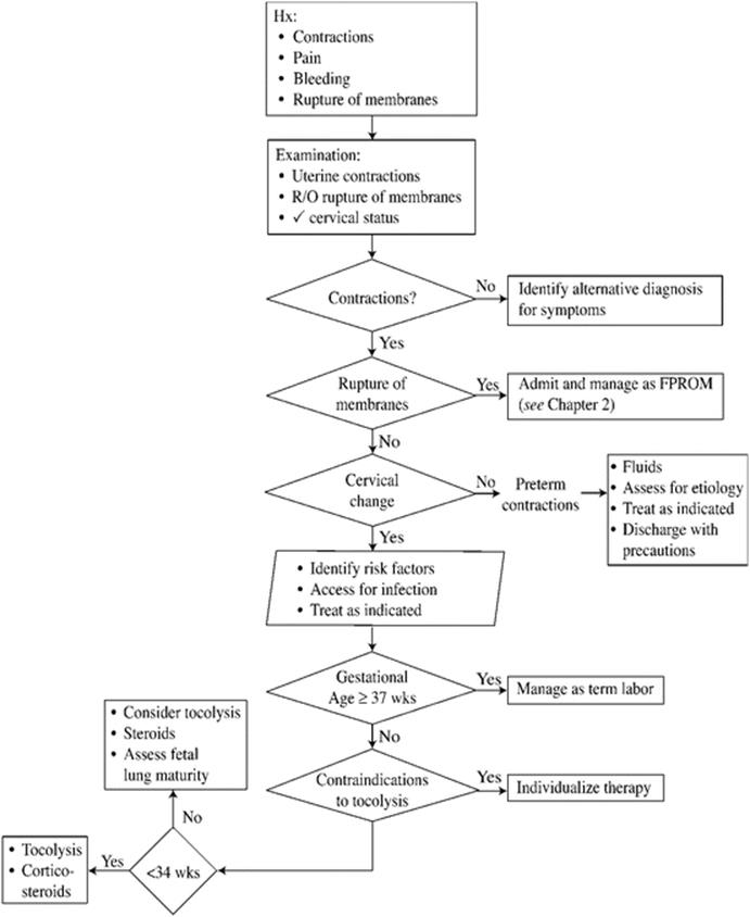
Fig. 7.1
Management of preterm labor
1.
2.
3.
4.
Decisions concerning appropriate management should be tailored to the individual patient. Despite the challenge and variability involved in managing preterm labor, a few general guidelines can be given.
Management Prior to 34 Weeks’ Gestation
In general, fetal lung maturity cannot be assumed in infants prior to 34 weeks’ gestation. For this reason, tocolysis is generally recommended. Although the efficacy and duration of such therapy is limited, a brief delay in delivery allows for administration of corticosteroids to enhance fetal lung maturity. All patients should be screened for contraindications to tocolysis (see Table 7.2). Contraindications to tocolysis include underlying medical contraindications (cardiac disease, renal insufficiency, pyelonephritis, pulmonary hypertension, untreated diabetes mellitus, and electrolyte abnormalities) and obstetrical contraindications (fetal stress, chorioamnionitis, eclampsia, fetal demise, and hemodynamic instability). Tocolytic options include the following:
Table 7.2
Contraindications to tocolysis
|
Medical contraindications |
|
Cardiac disease |
|
Renal insufficiency |
|
Pyelonephritis |
|
Pulmonary hypertension |
|
Untreated diabetes mellitus |
|
Electrolyte abnormalities |
|
Obstetrical contraindications |
|
Fetal stress |
|
Chorioamnionitis |
|
Eclampsia |
|
Fetal demise |
|
Hemodynamic instability |
1.
2.
3.
4.
5.
Patients should be admitted and placed on bed rest. If significant cervical dilation has occurred, patients may be placed in a head-down position. Routine management includes monitoring of fluid status, hemodynamic status, and fetal well-being. If there is question concerning the gestational age or fetal lung status, fetal lung maturity testing may be considered. Mothers of infants at risk for fetal lung immaturity (24–35 weeks’ gestation) should be treated with 12 mg of betamethasone, intramuscularly. Two doses should be given 24 h apart. Patients with evidence of contributory infection should be treated as appropriate for the infection.
Management at 34–37 Weeks
Fetal lung maturity in this range is highly variable and decisions to initiate tocolysis must be individualized. When time permits, assessment of fetal lung maturity may assist in decisions concerning tocolysis versus expectant management.
Assessment of Fetal Lung Maturity
Delivery of an infant prior to fetal lung maturation is associated with considerable neonatal morbidity and mortality. For this reason, assessment of fetal lung maturity is critical in all instances where gestational age cannot be firmly established or when prenatal complications require consideration of an early delivery.
Confirmation of the gestational age is critical. Gestational age can be confirmed by review of the last menstrual period, early obstetrical ultrasound results, and key developmental milestones such as quickening and fetal heart tones. Although these data may allow for accurate gestational dating when available, not all data will be available in all cases. Even with such data, a more accurate assessment of fetal lung maturity may be necessary to guide management decisions. A variety of options are available to assist in this assessment.
Lecithin-to-Sphingomyelin Ratio
As fetal lung maturity progresses, pulmonary secretions are accumulated in the amniotic fluid allowing for assessment of fetal lung maturity based on amniotic fluid sampling. Lecithin and sphingomyelin are present in approximately equal quantities until approximately 8 weeks prior to the EDD. Beginning at this point, lecithin concentrations increase and sphingomyelin concentrations remain stable. As the fetus nears maturation, therefore, the ratio of lecithin to sphingomyelin will increase. Although the exact interpretation of the results may be site dependent, a lecithin-to-sphingomyelin ratio of 2:1 or greater is associated with generally favorable neonatal pulmonary outcomes.
Phosphatidylglycerol
The presence of blood or meconium in the amniotic fluid may alter the results of the lecithin-to-sphingomyelin ratio. For this reason, alternative tests have been developed that are not sensitive to the presence of these substances. One such test is phosphatidylglycerol, a component of surfactant that is present in amniotic fluid and increases in quantity as fetal lung maturity advances. Amniotic fluid samples may be tested for phosphatidylglycerol alone or in conjunction with lecithin–sphingomyelin testing. The presence of phosphatidylglycerol is associated with more advanced fetal lung maturity and therefore with generally improved neonatal pulmonary outcomes. The results may be reported either qualitatively or quantitatively.
Additional Tests
Because of the importance of documenting fetal lung maturity and the complexity/limitations of traditionally available tests, a variety of alternative tests are now available. FLM-TDx II is a simple and reliable test (although it can be affected by blood or meconium). A value of >55 mg/g of albumin is considered indicative of fetal lung maturity. Lamellar body count (affected by blood but not meconium) with a value >50,000 is also indicative of fetal lung maturity.