
Paul Lyons1
(1)
Department of Family Medicine, University of California, Riverside, Riverside, CA, USA
Key Points
1.
2.
3.
4.
Background
Bleeding per vagina may occur at any point during the course of pregnancy and always warrants careful attention to identification and management of the underlying etiology. Bleeding that occurs late in pregnancy (generally within the third trimester) is covered in Chap. 10. Bleeding that occurs early in pregnancy (generally within the first trimester) is of particular concern because of concerns with pregnancy viability and possible ectopic pregnancy with its associated morbidity and mortality. A careful review of common etiologies combined with a careful history, directed physical examination, and selected diagnostic studies will allow the provider to identify the underlying cause and initiate appropriate management in a timely manner.
Early pregnancy bleeding per vagina is a relatively common presentation. Most cases of early bleeding are mild, self-limited, and of indeterminate etiology. Among the cases in which an etiology can be determined most are caused by either spontaneous abortion or ectopic pregnancy. Approximately 5–15 % of all pregnancies will end in a clinically recognized spontaneous abortion (involuntary expulsion prior to 20 weeks’ gestation). A considerably larger percentage (up to one-third) will end in an unrecognized abortion that is perceived to be menstrual bleeding. Although less common than spontaneous abortion, ectopic pregnancy is responsible for approximately 15 % of all maternal deaths. Although the differential diagnosis for early pregnancy bleeding includes several disparate conditions (see Table 9.1), the focus of initial evaluation is on identification of these two conditions. Less common causes of early pregnancy bleeding include trophoblastic disease and cervical pathology such as ulceration, infection, or cytopathology.
Table 9.1
Conditions associated with early pregnancy bleeding
|
Ectopic pregnancy |
|
Threatened abortion |
|
Incomplete abortion |
|
Complete abortion |
|
Trophoblastic disease |
|
Cervical polyps |
|
Cervical ulceration |
|
Cervical cytological abnormalities |
General Approach to Early Pregnancy Bleeding per Vagina
As noted, most cases of early pregnancy bleeding per vagina are of indeterminate cause. A significant minority are related to spontaneous abortion. A smaller but significant number are associated with ectopic pregnancy. Initial evaluation will be directed toward identification of patients with these two conditions, documentation of the viability of the pregnancy, assessment of the medical stability of the mother, and reassurance for patients without evidence of either condition. Figure 9.1 outlines a general approach to the evaluation and management of early pregnancy bleeding.
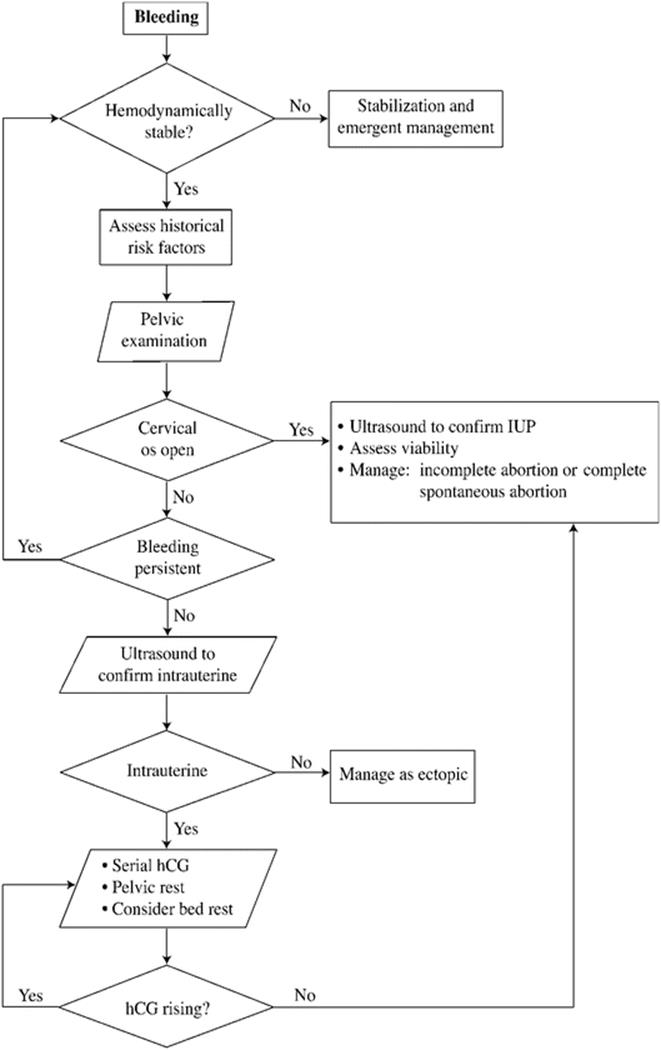
Fig. 9.1
Evaluation and management of early pregnancy bleeding
History
A careful history should be obtained of the bleeding itself as well as associated symptoms. In regard to the bleeding itself, information should be obtained concerning the following:
1.
2.
3.
4.
5.
6.
Particular attention should be paid to trauma, activity (including sexual activity), and associated symptoms such as pain, dizziness, or weakness. The patient’s past history should be reviewed for risk factors such as smoking, multiple sexual partners, past history of pelvic inflammatory disease, or intrauterine device use. Prior gynecological surgery should be reviewed, including tubal ligation and uterine surgery (including myomectomy). Any prior diagnosis of anatomic abnormalities of the genital tract should be noted. The patient’s obstetrical history should be reviewed for prior ectopic pregnancies and/or spontaneous abortions. The prenatal course should be reviewed for any high-risk conditions that may have been previously noted, including a family history of significant genetic abnormalities or significant toxic or infectious exposures. A note should also be made of the patient’s gestational age and the means used to determine the estimated delivery date.
Physical Examination
Physical examination begins with a rapid assessment of the hemodynamic stability of the mother. Depending on the gestational age of the patient, assessment of fetal heart rate may also be indicated. The mother’s blood pressure (BP) and pulse should be recorded including orthostatic measurements. (To measure orthostatic—positional—changes in BP and pulse, patients should first be placed in a recumbent position. BP and pulse are measured in the usual manner. The patient is then raised to a sitting position and the pulse and BP are measured a second time. A note is made of a significant drop in BP or a significant rise in pulse.) The general assessment of the patient should include obvious signs of distress or discomfort. Abdominal examination should include assessment of uterine size. A pelvic examination should be performed. A note should be made of blood (location, at the cervical os, pooled in the vaginal vault; quantity: from scant discoloration to copious active bleeding; character, bright red, clotted, brown). Cervical status should be noted including dilation. The presence of other findings including discharge, amniotic fluid, or cervical lesions such as polyps or erosions should be documented. A bimanual examination should be performed to assess for adnexal mass or tenderness. The uterus should be assessed for size and tenderness.
Ultrasound
Ultrasound examination may be critical in the assessment of early pregnancy bleeding. Confirmation of an intrauterine pregnancy is possible as early as 5 weeks’ gestation. In general, the presence of an intrauterine pregnancy significantly reduces but does not eliminate the possibility of ectopic pregnancy. Ultrasound examination is also diagnostic of trophoblastic disease. The absence of fetal heart activity may be indicative of a threatened or incomplete abortion. Transvaginal ultrasound is study of choice for early pregnancy assessment. The absence of an intrauterine gestational sac after 6 weeks from the last menstrual period is strongly suggestive of nonviable pregnancy (ectopic or aborted). Transabdominal ultrasound may be used to assess for intra-abdominal fluid including blood.
Laboratory Studies
Early pregnancy bleeding per vagina may represent a significant obstetrical emergency in the case of an ectopic pregnancy that has ruptured. Blood loss may be rapid and difficult to control. For this reason, laboratory studies are ordered to establish baseline hematological status and in anticipation of a potential need for transfusion. Studies should include complete blood count with platelets, blood type, and crossmatch. If not previously documented Rh status should be confirmed. The second key function of laboratory surveillance is to document the viability of the pregnancy. For this purpose, quantitative β-human chorionic gonadotropin (β-hCG) should be obtained.
Ectopic Pregnancy
Background
Ectopic pregnancy is diagnosed when pregnancy implantation occurs in a location outside the uterine cavity. The exact location can be variable and may include within the fallopian tubes (98 %) or in an intraperitoneal/extrauterine location. Because blood supply and/or space is limited outside the uterine cavity, these pregnancies are capable of growing for a brief period but will eventually outgrow their space or blood supply. If the pregnancy implants in a location (such as the fallopian tube) where space is limited, rupture may occur with associated intraperitoneal bleeding.
Risk factors associated with an increased risk for ectopic pregnancy include prior history of ectopic pregnancy, prior history of pelvic inflammatory disease, prior pelvic surgery (including tubal ligation and reversal of tubal ligation), multiple sexual partners (probably related to an increased risk for unrecognized pelvic inflammatory disease and subsequent scarring), and smoking.
Diagnosis
History
Patients who present with early pregnancy bleeding per vagina should be carefully screened for risk factors that increase the probability of ectopic pregnancy as noted previously. Patients with ectopic pregnancy may report bleeding, abdominal pain, and symptoms of peritonitis, or it may be relatively asymptomatic. The bleeding may be variable depending on the location of the pregnancy. Although copious bleeding should prompt evaluation for ectopic pregnancy, providers should maintain a high index of suspicion as the absence of clinically apparent bleeding does not eliminate the possibility of ectopic pregnancy. The onset of symptoms may be acute but is often subacute with gradual worsening of symptoms that may be initially quite mild.
Physical Examination
The physical examination should include those elements noted previously to assess maternal and fetal well-being, including vital signs, abdominal examination, and fetal heart rate monitoring when appropriate. The physical examination may be quite variable. For some patients, the findings on physical examination are all normal. For others with rupture and significant intraperitoneal bleeding, findings may be indicative of acute peritonitis including diminished bowel sounds and a rigid, tender abdomen with rebound and guarding. A pelvic examination should be performed to assess for the presence of blood and/or products of conception, cervical status, uterine tenderness, adnexal tenderness, and adnexal mass.
Ultrasound Examination
Although history and physical examination may contribute to the diagnosis of ectopic pregnancy, all patients with early pregnancy bleeding per vagina should be assumed to have ectopic pregnancy until it is ruled out. For this reason, all patients should have ultrasound confirmation of the presence, viability, and location of the developing fetus. Ultrasound may demonstrate a gestational sac in the extrauterine space. Alternately, the ultrasound may show no evidence of a gestational sac despite elevated β-hCG levels. Transvaginal ultrasound should demonstrate intrauterine pregnancy when hCG levels reach approximately 6000 mIU/mL. Ectopic pregnancy should be strongly suspected if the ultrasound does not demonstrate either characteristic findings of trophoblastic disease or intrauterine pregnancy when β-hCG levels are greater than 2000 mIU/mL.
Laboratory Studies
Supportive laboratory studies include serum progesterone levels and serial quantitative β-hCG. Progesterone levels are rarely less than 5 ng/mL in viable pregnancies. Conversely, less than 1 % of ectopic pregnancies will demonstrate serum progesterone levels of more than 25 ng/mL. Serum β-hCG may also add useful data. Ectopic pregnancy is rarely associated with β-hCG levels greater than 50,000 mIU/mL. If initial findings are equivocal, serial β-hCG levels are very helpful. In normal pregnancies, serum β-hCG doubles every 2–3 days. An increase in β-hCG less than 50 % in 48 h is considered abnormal and should warrant further evaluation.
Management
Management of ectopic pregnancy depends considerably on the stability of the patient. Following diagnosis of ectopic pregnancy, patients should be assessed for clinical stability. Patients with hemodynamic instability or evidence of peritonitis should be considered surgical emergencies. For patients who are stable, evaluation for surgical or medical management may be performed. The purpose of management is threefold: (a) to minimize morbidity and mortality for the mother, (b) to remove the ectopic pregnancy, and (c) to maximize future potential fertility options.
In addition to the conditions just listed, indications for surgical intervention include anemia, gestational sac greater than 4 cm on ultrasound evaluation, pain of more than 24 h duration, suspected heterotopic pregnancy, or uncertainty of diagnosis requiring laparoscopic confirmation.
Indications for medical management include gestational sac smaller than 4 cm on ultrasound examination and hemodynamic stability.
Surgical Management
Patients with clinical evidence of hemodynamic instability or peritonitis should be treated as a surgical emergency. Providers should assume that blood products and fluid resuscitation will be necessary and should be prepared to provide both. Patients should be crossmatched for at least four units of blood and should have at least two large bore intravenous access sites established as quickly as possible. A variety of surgical options exist depending on the clinical presentation of the patient, the desire to preserve future fertility, and the experience of the operator. These options include salpingostomy, segmental resection, fimbrial expression, salpingectomy (in cases with significant bleeding, significant tubal damage, or recurrent ectopic pregnancies at the same location), and laparotomy (in cases with severe hemodynamic stability).
Medical Management
The use of methotrexate has emerged as a medical alternative to surgical management for appropriate patients. The success rate with single-dose methotrexate regimens is 80–90 %. One such regimen is 50 mg/m2 of methotrexate via intramuscular injection. Serum β-hCG levels are obtained on days 1, 4, and 7. A second dose is administered on day 7 if serum β-hCG has not demonstrated a significant (~15 %) decrease. Serial weekly serum β-hCG levels should be obtained until levels are less than 15 mIU/mL.
Spontaneous Abortion
Background
Among cases of early pregnancy bleeding with a known etiology, spontaneous abortion is by far the most common. As noted, 5–15 % of all pregnancies will end with a clinically apparent spontaneous abortion; up to one-third of all pregnancies end in spontaneous abortion. Although generally spontaneous abortion is not as medically concerning as ectopic pregnancy, the significance of spontaneous abortion for the patient (and her family) will often be quite high. For this reason, appropriate diagnosis and management of spontaneous abortion is critical.
Risk factors for spontaneous abortion include genetic abnormalities, toxic exposures, structural abnormalities (such as atypical uterine anatomy), smoking, and toxic exposure. Most cases of spontaneous abortion occur without a clinically apparent risk factor.
Spontaneous abortion may be divided into three general subtypes: threatened, incomplete, and complete.
Threatened abortion is defined as bleeding per vagina before 20 weeks without passage of tissue or premature rupture of membranes. Threatened abortion may present with or without cervical dilation. Bleeding per vagina prior to 20 weeks’ gestation in conjunction with cervical dilation is referred to as inevitable abortion.
Incomplete abortion is defined as bleeding and incomplete passage of the products of conception. This condition may be associated with hemodynamic instability in which case it should be considered an obstetrical emergency and should include all the usual elements associated with management of such unstable patients.
Complete abortion is defined as bleeding with complete passage of all products of conception. The distinction between incomplete and complete abortion is not always clear, and care should be exercised to confirm the passage of all products prior to making the diagnosis of complete abortion.
Diagnosis
It should be recognized that patients will not present with a chief complaint of spontaneous abortion; they will present with a complaint of bleeding per vagina. For this reason, the general history for these patients will mirror that of patients with ectopic pregnancy.
History
History should include the elements described earlier, including a history of the bleeding, associated symptoms, a review of historical risk factors, and symptoms of hemodynamic instability. Particular attention should be paid to reports of passage of tissue or “clots.” A past history of spontaneous abortion should raise providers’ suspicion of abortion.
Physical Examination
Physical examination will begin with assessment of the hemodynamic stability of the patient. Pelvic examination will assess cervical dilation, bleeding, and/or products of conception. To assess for other potential causes of bleeding, providers should make note of adnexal tenderness or mass, cervical lesions, discharge, or other abnormalities.
Ultrasound Examination
The use of ultrasound in suspected abortion may provide limited but useful information. Ultrasound examination may demonstrate the presence or absence of a gestational sac, the presence or absence of fetal cardiac activity, and the evidence of retained products of conception.
Laboratory Studies
Laboratory studies are generally limited for patients who are clinically stable. Serial serum β-hCG levels should be obtained to document continued viability (for threatened abortion) or appropriate decline in levels (incomplete or complete abortion). For patients who are clinically unstable, laboratory studies are the same as noted for ectopic pregnancy.
Management
Threatened Abortion
Effective management of threatened abortion is quite limited. Although few proven interventions exist, recommendations include bed rest, abstention from intercourse, and close monitoring for persistent bleeding. As noted earlier, documentation of serum β-hCG levels may be helpful.
Incomplete Abortion
By definition, patients with incomplete abortion no longer have a viable pregnancy and have not yet passed all of the products of conception. Management requires (a) assessment of hemodynamic stability, (b) removal of retained products of conception, and (c) documentation of declining serum β-hCG levels to confirm adequacy of treatment. For hemodynamically unstable patients, management includes fluids, blood, and rapid surgical intervention. The use of oxytocin (30–40 U/L of fluid) may help control bleeding. For stable patients, surgical evacuation should be performed and serial serum β-hCG levels obtained. Patients who are Rh negative should receive rhogam at the time of dilation and evacuation to prevent isoimmunization.
Complete Abortion
Management of patients with confirmed complete abortion is relatively straightforward. No intervention is required and serial serum β-hCG levels should be sufficient to document adequacy of management. As previously noted, however, providers should exercise caution as the distinction between complete and incomplete abortions is not always clinically apparent.