
John E. Schjenken1, 2 and Sarah A. Robertson1, 2
(1)
Robinson Research Institute, University of Adelaide, Adelaide, SA, 5005, Australia
(2)
School of Paediatrics and Reproductive Health, University of Adelaide, Adelaide, SA, 5005, Australia
Sarah A. Robertson
Email: sarah.robertson@adelaide.edu.au
Abstract
Carriage of sperm is not the only function of seminal fluid in mammals. Studies in mice show that at conception, seminal fluid interacts with the female reproductive tract to induce responses which influence whether or not pregnancy will occur, and to set in train effects that help shape subsequent fetal development. In particular, seminal fluid initiates female immune adaptation processes required to tolerate male transplantation antigens present in seminal fluid and inherited by the conceptus. A tolerogenic immune environment to facilitate pregnancy depends on regulatory T cells (Treg cells), which recognise male antigens and function to suppress inflammation and immune rejection responses. The female response to seminal fluid stimulates the generation of Treg cells that protect the conceptus from inflammatory damage, to support implantation and placental development. Seminal fluid also elicits molecular and cellular changes in the oviduct and endometrium that directly promote embryo development and implantation competence. The plasma fraction of seminal fluid plays a key role in this process with soluble factors, including TGFB, prostaglandin-E, and TLR4 ligands, demonstrated to contribute to the peri-conception immune environment. Recent studies show that conception in the absence of seminal plasma in mice impairs embryo development and alters fetal development to impact the phenotype of offspring, with adverse effects on adult metabolic function particularly in males. This review summarises our current understanding of the molecular responses to seminal fluid and how this contributes to the establishment of pregnancy, generation of an immune-regulatory environment and programming long-term offspring health.
Keywords
Seminal fluidProstaglandin ETransforming growth factor-betaNatural killer cellsToll-like receptorsRegulatory T cellsCytokinesMaternal immune tolerance
Abbreviations
AI
Artificial Insemination
AMP
Antimicrobial peptide
APC
Antigen-presenting cells
ART
Assisted reproductive technologies
ASG
Accessory sex glands
BSP
Bovine seminal plasma protein
CCL
C-C motif chemokine ligand
COX2
Cyclooxygenase 2
CRISP3
Cysteine-rich secretory protein-3
CSF
Colony-stimulating factor
CXCL
C-X-C motif chemokine ligand
DAMP
Danger-associated molecular patterns
FOXP3
Forkhead box P3
GCSF
Granulocyte colony-stimulating factor
GMCSF
Granulocyte-macrophage colony-stimulating factor
GRO/KC
Growth regulated alpha
IFNG
Interferon gamma
IL
Interleukin
IVF
In vitro fertilisation
JAK/STAT
Janus kinase/Signal transducer and activator of transcription
LIF
Leukaemia inhibitory factor
MAPK
Mitogen-activated protein kinase
MCP1
Monocyte chemotactic protein 1
MHC
Major histocompatibility complex
MIP
Macrophage inflammatory protein
MMP
Matrix metalloproteinase
NK cells
Natural killer cells
OIF
Ovulation-inducing factor
P13K-AKT
Phosphatidylinositol-3 kinase-protein kinase B
PGE
Prostaglandin E
PSP
Porcine sperm adhesion proteins
sFlt1
Soluble fms-like tyrosine kinase-1
SVX
Seminal vesicle deficient
TGFB
Transforming growth factor beta
TIMP
Tissue inhibitor of matrix metalloproteinase
TLR
Toll-like receptor
TNF
Tumor Necrosis Factor
Treg cells
Regulatory T cells
VEGF
Vascular endothelial growth factor
6.1 Introduction
Over the last 20 years, it has become clear that the soluble fraction of the male ejaculate, known as seminal plasma, provides more than simply a transport medium for sperm. Compelling reports in a range of mammalian species demonstrate that seminal fluid interacts with female reproductive tract tissues to induce cellular and molecular changes that impact reproductive function. The post-copulatory environment activated by seminal fluid not only ensures fertilisation but also activates female responses that facilitate conception and sustain progression to pregnancy. Within seminal plasma, a range of soluble signalling factors including hormones and cytokines have been identified (Aumuller and Riva 1992; Maegawa et al. 2002; Mann 1964) and interfering with their function compromises early embryo development and affects the longer-term health of offspring (Bromfield et al. 2014; Poon et al. 2009). Recent advances in male–female seminal fluid signalling mainly focus on the mouse and human, but there is evidence of seminal fluid effects in every mammalian species examined, with considerable variation in the physiological origin, fate, and consequences of seminal fluid constituents between species (Schjenken and Robertson 2014; Robertson 2005). Remarkably, the biological mechanisms and effects have parallels with invertebrate species such as Drosophila, where male seminal fluid influences ovulation and egg storage, the production of antimicrobial proteins, female tract remodelling, and female receptivity to mating and related behaviours (Wolfner 2002; Chapman and Davies 2004). This conservation across taxa, with the tremendous variation across species in signalling proteins and their rapid evolution, implies there is substantial biological value in seminal fluid signalling that is driven by sexual selection with implications at a population level (McGraw et al. 2015).
In this review, we describe our current understanding of how seminal fluid acts in the female reproductive tract after coitus to promote pregnancy and reproductive success. We discuss the active components of seminal fluid and recent findings demonstrating that seminal fluid affects not only the peri-conception environment but also the course of gestation, such that alterations to seminal fluid composition impact fetal growth and offspring development. The majority of relevant studies concern the mouse and human, but since a comparative approach provides insight on mechanisms conserved across species, we note commonalities and differences between the signalling properties of seminal fluid in other mammalian and non-mammalian species. Finally, the clinical implications of these findings will be discussed with a particular focus on the use of seminal fluid or active components of seminal fluid in assisted reproductive technologies (ART), where the frequent absence of seminal plasma may be part of the explanation for their limited success.
6.2 Seminal Fluid Signalling in Invertebrate and Avian Species
Our knowledge of the effects of male accessory gland fluids on female reproductive physiology originated in invertebrates [reviewed in Wolfner (2002) and Gillott (2003)]. In flies, crickets, and other insects where intromission results in seminal fluid delivery to the female tract, the male accessory gland fluids have multiple functions, ultimately improving the likelihood of the male siring offspring. Studies in Drosophila are particularly informative and show that accessory gland proteins contribute to the induction of female refractoriness to subsequent mating, support sperm function and storage, induce ovulation and oviposition, regulate egg development, and alter behaviour, metabolism, immune function, and ultimately lifespan of the female. Successful fertilisation and transmission of the male germ line to the next generation depend on the male’s capacity to induce an adequate response in the female. Since most intromitting insects are polyandrous (females regularly mate with different males each reproductive cycle), the female tract response is part of a process through which different males compete to sire offspring, in so-called ‘sexual conflict’. The female has a high threshold for recognition and sufficient responsiveness to signals in seminal fluid—unless the seminal fluid elicits a sufficient response in the female reproductive tract, ovulation and progression of the reproductive cycle will not occur. Through this process of ‘cryptic female choice’, females interrogate the reproductive fitness of the prospective male partner and invest reproductive resources accordingly (Roldan et al. 1992; Eberhard 2009). This ensures optimal female investment of reproductive resources and ensures maximal progeny fitness since the next generation are sired only by the most competitive males.
Accessory gland proteins have been identified that contribute to various aspects of the female response. In Drosophila, a protein named Acp26Aa (otherwise known as ovulin) acts to trigger ovulation (Heifetz et al. 2000), while Acp36DE contributes to the formation of the anterior plug and facilitates sperm storage (Lung and Wolfner 2001; Chapman et al. 2000). There is a multi-step network of interactions between different seminal fluid proteins to induce behavioural responses lasting several days after mating (Ram and Wolfner 2009). Some male seminal fluid proteins utilise female reproductive tract proteases to achieve full activity, indicating a dynamic interaction between male and female reproductive biology for male effects to be fully exerted (Laflamme et al. 2014). Interestingly, Toll pathway and Imd pathway (which akin to mammalian Toll like receptors (TLRs), are activated by endogenous and pathogen-associated molecular structures) act to mediate the effects of seminal fluid signals to induce an innate immune response mediated by antimicrobial peptides (AMPs) in females (Peng et al. 2005). It is speculated that this response protects the female from injury and potential pathogen transfer at mating (Peng et al. 2005). Other studies show that despite the acute effect on AMP synthesis, females exposed to sperm and seminal fluid proteins have compromised humoral immune system activity and impaired immune defence against bacterial infection (Short et al. 2012). This trade-off between reproductive success and immune defence is common to many species of fruit flies, damselflies, beetles, crickets, and ant queens and is thought to lie at the heart of the well-known cost to longevity of reproductive success in insects (Short et al. 2012).
In birds, which are generally also polyandrous despite often being socially monogamous, there is also evidence of sperm competition and seminal plasma proteins interacting with the immune system may contribute to this. Fresh semen instilled into the chicken oviduct acts to induce local expression of proinflammatory cytokines (Das et al. 2009) and to recruit immune cells (Zheng et al. 2001). Since sperm not involved in fertilisation in the fowl oviduct are phagocytosed by immune cells (Koyanagi and Nishiyama 1981), the immune response induced by seminal fluid is speculated to contribute to the fate and survivability of sperm in the oviduct, as well as defence from infectious microorganisms (Das et al. 2008).
6.3 Seminal Fluid Signalling in Mammals
In mammalian species, various effects of male seminal fluid in the female reproductive tract are reported in humans, rodents, domestic and livestock animals, and all mammals examined to date (Schjenken and Robertson 2014; Robertson 2005). The first evidence of a female response to seminal fluid in mammals was reported in rabbits in 1952 (McDonald et al. 1952), where an influx of leukocytes into the female tract following semen exposure was observed. Since then, a leukocytic response to seminal fluid has been reported in pigs, sheep, cow, horses, donkeys and dogs (Schjenken and Robertson 2014). Typically the response is characterised by an influx of immune cells including macrophages and neutrophils into the cervical or uterine lumen, as well as changes in the leukocyte content of stromal tissues accompanied by the induction of expression of immune-regulatory genes (Lovell and Getty 1968; Troedsson et al. 2001; Mattner 1968; England et al. 2012).
The inflammatory response to seminal fluid is likely to have a central role in female tract processing of seminal material and recovery of tissue homeostasis after mating, which is common to all species. However the extent to which seminal fluid influences subsequent events to impact reproductive success presumably relates to extraordinary variation in reproductive strategies, the timing and nature of implantation and placentation, and the operation of different molecular pathways to induce and maintain pregnancy. Despite patchiness in the depth of investigation in different species, it is clear that the precise nature of the female response to seminal fluid—its tissue site, cellular composition, and the pattern of gene expression induced—varies between species according to the different reproductive physiology and anatomy of each, particularly the site of seminal fluid deposition and its access to different parts of the female tract.
In many livestock species, the success of artificial insemination (AI) with diluted semen suggests that seminal constituents other than sperm are not mandatory for pregnancy. Depending on the species, reproductive success and quality of the pregnancy can be compromised if females are not exposed to seminal plasma. In cattle, AI with diluted semen routinely achieves pregnancy at rates approaching natural service, despite very limited carry-over of seminal plasma (Lima et al. 2009). In other species such as rodents and pigs, effects ranging from reduced fertilisation and embryo implantation to altered growth of the placenta and fetus are observed when seminal signalling is perturbed. Moreover, given that some effects of seminal fluid manifest in offspring phenotype, there is the prospect that ignoring the effects of seminal plasma is potentially contributing to reduced fertility or declining reproductive fitness as seen in dairy cows (Lucy 2001), or the limited success of human in vitro fertilisation (IVF) programmes (Schieve 2002; Maher et al. 2003; Ceelen et al. 2007, 2008) (see later). Furthermore, long-held views that post-mating responses are detrimental to reproductive success in some species may be inappropriately founded. For example in horses, inflammation following seminal fluid exposure is linked with a pathological condition known as mating-induced endometritis, but recent studies contend that contrary to this, inflammation induced by seminal fluid facilitates resolution of the endometrium to a receptive state (Katila 2012).
Studies in rodents show clearly that seminal plasma increases the likelihood of conception and embryo implantation, through influencing sperm survival and competence, development of the pre-implantation embryo, and receptivity of the uterine endometrium to embryo implantation. Experiments in which the seminal vesicle, prostate, or coagulating glands are surgically removed from mice, rats, and hamsters prior to mating each show that seminal vesicle fluid is the most vital non-sperm component of the ejaculate (Pang et al. 1979; O et al. 1988; Queen et al. 1981; Peitz and Olds 1986), and in hamsters, this is accompanied by a slower cleavage rate in pre-implantation embryos and higher fetal loss after implantation (O et al. 1988). In mice, fetal loss and abnormality is much greater when embryos are transferred into recipients not exposed to male fluids (Watson et al. 1983). In rats, implantation rates and fetal growth are similarly impaired unless females are inseminated prior to embryo transfer (Carp et al. 1984).
Research in pigs also indicates that pregnancies conceived in the presence of seminal plasma can have better outcomes. Comparisons made when AI was first implemented suggested that conception rates and litter sizes were compromised by AI compared with natural mating (Claus 1990; Skjervold 1975). However with current AI practice in modern, large-scale facilities, farrowing rates, and litter sizes are comparable to natural service. This suggests that seminal fluid stimulation of the female tract is not essential, particularly when insemination occurs close to the time of ovulation, and when sperm are deposited high in the reproductive tract (Vazquez et al. 2005). However, in herds with poorer reproductive performance, seminal plasma can improve reproductive outcomes. Increases in farrowing rate from 70 % to 81 % were reported when seminal plasma was administered prior to natural service, and seminal plasma treatment increased both farrowing rate and litter size when given together with AI plus natural service (Flowers and Esbenshade 1993). Uterine infusion with heat-killed semen in the previous estrus can also increase litter size and improve farrowing rate (Murray et al. 1983, 1986), and similar effects result from mating with vasectomised boars in previous estrous periods (Flowers and Esbenshade 1993), suggesting that some benefit persists into subsequent cycles. Similarly when fertility is impaired by less than optimal female condition, addition of seminal plasma to sperm at AI may promote both conception rate and farrowing rate (Rozeboom et al. 2000).
6.4 Seminal Fluid and the Peri-conception Inflammatory Response
The molecular basis of the female response to seminal fluid mostly comes from laboratory studies using the mouse as a model (Robertson 2005; Robertson et al. 1996; Schjenken et al. 2015). At coitus in the mouse, seminal fluid is deposited in the uterus and disseminates throughout the female reproductive tract, where it interacts with epithelial cells of the uterine lumen to activate gene expression and synthesis of several proinflammatory cytokines and chemokines. Early studies focused on granulocyte-macrophage colony-stimulating factor (GMCSF) as a key element of the female response. More recently, several other cytokines and chemokines including interleukin-6 (IL6), tumor necrosis factor (TNF), C-X-C motif chemokine ligand-1 (CXCL1, also known as growth regulated alpha protein, GRO or KC), CXCL2 (macrophage inflammatory protein-2, MIP2), C-C motif chemokine ligand-3 (CCL3, macrophage inflammatory protein-1 alpha, MIP1A), and granulocyte colony-stimulating factor (GCSF) have been identified as elevated in the mouse uterus during the immediate post-coital phase (Schjenken et al. 2015; Sanford et al. 1992; Robertson et al. 1998; Pollard et al. 1998; Johansson et al. 2004). Seminal plasma is required for much of this response as coitus with males rendered seminal fluid deficient, by surgery to remove seminal vesicles and vasectomy, results in either no induction or substantially reduced cytokine induction (Schjenken et al. 2015). In contrast, delivery of seminal plasma by vasectomised males induces a response comparable to that induced by intact males (Robertson et al. 1996).
In response to the seminal fluid-induced cytokine and chemokine influx, immune cells including macrophages, granulocytes, and dendritic cells are recruited into the endometrial stroma (Robertson et al. 1992, 1996, 1998). Large populations of neutrophils are also observed to migrate into the luminal cavity (De et al. 1991). This leukocytic response is transient and is resolved prior to embryo implantation, when inflammatory cytokine release declines due to rising levels of progesterone. The effects of seminal fluid extend the full depth of the reproductive tract to the oviduct (Jasper et al. 2005) and to the ovary (Gangnuss et al. 2004) where cytokine synthesis and leukocyte numbers also increase.
Similar cytokine responses are observed in the human, where the cervix is the site of seminal fluid deposition (Pudney et al. 2005). In vitro and in vivo studies of the effect of seminal fluid on the cervix and vagina have shown an induction of cytokines and chemokines including GMCSF, IL1A, IL6, IL8, CCL2 (monocyte chemotactic protein-1 (MCP1)), and CCL20 (MIP3A) (Sharkey et al. 2007, 2012a, b). As with the mouse, the seminal plasma fraction appears to play a critical role in this response (Sharkey et al. 2007, 2012a), as no response is observed when barrier contraceptives are used (Sharkey et al. 2012b) while a significant induction mimicking the in vivo response is observed in cervical cells exposed to seminal fluid in vitro (Sharkey et al. 2007). Seminal fluid-induced cytokines initiate the recruitment of leukocytes into the epithelial layers and deeper stromal tissues with macrophages, dendritic cells, and memory T cells also being detected (Sharkey et al. 2012b). Even amongst proven fertile men, the cytokine response elicited by seminal plasma is highly variable (Sharkey et al. 2007) and the seminal fluid composition can vary greatly between individual men depending on fertility status or infection (Owen and Katz 2005). Thus there is the real prospect that coitus induces quite variable cytokine responses in women depending on seminal fluid composition, but how this might relate to fertility or subfertility remains to be determined.
In women, seminal fluid is largely retained in the cervix but effects of seminal fluid constituents may extend to the higher reproductive tract. In vitro experiments show that human endometrial epithelial cells can respond to seminal plasma with upregulated expression of IL1B, IL6, and leukaemia inhibitory factor (LIF) (Gutsche et al. 2003). It has been demonstrated that moieties in seminal plasma can bind to the sperm surface (Chu et al. 1996) and in the subcellular or sperm-bound form would be transported into the higher reproductive tract by uterine peristalsis, as demonstrated using hysterosalpingo scintigraphy which indicates rapid and extensive transport of particulate material from the cervix to the higher tract in women (Kunz et al. 1996). The ‘first uterine pass effect’, which is the vaginal to uterine transport of molecules (Bulletti et al. 1997), also supports a uterine response to semen exposure.
Studies in the pig, horse, and sheep all demonstrate the induction of endometrial cytokine expression following seminal plasma exposure with GMCSF, IL6, MCP1, and IL10 being induced in the pig (O’Leary et al. 2004), IL1B, IL6, TNF, and cyclooxygenase 2 (COX2) in the horse (Palm et al. 2008), and GMCSF and IL8 in the sheep (Scott et al. 2009). Studies in the cat and camel provide evidence for a similar function for seminal fluid as in other mammalian species (Lockett et al. 2010; Li and Zhao 2004).
6.5 Function of Seminal Fluid Response at Coitus
The consequences of seminal fluid-induced cytokine production and leukocyte recruitment in the female reproductive tract in turn have a range of effects on conception and pregnancy. In different species, effects include clearing the uterine cavity of microorganisms introduced at mating (Robertson et al. 1999), induction of ovulation and promoting corpus luteum formation (Gangnuss et al. 2004), supporting development of the pre-implantation embryo (Robertson et al. 2001), inducing endometrial expression of embryo attachment molecules and angiogenic factors to promote uterine receptivity (Jasper et al. 2011), and priming the female immune response to paternal antigens (Robertson and Sharkey 2001; Moldenhauer et al. 2009) to promote T cell-mediated immune tolerance (Moldenhauer et al. 2009) (Fig. 6.1). When seminal fluid signals operate optimally, the result is to facilitate fertilisation, embryo development, and implantation. But depending on the balance of cytokines produced and immune cells recruited, the female response to seminal fluid may also induce mechanisms that constrain embryo implantation and progression of the reproductive cycle (Robertson 2010). In this way, it seems biologically plausible that the mammalian response to seminal fluid provides a mechanism of post-copulatory selection, or cryptic female choice, analogous to that described in invertebrate species (Eberhard 2009).
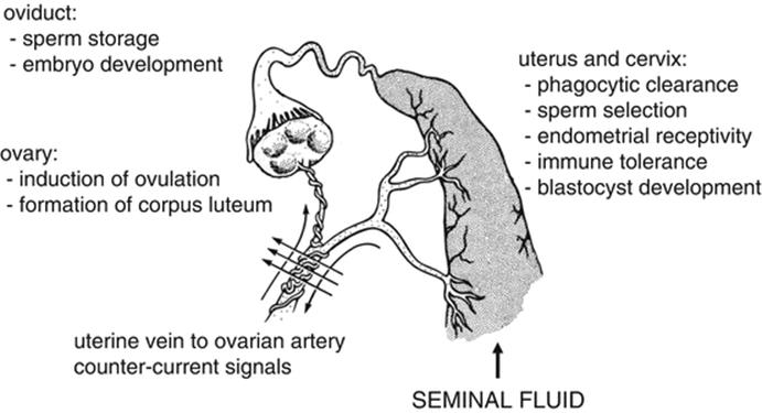
Fig. 6.1
The function of seminal fluid in the female reproductive tract following coitus. Seminal fluid exposure at coitus induces female responses that have consequences for conception and pregnancy. In the uterus, seminal fluid mediated effects include the phagocytic clearance of superfluous sperm and microorganisms introduced at mating, sperm selection, preparation of a receptive endometrium, promotion of blastocyst development, and regulation of the maternal immune response towards paternal antigens. In addition to effects in the uterus, seminal fluid has also been demonstrated to have effects in the higher reproductive tract. In the oviduct, components of seminal fluid influence sperm storage as well as promote the development of the embryo through generation of embryotrophic cytokines. Further effects are seen in the ovary, where seminal fluid exposure contributes to the induction of ovulation and the promotion of corpus luteum formation, potentially via a countercurrent mechanism between the uterine vein and ovarian artery. Figure is updated from (O’Leary et al. 2002)
6.6 Clearance of Microorganisms and Superfluous Sperm
A major component of the female immune response to seminal fluid is the recruitment of neutrophils, which are prevalent in the uterine luminal cavity during the initial acute inflammatory phase following mating (De et al. 1991; Pandya and Cohen 1985). These neutrophils are believed to influence the capacity of sperm to reach the oocyte by removing superfluous sperm, microorganisms, and seminal debris (Robertson 2007). At coitus, commensal microorganisms from male or female tissues or sexually transmitted infections have the opportunity to enter the normally sterile higher reproductive tract where they can be detected in the immediate period post-coitus with sterility being recovered within 24 h (Robertson et al. 1999).
In addition to a function for neutrophils and other phagocytes in the rapid clearance of microorganisms introduced at coitus, there is also evidence that these cells may respond to seminal fluid exposure and act to phagocytose non-fertilising sperm. This process was originally postulated to act to filter out morphologically abnormal spermatozoa (Tomlinson et al. 1992). However, both abnormal and apparently viable and morphologically normal spermatozoa can undergo phagocytosis, suggesting that sperm selection may occur on the basis of morphological or antigenic parameters other than the ability to fertilise (Robertson 2005), and conferring female immune cells with the capacity to influence sperm selection through the recognition of surface markers that indicate faulty or damaged sperm (Sutovsky and Lovercamp 2010). The means by which phagocytes could discriminate between gametes remains to be determined, but if a biologically plausible mechanism were identified, this could explain observations of active female tract selection of male gametes (Robertson 2007).
6.7 Endometrial Receptivity and Embryo Implantation
The immune cells recruited into the endometrium in response to seminal fluid contribute to restructuring the endometrial environment to facilitate embryo implantation and support the first phase of placental development (Robertson 2005). Of the leukocytes present in the female reproductive tract following coitus, macrophages are postulated to play a critical role in the tissue remodelling process through the regulation of angiogenesis. Macrophages are a potent source of vascular endothelial growth factor (VEGF) and other key angiogenic factors and vascular permeability agents (Yoshida et al. 1997). The expression of these angiogenic factors is tightly regulated through the estrous cycle and early pregnancy (Ma et al. 2001) with a reduction of VEGF mRNA expression in hamsters after coitus occurring when seminal fluid signalling is perturbed by mating with accessory gland-deficient males (Chow et al. 2003). Evidence of a role for seminal fluid in angiogenesis is also demonstrated by studies in the horse and pig which show an increase in uterine vasodilation and oedema in the days following coitus (O’Leary et al. 2004; Bollwein et al. 2003).
Macrophage secreted products may also target the extracellular matrix of the endometrial stroma, which is remodelled during the decidualisation period (Aplin 2002). In particular, macrophage-secreted matrix metalloproteinases (MMPs) and tissue inhibitors of MMPs (TIMPs) coordinate their expression to contribute to this remodelling process (Robertson 2005). In rodents, MMPs and TIMPs are induced during the pre-implantation period (Das et al. 1997; Feng et al. 1998), with seminal plasma postulated to contribute to their induction as the absence of accessory gland fluid at coitus in the hamster is associated with a reduction in MMP2 at the implantation site (Chow et al. 2003).
In addition to macrophages, seminal fluid-induced dendritic cells may contribute to tissue remodelling in preparation for implantation. Uterine dendritic cells have been demonstrated to be crucial in decidua formation and maternal receptivity. Depletion of uterine dendritic cells around the period of implantation leads to impaired decidual formation through perturbed angiogenesis. This process was postulated to be regulated by dendritic cell soluble fms-like tyrosine kinase-1 (sFlt1) and transforming growth factor beta (TGFB) which promote coordinated blood vessel maturation (Plaks et al. 2008).
Embryo attachment and implantation is a process requiring specific changes in the expression of integrins and mucins which function to allow close apposition and then adhesion between the blastocyst and implantation site (Aplin 1997). Fucosyltransferases, which modulate the expression of fucosylated structures involved in the attachment and implantation process, are deficient when seminal plasma signalling is perturbed by surgical removal of the seminal vesicle from males (Jasper et al. 2011), suggesting that seminal plasma exposure contributes to this process.
Macrophages are implicated as cellular mediators transmitting the effects of seminal fluid on endometrial receptivity. They are postulated to secrete molecules that target luminal epithelial cells to induce modifications to proteins involved in embryo attachment (Robertson 2005). In support of this, mouse macrophage-derived LIF and IL1B induce fucosyltransferases Fut2 and Fut4 required for embryo attachment and implantation (Jasper et al. 2011), and activity of fucosyltransferases is impaired when females are mated with seminal fluid-deficient males (Jasper et al. 2011). Studies in human tissues indicate a similar mechanism, where leukocytes influence the adhesive properties of uterine epithelial cells in vitro (Kosaka et al. 2003). In the cow, intrauterine administration of leukocytes may operate through a similar pathway to increase embryo implantation rates (Ideta et al. 2010).
6.8 Synthesis of Embryotrophic Cytokines
A key role of the female tract response to seminal fluid is the synthesis of embryotrophic cytokines that control the development of the pre-implantation embryo. Alterations to embryo programming may have a major influence on long-term offspring health as small perturbations in blastomere number and inner cell mass/trophectoderm allocation in the blastocyst are associated with an altered growth trajectory of the fetus and resulting offspring (Thompson et al. 2002). Embryos express cytokine receptors from conception until implantation with seminal fluid-induced cytokines GMCSF, IL6, LIF, and MCSF all exerting direct effects on blastocyst cell number and viability, gene expression, and developmental competence (Robertson 2005). Prominent cytokine receptor pathways demonstrated to be involved in pre-implantation embryo development include the phosphatidylinositol-3 kinase-protein kinase B (PI3K-AKT), Janus kinase/signal transducer and activator of transcription (JAK/STAT), and mitogen-activated protein kinase (MAPK) pathways. Depending on the cytokine, ligation may affect one or more of these signal transduction pathways to modify gene expression, metabolism, and differentiation, alter the epigenome, suppress the cell stress response, and ultimately impact blastomere survival (Robertson et al. 2011). Cytokine effects have profound impact on embryo developmental competence and programming future developmental trajectory, providing a pathway whereby the peri-conception environment affects the future growth of the fetus and phenotype of offspring after birth (Robertson et al. 2011; Sjoblom et al. 2005).
GMCSF, CSF1, IL6, and LIF are well-described examples of key embryotrophic cytokines induced in the uterus and oviduct by seminal fluid contact (Bromfield et al. 2014). In mice, GMCSF targets the pre-implantation embryo to promote blastocyst formation and increase the number of viable blastomeres by inhibiting apoptosis and facilitating glucose uptake (Robertson et al. 2001) and GMCSF deficiency compromises blastocyst development by inducing stress response and apoptotic gene pathways (Robertson et al. 2001; Chin et al. 2009). In cattle embryos, GMCSF regulates genes involved in de novo methylation to influence epigenetic reprogramming (Loureiro et al. 2009). IL6 regulates STAT3-dependent miRNAs that function to protect embryos from apoptosis (Shen et al. 2009) while neutralisation of LIF affects embryo implantation rates and fetoplacental development (Mitchell et al. 2002). Addition of CSF1 to embryo culture media improves embryo development (Pampfer et al. 1991) and reduced fertility in CSF1-deficient mice may be due to adverse effects in blastocysts (Cohen et al. 1997a). The contribution of seminal plasma to embryo development is illustrated by findings that coitus with seminal plasma-deficient males is associated with reduced cytokine synthesis and this is linked with a significant reduction in the rate of cleavage to the 2-cell stage in zygotes and reduction in the development of blastocysts from 2-cell embryos (Bromfield et al. 2014).
In contrast to cytokines and growth factors that exert a positive influence on embryonic development, factors that have profound inhibitory effects on embryo development have also been identified. Increased levels of apoptosis-inducing cytokines TNF, interferon gamma (IFNG), and TNF-related apoptosis-inducing ligand (TRAIL) in the female reproductive tract following coitus are emerging as factors that cause pre-implantation embryonic loss or impair embryo development. TNF acts in vitro to increase the percentage of apoptotic blastomeres in the mouse (Fabian et al. 2007), while IFNG inhibits the embryotrophic factor GMCSF and also exerts direct inhibitory effects in mouse embryo culture (Robertson et al. 1992; Haimovici et al. 1988). TRAIL induces apoptosis in the pre-implantation embryo (Riley et al. 2004). Like the embryotrophic cytokines, these cytokines are regulated by seminal fluid and particularly oviduct TRAIL expression is suppressed in response to seminal fluid factors (Bromfield et al. 2014).
In the human, the embryotrophic cytokines GMCSF, IL6, and LIF are induced in cervical and endometrial epithelial cells in response to seminal plasma exposure (Sharkey et al. 2012a, b; Gutsche et al. 2003), but to date whether this occurs in the higher reproductive tract in vivo has not been confirmed. These cytokines are predicted to have a similar role in the human as in vitro culture of human embryos in GMCSF improves developmental outcomes through an increase in embryos reaching blastocyst stage, with earlier blastulation and increased inner cell mass and trophectoderm cell number (Sjoblom et al. 1999), followed by improved implantation rates and progression to healthy birth after IVF treatment (Ziebe et al. 2013). Due to the challenges in accessing appropriate tissues, it has not been possible to ascertain the extent to which expression of embryotrophic and embryotoxic cytokines responds to coital activity to influence human embryo development and implantation after natural conception.
The capacity for seminal fluid to alter the balance of embryotrophic and embryotoxic cytokines provides a mechanism through which the female reproductive tract can reflect signals from the male, to either support or impair progression of pre-implantation embryo development, and impart longer-term programming. The capacity to liberate embryotoxic cytokines such as TRAIL in the absence of sufficient seminal fluid suppression raises the prospect of maternal tract ‘quality control’. If proven to occur, this would be relevant in couples where altered seminal fluid composition provides insufficient signalling factors (Sharkey et al. 2007) or where sexually transmitted infection alters the balance of seminal fluid signals to induce a proinflammatory phenotype (Rasmussen et al. 1997). These changes may affect progression of these embryos as well as imparting effects on offspring phenotype and health in adult life (Bromfield et al. 2014; Sjoblom et al. 2005).
6.9 Maternal Immune Tolerance
A critical component of the female tract response to seminal fluid which is essential for ongoing pregnancy success is programming of the maternal immune system to respond to the presence of the genetically disparate fetus. Immune tolerance must exist from the very earliest time the embryo contacts the maternal tissues at implantation. A combination of strategies contributes to the activation of maternal immune tolerance, with seminal fluid providing antigens and cytokines that, in the correct immune environment, drive production of regulatory T cells (Trowsdale and Betz 2006; Robertson et al. 2009a).
Seminal fluid contains several antigens specific to individual males including class Ia, Ib, and II major histocompatibility complex (MHC) (Hutter and Dohr 1998) that can be presented by antigen-presenting cells (APCs) such as macrophages and dendritic cells recruited into the endometrium following exposure to seminal fluid (Robertson et al. 1996; McMaster et al. 1992). These dendritic cells and macrophages in the presence of seminal fluid factors prostaglandin E (PGE) and TGFB differentiate into cells that mediate tolerogenic immune responses (Blois et al. 2007; Jaiswal et al. 2012). However, not all APCs are tolerogenic as the immune environment at this stage of pregnancy is highly varied with both tolerogenic and immunogenic cells present, demonstrating differing roles for these cells during peri-conception (Jaiswal et al. 2012). The APCs are thought to take up paternal antigens and either traffic to the uterine draining lymph nodes or interact locally with resident uterine T cells driving activation and expansion of clonal subsets of Treg cells which recognise and appropriately respond to paternal/fetal antigens (Robertson et al. 2009a). An antigen-specific T cell response has been demonstrated using a T cell receptor transgenic model with OVA as a model paternal antigen (Moldenhauer et al. 2009). The concept of seminal fluid-induced tolerance of paternal antigens is supported by experiments demonstrating prolonged survival of skin grafts of paternal origin in mated female mice (Lengerova and Vojtiskova 1966) and studies demonstrating tolerance towards tumor cells expressing the same MHC as the donor male (Robertson et al. 2009b).
Treg cells operate as potent suppressors of inflammation and cell-mediated immunity (Rudensky 2011). These cells act via a variety of mechanisms, generally involving the suppression of cytokine production and effector function in T cells, B cells, natural killer (NK) cells, dendritic cells, and macrophages (Sakaguchi 2000; Shevach 2002). In pregnancy, Treg cells play a critical role in mediating immune tolerance required for embryo implantation, with several studies demonstrating in the mouse that allogenic mating leads to fetal rejection unless sufficient Treg cells are present in the endometrium in the pre- and peri-implantation phase (Aluvihare et al. 2004). In humans, Treg cells play a similar regulatory role, with an increase in pregnant women of circulating CD4+ CD25+ cells highly enriched for the signature Treg transcription factor forkhead box P3 (FOXP3) in early pregnancy. The number of these regulatory cells peaks during the second trimester before declining at term (Somerset et al. 2004).
The presence of TGFB and PGE in seminal fluid confers immune-deviating activity that drives immune cells into tolerogenic phenotypes. Both factors have been linked with the induction of naïve T cells into suppressor T cells expressing Foxp3 (Chen et al. 2003; Baratelli et al. 2005) and exogenous TGFB delivery at conception is shown to boost vaginal Treg cell numbers which acts to reduce fetal loss in the abortion prone CBA/J x DBA/2 J model (Clark et al. 2008). Because of poor availability of suitable reagents for Treg cells, they have not been extensively studied in other species, other than in the cat where there is evidence that an increase in Treg cells in early pregnancy occurs after seminal fluid exposure (Lockett et al. 2010).
6.10 Ovulation and Corpus Luteum Formation
As well as the endometrium, seminal fluid exposure at coitus has effects in the ovary. This effect was first described in camelids, where effects of seminal plasma on induction of ovulation are well known (Li and Zhao 2004). It is challenging to conceive how seminal fluid moieties could reach the ovary via the oviduct. An alternative pathway for seminal fluid effects on ovarian function may be due to a unique countercurrent exchange mechanism through which prostaglandins and other small molecules can be transmitted from the uterine vein to the ovarian artery (Krzymowski et al. 1989). In the presence of a mature follicle, ovulation can be induced by natural coitus, artificial insemination, or intramuscular injection of seminal plasma, but not washed sperm. The ovulation-inducing factor in camelid seminal plasma has been identified as the highly conserved beta-nerve growth factor, which acts via an endocrine mechanism targeting the hypothalamic–pituitary axis to induce LH release and ovulation (Ratto et al. 2012). This so-called ovulation-inducing factor (OIF) is present in other species demonstrating induced ovulation (koalas and rabbits). Recent studies indicate that OIF in seminal plasma is conserved among species, including cattle, horses, pigs, mice, and other species considered to be spontaneous ovulators (Bogle et al. 2011; Ratto et al. 2006). Additional studies showing that OIF can induce ovulation in prepubertal mice (Bogle et al. 2011) and alter ovarian follicular wave dynamics in cows (Tanco et al. 2012) imply this factor may have broader relevance than originally thought (Ratto et al. 2012). Seminal plasma-mediated effects on ovarian function are clearly demonstrated in pigs, where a reduction in the interval between LH surge and ovulation, followed by elevated plasma progesterone concentration over the pre-implantation phase, occurs when seminal plasma is instilled into the uterus just prior to ovulation (Waberski et al. 1997; O’Leary et al. 2001).
As well as endocrine actions, the effects of seminal fluid on ovarian function in rodents and pigs may be mediated through local mechanisms involving stimulation of macrophage populations. Ovarian macrophages contribute to regulation of follicle development, ovulation, and post-ovulatory tissue remodelling associated with the conversion of the ruptured follicle into the corpus luteum (Cohen et al. 1997b; Care et al. 2013). Ovarian macrophages are present in the theca of developing follicles and their numbers increase at ovulation (Brannstrom et al. 1993). These macrophages support follicle growth, ovulation, and vascular homeostasis (Brannstrom et al. 1993; Wu et al. 2004; Van der Hoek et al. 2000; Turner et al. 2011). In both rodents and humans, macrophages migrate into the developing corpora lutea immediately after ovulation (Cohen et al. 1997b; Hellberg et al. 1991; Brannstrom et al. 1994a, b) where they influence steroidogenic function of luteal cells as well as tissue remodelling after luteal regression (Kirsch et al. 1981; Nelson et al. 1992).
In mice, seminal plasma influences macrophage recruitment into the ovary, as mating with seminal plasma-deficient males results in fewer macrophages in the corpus luteum compared to mating with seminal plasma sufficient males (Gangnuss et al. 2004). Cytokines that are induced by seminal fluid in the uterus such as MCP1, MCSF, and GMCSF (Robertson 2005) also act as chemoattractants to recruit macrophages into the ovary (Cohen et al. 1997b; Jasper et al. 2000; Townson et al. 1996), although whether these cytokines are induced in the ovary by seminal fluid remains to be examined. Serum progesterone levels on days 1, 2, and 4 of pregnancy in mice are not different with seminal plasma deficiency (Gangnuss et al. 2004). However, a threshold level of macrophage activity is essential for normal corpus luteum development as macrophage depletion disrupts the luteal microvascular network and alters expression of VEGFs required to support luteal angiogenesis in the peri-implantation period (Care et al. 2013). The result is increased inflammatory and apoptotic gene expression, decreased steroidogenic gene expression, and infertility which can be rescued by exogenous progesterone administration (Care et al. 2013).
6.11 Active Factors in Seminal Fluid
To further understand the function of seminal fluid at coitus, studies have aimed to identify the components of the ejaculate that induce the female tract response. Early studies in the mouse demonstrated through removal of the male accessory glands that the seminal plasma fraction contains many of the active factors, as its absence resulted in a reduced production of GMCSF and reduced leukocyte infiltration in the endometrium (Robertson et al. 1996). Further analysis of the high molecular weight component of mouse seminal fluid that induced these changes was able to identify TGFB as the principal trigger (Tremellen et al. 1998). These findings have since been confirmed in the human, where high levels of TGFB in seminal plasma contribute to the peri-conception inflammatory response by inducing GMCSF, IL1B, IL6, and LIF in human cervical epithelial and endometrial epithelial cells in vitro (Sharkey et al. 2012a; Gutsche et al. 2003). Secreted TGFB is initially produced in a precursor dimeric form where it is activated in the female tract after insemination as a consequence of the low pH, enzymatic activation with plasmin or interaction of the latent TGFB with thrombospondin-1 or avb6 integrins (Robertson 2005). In the mouse there is approximately 30 ng/ml TGFB1 in the ejaculate (Robertson et al. 2002) while in the human the TGFB concentrations are 219 ng/ml TGFB1, 5 ng/ml TGFB2, and 172 ng/ml TGFB3 (Sharkey et al. 2012a). TGFB is also detected in the seminal plasma of pigs (O’Leary et al. 2011) and sheep (Scott et al. 2006), with the pig carrying similar concentrations [150 ng/ml TGFB1 (O’Leary et al. 2011)] to that of the human and mouse. There is no correlation between TGFB and fertility (Loras et al. 1999), but studies in the seminal plasma of fertile men show that there are substantial differences in TGFB concentrations between individuals (Sharkey et al. 2012a).
In addition to TGFB, we have recently identified that ligands for TLR4 contribute to the peri-conception inflammatory response in the mouse. In these studies, the induction of colony-stimulating factor 3 (Csf3), Cxcl1, and Cxcl2was shown to require the presence of TLR4 while the TLR4 signalling pathway may also contribute to the induction of Il1a, Tnf, and Csf2 (Schjenken et al. 2015). While it has been speculated that bacterial products including endotoxin may contribute to the capacity of seminal fluid to interact with the female reproductive tract (Schaefer et al. 2004), the amount of endotoxin detectable in the female reproductive tract of mice following coitus (Schjenken et al. 2015) and in human seminal fluid (Sharkey, in preparation) is insufficient to explain the induction of TLR4 regulated cytokines. This suggests that other TLR4 ligands such as the endogenous danger-associated molecular patterns (DAMPs) contribute to eliciting the female response; however, these studies are yet to be completed. DAMPS have previously been identified in seminal fluid with beta-defensin-2 (Narciandi et al. 2014), fibronectin (Lilja et al. 1987), heat shock proteins (Rego et al. 2014; Pilch and Mann 2006), hyaluronidase (Geipel et al. 1992; Shimada et al. 2008), and S100 proteins (Rego et al. 2014; Donato et al. 2013) all being present.
In line with the functions of seminal fluid, activation of TLR4 signalling may contribute to tolerogenic immune responses (Conroy et al. 2008), as studies have demonstrated that CD45RBlow CD25+ regulatory T cells can express TLR4 and enhance suppressive function following TLR4 ligation (Caramalho et al. 2003), dendritic cells from TLR4-deficient mice have reduced capacity to produce IL10 in response to TLR4 ligation and have impaired expansion of Treg cells (Higgins et al. 2003), and Treg cells cultured in the presence of dendritic cells can induce tryptophan catabolism which enhances tolerogenic dendritic cell production (Fallarino et al. 2015). More detailed studies are required to understand how disruption of TLR4 signalling may affect the female response to seminal fluid in the mouse and other species and how in turn offspring health might be affected.
In humans, PGE in seminal fluid are postulated to act as signalling agents, most notably those in the 19-hydroxy (19-OH) form (Templeton et al. 1978). While PGE is detectable in equine seminal plasma (Claus et al. 1992), it is undetectable in rodent and porcine samples. In the human, extremely high levels of PGE are detected in seminal fluid, with 300 μM of the 19-OH form detectable (Templeton et al. 1978). Due to the immunosuppressive nature of 19-OH PGE, it was postulated that seminal prostaglandins may protect sperm from immunological damage in the male and protect the female against sensitisation to sperm antigens (James and Hargreave 1984; Alexander and Anderson 1987). In vitro experiments using human cervical explants stimulated with 19-OH PGE demonstrated an induction of IL8 and suppression of the anti-inflammatory molecule, secretory leukocyte protease inhibitor (Denison et al. 1999). Seminal plasma PGE2 has also been linked to facilitate tumorigenesis-angiogenesis in reproductive tract cells through activation of fibroblast growth factor 2, COX2, and VEGF expression and E series prostanoid-2 and -4 receptor, EGF receptor, and ERK1/2 signalling pathways (Battersby et al. 2007; Muller et al. 2006). Further, PGE has been demonstrated in the human to induce a regulatory phenotype in naïve CD4+CD25− T cells, enhance the in vitro inhibitory function of Treg cells, and induce expression of Foxp3 (Baratelli et al. 2005) which is consistent with the female tract response to seminal fluid.
Other factors postulated to contribute to seminal fluid signalling include IL8, which is present in seminal plasma of healthy human donors at 1456 pg/ml. Treatment of human endometrial epithelial cells in vitro with recombinant IL8 at physiological levels can induce IL1B, IL6, and LIF (Gutsche et al. 2003). Outside of mice and humans, other novel seminal fluid signalling molecules that have been examined are porcine sperm adhesion proteins (PSP)1 and PSP2, which contribute in part to the influx of neutrophils in pigs and can preserve sperm viability, motility, and mitochondrial activity (Rodriguez-Martinez et al. 2010; Caballero et al. 2006), and cysteine-rich secretory protein-3 (CRISP3) in horses, which mediates the interaction between sperm and neutrophils (Doty et al. 2011). Positive correlations between specific seminal plasma proteins and fertility have been observed in the horse (Brandon et al. 1999) and bull (Killian et al. 1993).
Despite the evidence showing an integral role for seminal plasma in the signalling process, it is becoming apparent that not all seminal fluid effects can be accounted for by the soluble fraction, raising the prospect that sperm also communicate with female reproductive tract cells. In domestic animal species, there is extensive evidence that sperm is critical to the inflammatory response and may be required to interact with seminal plasma to regulate the influx and function of immune cells (Schjenken and Robertson 2014). In the human and mouse, while the literature has focused on seminal plasma, there is some evidence for a sperm signalling component. In the mouse, sperm has been shown to contribute to the peri-conception immune environment as Foxp3 expression requires the presence of both sperm and seminal plasma for its complete induction (Guerin et al. 2011) while in the human, AI of human sperm into the cervix results in an influx of leukocytes (Pandya and Cohen 1985; Thompson et al. 1992). A function for sperm in the signalling process is further supported by studies in the mouse showing that sperm can form intimate associations with cells of the female reproductive tract following coitus (Reid 1965). Non-fertilising sperm can be taken up by female tissues, potentially by phagocytosis, where sperm-associated transcripts can be identified in the uterus and other distal sites, including the uterine draining lymph nodes for several days post-coitus (Watson et al. 1983). Similar interactions between sperm and the female reproductive tract can be seen in other species, including human, pigs, cow, dog, cats, bats, lizards, and even some marsupials (Suarez and Pacey 2006; Murakami et al. 1985; Rasweiler 1987; Nogueira et al. 2011; Pacey et al. 1995; Taylor et al. 2008; Rijsselaere et al. 2004; Ignotz et al. 2001; Breed et al. 1989). Seminal plasma proteins contribute to these interactions as bovine seminal plasma (BSP) proteins coat bull sperm and mediate interactions between sperm and oviductal epithelial cells to assist in the formation of sperm storage reservoirs, which function to maintain fertility of sperm until ovulation (Suarez 2008). In addition to the formation of storage reservoirs, these interactions assist in the clearance of sperm at coitus. Additionally, these interactions may potentially be a mechanism by which sperm communicates with the female reproductive tract to transmit antigen and facilitate pregnancy success; however, this remains to be determined.
In contrast to signalling molecules that promote successful pregnancy, there is also evidence that seminal fluid may contain factors that inhibit seminal fluid signalling. The type 1 cytokine IFNG can be detected in the seminal fluid of both mouse (~50 pg/ml) (Gopichandran et al. 2006) and human (range: 0–130 pg/ml) (Politch et al. 2007) and in the mouse has been shown to inhibit the tolerance inducing properties of TGFB during early pregnancy (Glynn et al. 2004). Whether IFNG levels are altered in the seminal fluid of infertile or subfertile men remains to be determined.
While several active factors that contribute to seminal fluid signalling have been identified, future studies will be required to understand in more detail how these factors interact to regulate the female response. In addition, if sperm contain signalling factors as has been postulated, studies will be required to elucidate these factors and their function. Additional factors beyond those discussed here no doubt exist. Exciting candidates include CD52, transglutaminase 4, and seminal vesicle secretion 2. CD52 is present in soluble form in seminal plasma and on the sperm surface (Koyama et al. 2009), which is recently reported to be a potent immune-regulatory signal driving induction of antigen-activated suppressor T cells, distinct from FOXP3+ Treg cells, from CD4 precursors (Bandala-Sanchez et al. 2013). Transglutaminase 4 and seminal vesicle secretion 2 are proteins which are known to play a critical role in copulatory plug formation (Dean 2013; Kawano et al. 2014). Interestingly, the absence of these seminal proteins disrupts not only plug formation but also fertility. Sperm from ejaculates lacking transglutaminase 4 fertilised at normal rates but were significantly less likely to give birth to a litter (Dean 2013). Sperm from seminal vesicle secretion 2-deficient mice are disrupted by uterine-derived cytotoxic factors (Kawano et al. 2014), suggesting that seminal plasma plays a protective role in sperm function.
Consideration of species-specific differences is integral as it is highly likely, due to the different sites of semen deposition, that different species will utilise different active factors. Within a species, fluctuations in the seminal fluid profile between individuals, or even within an individual, may influence the female response leading to altered outcomes for offspring depending on male health and environmental exposures.
6.12 Seminal Fluid and Programming of Offspring Health
The studies detailed above document a complex role for seminal fluid in preparing the female reproductive tract for pregnancy, particularly through regulating the maternal immune response to tolerate exposure to paternal antigens. These changes not only influence the period of early pregnancy but also have the potential to influence fetal development and offspring health. It is well documented that the in utero environment is a major determinant in programming how adult individuals respond to stressors and challenge, and the risk of disease later in life (Godfrey et al. 2010). Increasingly the peri-conception period is identified as the most sensitive for imprinting later life consequences, and the health and experiences of both the mother and father even prior to conception contribute through effects transmitted by the male and female gametes (Lane et al. 2014). There is evidence that altered conditions at conception can induce adaptations to protect the fetus from immediate effects, but a consequence of these changes is an increased risk of later metabolic disease (Barker and Clark 1997; Hanson and Gluckman 2005). Studies on the paternal contribution towards health and disease have identified smoking, age, environmental exposures, and obesity being linked to later offspring illness (Fullston et al. 2012), with sperm transmission of epigenetic mechanisms including non-coding RNAs (Fullston et al. 2012; Liu et al. 2012) being postulated to contribute to these changes.
As well as a contribution from sperm, there is the prospect that seminal fluid can contribute at fertilisation to influence the peri-conception environment in turn affecting later development and offspring health (Lane et al. 2014). Some evidence supporting a role for seminal plasma through its influence over the female reproductive tract at coitus is emerging. In mice, Bromfield et al. (Bromfield et al. 2014) demonstrate that conception in the absence of seminal plasma using mice surgically rendered seminal vesicle deficient (SVX) leads to a reduction in fecundity and altered fetal and neonatal outcomes. Mice mated with SVX males had a significant reduction in progression to pregnancy and fewer implanting embryos compared to intact mated females. Changes in development were observed as early as the eight-cell stage, where most embryos from SVX fathers were developmentally delayed. Prior to birth, changes in phenotype were evident as placental hypertrophy. Male offspring were observed to have altered growth trajectory and metabolic parameters with evidence of obesity, distorted metabolic hormones, reduced glucose tolerance, and hypertension. Embryo transfer experiments confirmed the role of seminal plasma in programming altered phenotype as increased adiposity was also observed in adult male progeny when normal 2-cell embryos were transferred to females mated with SVX males (Bromfield et al. 2014).
Similar studies have been conducted in the golden hamster, where ablation of the accessory sex glands (ASG), which produce seminal plasma, delayed entry of the zygote into the first cell cycle (Ying and Chow 1998), reduced pre-implantation embryonic cell number (Chan et al. 2001), resulted in an early transit of the embryo into the uterus from the oviduct (Wong et al. 2008), decreased the rate of implantation, and ultimately resulted in embryo death (Jiang et al. 2001). The offspring of fathers deficient in seminal plasma exhibited altered postnatal growth and elevated anxiety (Wong et al. 2007). It is postulated that these changes may be associated with epigenetic mechanisms as cleavage stage embryos from females mated with ASG-deficient males showed reduced acetylation and altered methylation kinetics which were associated with dysregulated expression of the paternally expressed Igf2 and Dlk1(Jiang et al. 2001).
In the human, there is no clear direct evidence implicating altered seminal fluid signalling activity with long-term health of offspring. However, there are clinical observations that are consistent with a similar function (discussed below). This means that alterations to the content of seminal fluid outside of the normal range may not manifest as impaired fertility, but could potentially alter fetal development and impart long-term consequences for offspring health. Carefully designed studies will be required to evaluate this, as effects on offspring can be subtle and require large populations to detect. Additionally, it will be important to distinguish effects of seminal fluid factors exerted via the female reproductive tract from those exerted through effects on sperm fertilising capacity and epigenetic status.
6.13 Clinical Implications of Altered Seminal Fluid Signalling
These considerations raise the question of the extent to which seminal fluid exposure may influence pregnancy outcomes in humans. The success of IVF even in women without male partners demonstrates that seminal plasma is not an absolute requirement for pregnancy. Rather, evidence suggests that rather than being an absolute prerequisite, seminal plasma influences the success and quality of pregnancy outcome (Robertson 2005). There are few definitive studies in humans focussed on evaluating effects of seminal fluid, but clinical studies point to a consistent beneficial effect in improving the chance of conception and protecting from immune-mediated gestational disorders such as preeclampsia.
Preeclampsia is characterised by a significant reduction in peripheral blood CD4+CD25high T regulatory cells and decidual Foxp3-positive cells within the CD3+ population (Sasaki et al. 2007). The altered Treg cell numbers may in part be due to altered seminal fluid function, with extensive evidence pointing to an important role for TGFB in regulating human Treg cell reactivity and autoimmunity (Rubtsov and Rudensky 2007). Clinical studies show that preeclampsia is more common in situations where limited exposure to the conceiving partners seminal fluid has occurred—including first conception, after a short period of cohabitation, when barrier contraception has been used, or when multiparous mothers conceive with a new male partner [reviewed in Schjenken and Robertson (2014)]. Treg cell numbers and function are found to be altered in the peripheral blood and decidua of pregnant women who have spontaneous abortions compared to induced abortions (Sasaki et al. 2004; Arruvito et al. 2007), while FOXP3expression, used as a surrogate measure of Treg cells, is reduced in the endometrium of women with primary unexplained infertility (Jasper et al. 2006).
Data from ART further support a protective function for seminal fluid. The absence of seminal plasma in the in vivo setting has been associated with higher rates of implantation failure and decreases in embryo quality [reviewed in Schjenken and Robertson (2014)]. Exposure to semen around the time of embryo transfer is demonstrated to improve embryo viability 6–8 weeks post-transfer (Tremellen et al. 2000). Given the emerging recognition that IVF is linked with reduced birth weights and impaired metabolic health outcomes for IVF children (Hart and Norman 2013), and the overlap between these changes and the phenotypes of offspring conceived without seminal fluid in mouse studies (Bromfield et al. 2014), it would seem prudent to more formally investigate the impact of seminal fluid on perinatal outcome and offspring health in human IVF. One difficulty with advising couples to have coitus at the time of embryo transfer is the small but real prospect of multiple pregnancy due to natural conception, but this would be expected to be a small risk in couples experiencing long-term infertility.
6.14 Conclusions
Seminal fluid signalling appears to play an integral role in eliciting inflammatory changes in the female reproductive tract following coitus in all species examined so far, despite differences in sites of semen deposition and reproductive physiology. This implies that the capacity for seminal fluid to exert effects in the female reproductive tract has biological value beyond the support of sperm. The enormous variation between mammalian species in seminal fluid volume, tissue origin and composition, and actions in the female implies rapid evolution that is driven by sexual selection, analogous to that which occurs in invertebrate species. For sexual selection to occur there must be variation in the female response that impacts the likelihood of progression to pregnancy. The array of effects induced by seminal fluid that impact conception, embryo development, implantation, and fetal growth together impact fertility and fecundity and ultimately influence offspring survival and health. Signalling factors within seminal fluid, in particular in the seminal plasma fraction, have been identified, but there are clearly other, as yet unknown factors that contribute to signalling in the female tract, potentially including sperm.
Despite the advances that have been made in understanding the contribution of seminal fluid, there is still much to learn. In particular, the contribution of seminal fluid to offspring phenotype requires further study and the knowledge to be gained will have major implications in human and animal ART, where seminal fluid signalling is often overlooked or avoided. In humans there are substantial variations in seminal fluid profiles, but the implications and causes of this are not known. Whether these variations, or the presence of conditions such as reproductive tract infection, affect the capacity of seminal fluid to induce the appropriate response and ultimately affect male fertility, pregnancy health, or even offspring phenotype remains to be understood. Ultimately, a better understanding of seminal fluid signalling may improve success rates and outcomes of human IVF, and yield novel therapies for infertility and pathologies of pregnancy.
References
Alexander NJ, Anderson DJ (1987) Immunology of semen. Fertil Steril 47:192–204PubMed
Aluvihare VR, Kallikourdis M, Betz AG (2004) Regulatory T cells mediate maternal tolerance to the fetus. Nat Immunol 5:266–271PubMed
Aplin JD (1997) Adhesion molecules in implantation. Rev Reprod 2:84–93PubMed
Aplin JD (2002) Endometrial extracellular matrix. In: Glasser SR, Aplin J, Guidice LC, Tabibzadeh S (eds) The endometrium. Taylor and Francis, London and New York, pp 294–307
Arruvito L, Sanz M, Banham AH, Fainboim L (2007) Expansion of CD4 + CD25 + and FOXP3+ regulatory T cells during the follicular phase of the menstrual cycle: implications for human reproduction. J Immunol 178:2572–2578PubMed
Aumuller G, Riva A (1992) Morphology and functions of the human seminal vesicle. Andrologia 24:183–196PubMed
Bandala-Sanchez E, Zhang Y, Reinwald S, Dromey JA, Lee BH, Qian J, Bohmer RM, Harrison LC (2013) T cell regulation mediated by interaction of soluble CD52 with the inhibitory receptor Siglec-10. Nat Immunol 14:741–748PubMed
Baratelli F, Lin Y, Zhu L, Yang S-C, Heuze-Vourc'h N, Zeng G, Reckamp K, Dohadwala M, Sharma S, Dubinett SM (2005) Prostaglandin E2 induces FOXP3 gene expression and T regulatory cell function in human CD4+ T cells. J Immunol 175:1483–1490PubMed
Barker DJ, Clark PM (1997) Fetal undernutrition and disease in later life. Rev Reprod 2:105–112PubMed
Battersby S, Sales KJ, Williams AR, Anderson RA, Gardner S, Jabbour HN (2007) Seminal plasma and prostaglandin E2 up-regulate fibroblast growth factor 2 expression in endometrial adenocarcinoma cells via E-series prostanoid-2 receptor-mediated transactivation of the epidermal growth factor receptor and extracellular signal-regulated kinase pathway. Hum Reprod 22:36–44PubMedCentralPubMed
Blois SM, Kammerer U, Alba Soto C, Tometten MC, Shaikly V, Barrientos G, Jurd R, Rukavina D, Thomson AW, Klapp BF, Fernandez N, Arck PC (2007) Dendritic cells: key to fetal tolerance? Biol Reprod 77:590–598PubMed
Bogle OA, Ratto MH, Adams GP (2011) Evidence for the conservation of biological activity of ovulation-inducing factor in seminal plasma. Reproduction 142:277–283PubMed
Bollwein H, Sowade C, Stolla R (2003) The effect of semen extender, seminal plasma and raw semen on uterine and ovarian blood flow in mares. Theriogenology 60:607–616PubMed
Brandon CI, Heusner GL, Caudle AB, Fayrer-Hosken RA (1999) Two-dimensional polyacrylamide gel electrophoresis of equine seminal plasma proteins and their correlation with fertility. Theriogenology 52:863–873PubMed
Brannstrom M, Mayrhofer G, Robertson SA (1993) Localization of leukocyte subsets in the rat ovary during the periovulatory period. Biol Reprod 48:277–286PubMed
Brannstrom M, Giesecke L, Moore IC, van den Heuvel CJ, Robertson SA (1994a) Leukocyte subpopulations in the rat corpus luteum during pregnancy and pseudopregnancy. Biol Reprod 50:1161–1167PubMed
Brannstrom M, Pascoe V, Norman RJ, McClure N (1994b) Localization of leukocyte subsets in the follicle wall and in the corpus luteum throughout the human menstrual cycle. Fertil Steril 61:488–495PubMed
Breed WG, Leigh CM, Bennett JH (1989) Sperm morphology and storage in the female reproductive tract of the fat-tailed dunnart, Sminthopsis crassicaudata (Marsupialia: Dasyuridae). Gamete Res 23:61–75PubMed
Bromfield JJ, Schjenken JE, Chin PY, Care AS, Jasper MJ, Robertson SA (2014) Maternal tract factors contribute to paternal seminal fluid impact on metabolic phenotype in offspring. Proc Natl Acad Sci USA 111:2200–2205PubMedCentralPubMed
Bulletti C, De Ziegler D, Giacomucci E, Polli V, Rossi S, Alfieri S, Flamigni C (1997) Vaginal drug delivery: the first uterine pass effect. Ann N Y Acad Sci 828:285–290PubMed
Caballero I, Vazquez JM, Garcia EM, Roca J, Martinez EA, Calvete JJ, Sanz L, Ekwall H, Rodriguez-Martinez H (2006) Immunolocalization and possible functional role of PSP-I/PSP-II heterodimer in highly extended boar spermatozoa. J Androl 27:766–773PubMed
Caramalho I, Lopes-Carvalho T, Ostler D, Zelenay S, Haury M, Demengeot J (2003) Regulatory T cells selectively express toll-like receptors and are activated by lipopolysaccharide. J Exp Med 197:403–411PubMedCentralPubMed
Care AS, Diener KR, Jasper MJ, Brown HM, Ingman WV, Robertson SA (2013) Macrophages regulate corpus luteum development during embryo implantation in mice. J Clin Invest 123:3472–3487PubMedCentralPubMed
Carp HJ, Serr DM, Mashiach S, Nebel L (1984) Influence of insemination on the implantation of transferred rat blastocysts. Gynecol Obstet Invest 18:194–198PubMed
Ceelen M, van Weissenbruch MM, Roos JC, Vermeiden JP, van Leeuwen FE, Delemarre-van de Waal HA (2007) Body composition in children and adolescents born after in vitro fertilization or spontaneous conception. J Clin Endocrinol Metab 92:3417–3423PubMed
Ceelen M, van Weissenbruch MM, Vermeiden JP, van Leeuwen FE, Delemarre-van de Waal HA (2008) Cardiometabolic differences in children born after in vitro fertilization: follow-up study. J Clin Endocrinol Metab 93:1682–1688PubMed
Chan OC, Chow PH, O WS (2001) Total ablation of paternal accessory sex glands curtails developmental potential in preimplantation embryos in the golden hamster. Anat Embryol (Berl) 204:117–122
Chapman T, Davies SJ (2004) Functions and analysis of the seminal fluid proteins of male Drosophila melanogaster fruit flies. Peptides 25:1477–1490PubMed
Chapman T, Neubaum DM, Wolfner MF, Partridge L (2000) The role of male accessory gland protein Acp36DE in sperm competition in Drosophila melanogaster. Proc Biol Sci 267:1097–1105PubMedCentralPubMed
Chen W, Jin W, Hardegen N, Lei KJ, Li L, Marinos N, McGrady G, Wahl SM (2003) Conversion of peripheral CD4+CD25- naive T cells to CD4+CD25+ regulatory T cells by TGF-beta induction of transcription factor Foxp3. J Exp Med 198:1875–1886PubMedCentralPubMed
Chin PY, Macpherson AM, Thompson JG, Lane M, Robertson SA (2009) Stress response genes are suppressed in mouse preimplantation embryos by granulocyte-macrophage colony-stimulating factor (GM-CSF). Hum Reprod 24:2997–3009PubMed
Chow PH, Jiang HY, Poon HK, Lee KH, O WS (2003) Embryos sired by males without accessory sex glands induce failure of uterine support: a study of VEGF, MMP and TGF expression in the golden hamster. Anat Embryol 206:203–213PubMed
Chu TM, Nocera MA, Flanders KC, Kawinski E (1996) Localization of seminal plasma transforming growth factor-beta1 on human spermatozoa: an immunocytochemical study. Fertil Steril 66:327–330PubMed
Clark DA, Fernandes J, Banwatt D (2008) Prevention of spontaneous abortion in the CBA x DBA/2 mouse model by intravaginal TGF-beta and local recruitment of CD4+8+ FOXP3+ cells. Am J Reprod Immunol 59:525–534PubMed
Claus R (1990) Physiological role of seminal components in the reproductive tract of the female pig. J Reprod Fertil Suppl 40:117–131PubMed
Claus R, Dimmick MA, Gimenez T, Hudson LW (1992) Estrogens and prostaglandin F2alpha in the semen and blood plasma of stallions. Theriogenology 38:687–693PubMed
Cohen PE, Hardy MP, Pollard JW (1997a) Colony-stimulating factor-1 plays a major role in the development of reproductive function in male mice. Mol Endocrinol 11:1636–1650PubMed
Cohen PE, Zhu L, Pollard JW (1997b) Absence of colony stimulating factor-1 in osteopetrotic (csfmop/csfmop) mice disrupts estrous cycles and ovulation. Biol Reprod 56:110–118PubMed
Conroy H, Marshall NA, Mills KH (2008) TLR ligand suppression or enhancement of Treg cells? A double-edged sword in immunity to tumours. Oncogene 27:168–180PubMed
Das SK, Yano S, Wang J, Edwards DR, Nagase H, Dey SK (1997) Expression of matrix metalloproteinases and tissue inhibitors of metalloproteinases in the mouse uterus during the peri-implantation period. Dev Genet 21:44–54PubMed
Das SC, Isobe N, Yoshimura Y (2008) Mechanism of prolonged sperm storage and sperm survivability in hen oviduct: a review. Am J Reprod Immunol 60:477–481PubMed
Das SC, Isobe N, Yoshimura Y (2009) Changes in the expression of interleukin-1beta and lipopolysaccharide-induced TNF factor in the oviduct of laying hens in response to artificial insemination. Reproduction 137:527–536PubMed
De M, Choudhuri R, Wood GW (1991) Determination of the number and distribution of macrophages, lymphocytes, and granulocytes in the mouse uterus from mating through implantation. J Leukoc Biol 50:252–262PubMed
Dean MD (2013) Genetic disruption of the copulatory plug in mice leads to severely reduced fertility. PLoS Genet 9:e1003185PubMedCentralPubMed
Denison FC, Grant VE, Calder AA, Kelly RW (1999) Seminal plasma components stimulate interleukin-8 and interleukin-10 release. Mol Hum Reprod 5:220–226PubMed
Donato R, Cannon BR, Sorci G, Riuzzi F, Hsu K, Weber DJ, Geczy CL (2013) Functions of S100 proteins. Curr Mol Med 13:24–57PubMedCentralPubMed
Doty A, Buhi WC, Benson S, Scoggin KE, Pozor M, Macpherson M, Mutz M, Troedsson MH (2011) Equine CRISP3 modulates interaction between spermatozoa and polymorphonuclear neutrophils. Biol Reprod 85:157–164PubMed
Eberhard WG (2009) Postcopulatory sexual selection: Darwin’s omission and its consequences. Proc Natl Acad Sci USA 106(Suppl 1):10025–10032PubMedCentralPubMed
England GC, Russo M, Freeman SL (2012) The bitch uterine response to semen deposition and its modification by male accessory gland secretions. Vet J 195:179–84PubMed
Fabian D, Juhas S, Il'kova G, Koppel J (2007) Dose- and time-dependent effects of TNFalpha and actinomycin D on cell death incidence and embryo growth in mouse blastocysts. Zygote 15:241–249PubMed
Fallarino F, Pallotta MT, Matino D, Gargaro M, Orabona C, Vacca C, Mondanelli G, Allegrucci M, Boon L, Romani R, Talesa VN, Puccetti P et al (2015) LPS-conditioned dendritic cells confer endotoxin tolerance contingent on tryptophan catabolism. Immunobiology 220:315–21PubMed
Feng J, Woessner JF Jr, Zhu C (1998) Matrilysin activity in the rat uterus during the oestrous cycle and implantation. J Reprod Fertil 114:347–350PubMed
Flowers WL, Esbenshade KL (1993) Optimizing management of natural and artificial matings in swine. J Reprod Fertil Suppl 48:217–228PubMed
Fullston T, Palmer NO, Owens JA, Mitchell M, Bakos HW, Lane M (2012) Diet-induced paternal obesity in the absence of diabetes diminishes the reproductive health of two subsequent generations of mice. Hum Reprod 27:1391–1400PubMed
Gangnuss S, Sutton-McDowall ML, Robertson SA, Armstrong DT (2004) Seminal plasma regulates corpora lutea macrophage populations during early pregnancy in mice. Biol Reprod 71:1135–1141PubMed
Geipel U, Kropf J, Krause W, Gressner AM (1992) The concentration pattern of laminin, hyaluronan, and aminoterminal propeptide of type III procollagen in seminal fluid. Andrologia 24:205–211PubMed
Gillott C (2003) Male accessory gland secretions: modulators of female reproductive physiology and behavior. Annu Rev Entomol 48:163–184PubMed
Glynn DJ, Sharkey DJ, Robertson SA (2004) Interferon-gamma inhibits female reproductive tract responsiveness to seminal plasma, 37th Annual Meeting of the Society for the Study of Reproduction. Biol Reprod 70:242
Godfrey KM, Gluckman PD, Hanson MA (2010) Developmental origins of metabolic disease: life course and intergenerational perspectives. Trends Endocrinol Metab 21:199–205PubMed
Gopichandran N, Ekbote UV, Walker JJ, Brooke D, Orsi NM (2006) Multiplex determination of murine seminal fluid cytokine profiles. Reproduction 131:613–621PubMed
Guerin LR, Moldenhauer LM, Prins JR, Bromfield JJ, Hayball JD, Robertson SA (2011) Seminal fluid regulates accumulation of FOXP3(+) regulatory T cells in the preimplantation mouse uterus through expanding the FOXP3(+) cell pool and CCL19-mediated recruitment. Biol Reprod 85:397–408PubMed
Gutsche S, von Wolff M, Strowitzki T, Thaler CJ (2003) Seminal plasma induces mRNA expression of IL-1beta, IL-6 and LIF in endometrial epithelial cells in vitro. Mol Hum Reprod 9:785–791PubMed
Haimovici F, Hill JA, Anderson DJ (1988) Variables affecting toxicity of human sera in mouse embryo cultures. J In Vitro Fert Embryo Transf 5:202–206PubMed
Hanson MA, Gluckman PD (2005) Developmental processes and the induction of cardiovascular function: conceptual aspects. J Physiol 565:27–34PubMedCentralPubMed
Hart R, Norman RJ (2013) The longer-term health outcomes for children born as a result of IVF treatment: part I–general health outcomes. Hum Reprod Update 19:232–243PubMed
Heifetz Y, Lung O, Frongillo EA Jr, Wolfner MF (2000) The Drosophila seminal fluid protein Acp26Aa stimulates release of oocytes by the ovary. Curr Biol 10:99–102PubMed
Hellberg P, Thomsen P, Janson PO, Brannstrom M (1991) Leukocyte supplementation increases the luteinizing hormone-induced ovulation rate in the in vitro-perfused rat ovary. Biol Reprod 44:791–797PubMed
Higgins SC, Lavelle EC, McCann C, Keogh B, McNeela E, Byrne P, O’Gorman B, Jarnicki A, McGuirk P, Mills KH (2003) Toll-like receptor 4-mediated innate IL-10 activates antigen-specific regulatory T cells and confers resistance to Bordetella pertussis by inhibiting inflammatory pathology. J Immunol 171:3119–3127PubMed
Hutter H, Dohr G (1998) HLA expression on immature and mature human germ cells. J Reprod Immunol 38:101–122PubMed
Ideta A, Sakai S, Nakamura Y, Urakawa M, Hayama K, Tsuchiya K, Fujiwara H, Aoyagi Y (2010) Administration of peripheral blood mononuclear cells into the uterine horn to improve pregnancy rate following bovine embryo transfer. Anim Reprod Sci 117:18–23PubMed
Ignotz GG, Lo MC, Perez CL, Gwathmey TM, Suarez SS (2001) Characterization of a fucose-binding protein from bull sperm and seminal plasma that may be responsible for formation of the oviductal sperm reservoir. Biol Reprod 64:1806–1811PubMed
Jaiswal MK, Mallers TM, Larsen B, Kwak-Kim J, Chaouat G, Gilman-Sachs A, Beaman KD (2012) V-ATPase upregulation during early pregnancy: a possible link to establishment of an inflammatory response during preimplantation period of pregnancy. Reproduction 143:713–725PubMed
James K, Hargreave TB (1984) Immunosuppression by seminal plasma and its possible clinical significance. Immunol Today 5:357–363
Jasper MJ, Robertson SA, Van der Hoek KH, Bonello N, Brannstrom M, Norman RJ (2000) Characterization of ovarian function in granulocyte-macrophage colony-stimulating factor-deficient mice. Biol Reprod 62:704–713PubMed
Jasper MJ, Bromfield JJ, Skinner R, Robertson SA (2005) 242. LIF expression is induced in the mouse oviduct following activation by seminal factors. Reprod Fertil Dev 17:96–96
Jasper MJ, Tremellen KP, Robertson SA (2006) Primary unexplained infertility is associated with reduced expression of the T-regulatory cell transcription factor Foxp3 in endometrial tissue. Mol Hum Reprod 12:301–308PubMed
Jasper MJ, Care AS, Sullivan B, Ingman WV, Aplin JD, Robertson SA (2011) Macrophage-derived LIF and IL1B regulate alpha(1,2)fucosyltransferase 2 (Fut2) expression in mouse uterine epithelial cells during early pregnancy. Biol Reprod 84:179–188PubMed
Jiang HY, O WS, Lee KH, Tang PL, Chow PH (2001) Ablation of paternal accessory sex glands is detrimental to embryo development during implantation. Anat Embryol (Berl) 203:255–263
Johansson M, Bromfield JJ, Jasper MJ, Robertson SA (2004) Semen activates the female immune response during early pregnancy in mice. Immunology 112:290–300PubMedCentralPubMed
Katila T (2012) Post-mating inflammatory responses of the uterus. Reprod Domest Anim 47(Suppl 5):31–41PubMed
Kawano N, Araki N, Yoshida K, Hibino T, Ohnami N, Makino M, Kanai S, Hasuwa H, Yoshida M, Miyado K, Umezawa A (2014) Seminal vesicle protein SVS2 is required for sperm survival in the uterus. Proc Natl Acad Sci USA 111:4145–4150PubMedCentralPubMed
Killian GJ, Chapman DA, Rogowski LA (1993) Fertility-associated proteins in Holstein bull seminal plasma. Biol Reprod 49:1202–1207PubMed
Kirsch TM, Friedman AC, Vogel RL, Flickinger GL (1981) Macrophages in corpora lutea of mice: characterization and effects on steroid secretion. Biol Reprod 25:629–638PubMed
Kosaka K, Fujiwara H, Tatsumi K, Yoshioka S, Higuchi T, Sato Y, Nakayama T, Fujii S (2003) Human peripheral blood mononuclear cells enhance cell-cell interaction between human endometrial epithelial cells and BeWo-cell spheroids. Hum Reprod 18:19–25PubMed
Koyama K, Hasegawa A, Komori S (2009) Functional aspects of CD52 in reproduction. J Reprod Immunol 83:56–59PubMed
Koyanagi F, Nishiyama H (1981) Fate of spermatozoa that do not participate in fertilization in the domestic fowl. Cell Tissue Res 214:89–95PubMed
Krzymowski T, Stefanczyk-Krzymowska S, Koziorowski M (1989) Counter current transfer of PGF2 alpha in the mesometrial vessels as a mechanism for prevention of luteal regression in early pregnancy. Acta Physiol Pol 40:23–34PubMed
Kunz G, Beil D, Deininger H, Wildt L, Leyendecker G (1996) The dynamics of rapid sperm transport through the female genital tract: evidence from vaginal sonography of uterine peristalsis and hysterosalpingoscintigraphy. Hum Reprod 11:627–632PubMed
Laflamme BA, Avila FW, Michalski K, Wolfner MF (2014) A Drosophila protease cascade member, seminal metalloprotease-1, is activated stepwise by male factors and requires female factors for full activity. Genetics 196:1117–1129PubMedCentralPubMed
Lane M, Robker RL, Robertson SA (2014) Parenting from before conception. Science 345:756–760PubMed
Lengerova A, Vojtiskova M (1966) Prolonged survival of syngenic male skin grafts in parous C57 B1 mice. Folia Biol 8:21
Li X, Zhao X (2004) Separation and purification of ovulation-inducing factors in the seminal plasma of the Bactrian camel (Camelus bactrianus). Vet Res Commun 28:235–245PubMed
Lilja H, Oldbring J, Rannevik G, Laurell CB (1987) Seminal vesicle-secreted proteins and their reactions during gelation and liquefaction of human semen. J Clin Invest 80:281–285PubMedCentralPubMed
Lima FS, Risco CA, Thatcher MJ, Benzaquen ME, Archbald LF, Santos JE, Thatcher WW (2009) Comparison of reproductive performance in lactating dairy cows bred by natural service or timed artificial insemination. J Dairy Sci 92:5456–5466PubMed
Liu WM, Pang RT, Chiu PC, Wong BP, Lao K, Lee KF, Yeung WS (2012) Sperm-borne microRNA-34c is required for the first cleavage division in mouse. Proc Natl Acad Sci USA 109:490–494PubMedCentralPubMed
Lockett NN, Scott VL, Boudreaux CE, Clay BT, Pruett SB, Ryan PL, Coats KS (2010) Expression of regulatory T cell (Treg) activation markers in endometrial tissues from early and late pregnancy in the feline immunodeficiency virus (FIV)-infected cat. Placenta 31:796–802PubMedCentralPubMed
Loras B, Vetele F, El Malki A, Rollet J, Soufir JC, Benahmed M (1999) Seminal transforming growth factor-beta in normal and infertile men. Hum Reprod 14:1534–1539PubMed
Loureiro B, Bonilla L, Block J, Fear JM, Bonilla AQ, Hansen PJ (2009) Colony-stimulating factor 2 (CSF-2) improves development and posttransfer survival of bovine embryos produced in vitro. Endocrinology 150:5046–5054PubMedCentralPubMed
Lovell JW, Getty R (1968) Fate of semen in the uterus of the sow: histologic study of endometrium during the 27 hours after natural service. Am J Vet Res 29:609–625PubMed
Lucy MC (2001) Reproductive loss in high-producing dairy cattle: where will it end? J Dairy Sci 84:1277–1293PubMed
Lung O, Wolfner MF (2001) Identification and characterization of the major Drosophila melanogaster mating plug protein. Insect Biochem Mol Biol 31:543–551PubMed
Ma W, Tan J, Matsumoto H, Robert B, Abrahamson DR, Das SK, Dey SK (2001) Adult tissue angiogenesis: evidence for negative regulation by estrogen in the uterus. Mol Endocrinol 15:1983–1992PubMed
Maegawa M, Kamada M, Irahara M, Yamamoto S, Yoshikawa S, Kasai Y, Ohmoto Y, Gima H, Thaler CJ, Aono T (2002) A repertoire of cytokines in human seminal plasma. J Reprod Immunol 54:33–42PubMed
Maher ER, Afnan M, Barratt CL (2003) Epigenetic risks related to assisted reproductive technologies: epigenetics, imprinting, ART and icebergs? Hum Reprod 18:2508–2511PubMed
Mann T (1964) The biochemistry of semen and the male reproductive tract. Wiley, New York
Mattner PE (1968) The distribution of spermatozoa and leucocytes in the female genital tract in goats and cattle. J Reprod Fertil 17:253–261PubMed
McDonald LE, Black WG, McNutt SH, Casida LE (1952) The response of the rabbit uterus to instillation of semen at different phases of the estrous cycle. Am J Vet Res 13:419–424PubMed
McGraw LA, Suarez SS, Wolfner MF (2015) On a matter of seminal importance. Bioessays 37:142–147PubMed
McMaster M, Newton R, Dey S, Andrews G (1992) Activation and distribution of inflammatory cells in the mouse uterus during the preimplantation period. J Immunol 148:1699–1705PubMed
Mitchell MH, Swanson RJ, Oehninger S (2002) In vivo effect of leukemia inhibitory factor (LIF) and an anti-LIF polyclonal antibody on murine embryo and fetal development following exposure at the time of transcervical blastocyst transfer. Biol Reprod 67:460–464PubMed
Moldenhauer LM, Diener KR, Thring DM, Brown MP, Hayball JD, Robertson SA (2009) Cross-presentation of male seminal fluid antigens elicits T cell activation to initiate the female immune response to pregnancy. J Immunol 182:8080–8093PubMed
Muller M, Sales KJ, Katz AA, Jabbour HN (2006) Seminal plasma promotes the expression of tumorigenic and angiogenic genes in cervical adenocarcinoma cells via the E-series prostanoid 4 receptor. Endocrinology 147:3356–3365PubMed
Murakami M, Nishida T, Shiromoto M, Iwanaga S (1985) Phagocytosis of spermatozoa and latex beads by the epithelial cell of the cat oviduct: combined SEM and TEM study. Arch Histol Jpn 48:519–526PubMed
Murray FA, Grifo P, Parker CF (1983) Increased litter size in gilts by intrauterine infusion of seminal and sperm antigens before mating. J Anim Sci 56:895–900PubMed
Murray FA, Grifo P, Parker CF (1986) Increased litter size in gilts by presensitization with boar semen. J Anim Sci 196(Suppl):189
Narciandi F, Lloyd A, Meade KG, O’Farrelly C (2014) A novel subclass of bovine β-defensins links reproduction and immunology. Reprod Fertil Dev 26:769–77PubMed
Nelson SE, McLean MP, Jayatilak PG, Gibori G (1992) Isolation, characterization, and culture of cell subpopulations forming the pregnant rat corpus luteum. Endocrinology 130:954–966PubMed
Nogueira KD, Araujo VA, Rodrigues Sartori SS, Neves CA (2011) Phagocytosis of spermatozoa by epithelial cells in the vagina of the lizard Hemidactylus mabouia (Reptilia, Squamata). Micron 42:377–80
O WS, Chen HQ, Chow PH (1988) Effects of male accessory sex gland secretions on early embryonic development in the golden hamster. J Reprod Fertil 84:341–344PubMed
O’Leary S, Robertson SA, Armstrong DT (2001) Seminal plasma treatment stimulates progesterone synthesis during early pregnancy in the pig. In: Proceedings of the 32nd annual conference of the society for reproductive biology, Gold Coast, QLD
O’Leary S, Robertson SA, Armstrong DT (2002) The influence of seminal plasma on ovarian function in pigs—a novel inflammatory mechanism? J Reprod Immunol 57:225–238PubMed
O’Leary S, Armstrong DT, Robertson SA (2011) Transforming growth factor-beta (TGFbeta) in porcine seminal plasma. Reprod Fertil Dev 23:748–758PubMed
O’Leary S, Jasper MJ, Warnes GM, Armstrong DT, Robertson SA (2004) Seminal plasma regulates endometrial cytokine expression, leukocyte recruitment and embryo development in the pig. Reproduction 128:237–247PubMed
Owen DH, Katz DF (2005) A review of the physical and chemical properties of human semen and the formulation of a semen simulant. J Androl 26:459–469PubMed
Pacey AA, Hill CJ, Scudamore IW, Warren MA, Barratt CL, Cooke ID (1995) The interaction in vitro of human spermatozoa with epithelial cells from the human uterine (fallopian) tube. Hum Reprod 10:360–366PubMed
Palm F, Walter I, Budik S, Kolodziejek J, Nowotny N, Aurich C (2008) Influence of different semen extenders and seminal plasma on PMN migration and on expression of IL-1beta, IL-6, TNF-alpha and COX-2 mRNA in the equine endometrium. Theriogenology 70:843–851PubMed
Pampfer S, Arceci RJ, Pollard JW (1991) Role of colony stimulating factor-1 (CSF-1) and other lympho-hematopoietic growth factors in mouse pre-implantation development. Bioessays 13:535–540PubMed
Pandya IJ, Cohen J (1985) The leukocytic reaction of the human uterine cervix to spermatozoa. Fertil Steril 43:417–421PubMed
Pang SF, Chow PH, Wong TM (1979) The role of the seminal vesicle, coagulating glands and prostate glands on the fertility and fecundity of mice. J Reprod Fertil 56:129–132PubMed
Peitz B, Olds CP (1986) Effects of seminal vesicle removal on fertility and uterine sperm motility in the house mouse. Biol Reprod 35:608–617PubMed
Peng J, Zipperlen P, Kubli E (2005) Drosophila sex-peptide stimulates female innate immune system after mating via the Toll and Imd pathways. Curr Biol 15:1690–1694PubMed
Pilch B, Mann M (2006) Large-scale and high-confidence proteomic analysis of human seminal plasma. Genome Biol 7:R40PubMedCentralPubMed
Plaks V, Birnberg T, Berkutzki T, Sela S, BenYashar A, Kalchenko V, Mor G, Keshet E, Dekel N, Neeman M, Jung S (2008) Uterine DCs are crucial for decidua formation during embryo implantation in mice. J Clin Invest 118:3954–3965PubMedCentralPubMed
Politch JA, Tucker L, Bowman FP, Anderson DJ (2007) Concentrations and significance of cytokines and other immunologic factors in semen of healthy fertile men. Hum Reprod 22:2928–2935PubMed
Pollard JW, Lin EY, Zhu L (1998) Complexity in uterine macrophage responses to cytokines in mice. Biol Reprod 58:1469–1475PubMed
Poon HK, Lee KH, Wong CL, O WS, Chow PH (2009) Absence of paternal accessory sex gland secretions disturbs epigenetic reprogramming and expression of Igf2 and Dlk1 in golden hamster embryos. Theriogenology 71:1367–1380PubMed
Pudney J, Quayle AJ, Anderson DJ (2005) Immunological microenvironments in the human vagina and cervix: mediators of cellular immunity are concentrated in the cervical transformation zone. Biol Reprod 73:1253–1263PubMed
Queen F, Dhabuwala CB, Pierrepoint CG (1981) The effect of removal of the various accessory sex glands on the fertility of male rats. J Reprod Fertil 62:423–436PubMed
Ram KR, Wolfner MF (2009) A network of interactions among seminal proteins underlies the long-term postmating response in Drosophila. Proc Natl Acad Sci USA 106:15384–15389PubMedCentralPubMed
Rasmussen SJ, Eckmann L, Quayle AJ, Shen L, Zhang YX, Anderson DJ, Fierer J, Stephens RS, Kagnoff MF (1997) Secretion of proinflammatory cytokines by epithelial cells in response to Chlamydia infection suggests a central role for epithelial cells in chlamydial pathogenesis. J Clin Invest 99:77–87PubMedCentralPubMed
Rasweiler JJ (1987) Prolonged receptivity to the male and the fate of spermatozoa in the female black mastiff bat, Molossus ater. J Reprod Fertil 79:643–654PubMed
Ratto M, Huanca W, Singh J, Adams GP (2006) Comparison of the effect of natural mating, LH, and GnRH on interval to ovulation and luteal function in llamas. Anim Reprod Sci 91:299–306PubMed
Ratto MH, Leduc YA, Valderrama XP, van Straaten KE, Delbaere LT, Pierson RA, Adams GP (2012) The nerve of ovulation-inducing factor in semen. Proc Natl Acad Sci USA 109:15042–15047PubMedCentralPubMed
Rego JP, Crisp JM, Moura AA, Nouwens AS, Li Y, Venus B, Corbet NJ, Corbet DH, Burns BM, Boe-Hansen GB, McGowan MR (2014) Seminal plasma proteome of electroejaculated Bos indicus bulls. Anim Reprod Sci 148:1–17PubMed
Reid BL (1965) Interaction between homologous sperm and somatic cells of the uterus and peritoneum in the mouse. Exp Cell Res 40:679–683PubMed
Rijsselaere T, Van Soom A, Van Cruchten S, Coryn M, Gortz K, Maes D, de Kruif A (2004) Sperm distribution in the genital tract of the bitch following artificial insemination in relation to the time of ovulation. Reproduction 128:801–811PubMed
Riley JK, Heeley JM, Wyman AH, Schlichting EL, Moley KH (2004) TRAIL and KILLER are expressed and induce apoptosis in the murine preimplantation embryo. Biol Reprod 71:871–877PubMed
Robertson SA (2005) Seminal plasma and male factor signalling in the female reproductive tract. Cell Tissue Res 322:43–52PubMed
Robertson SA (2007) Seminal fluid signaling in the female reproductive tract: lessons from rodents and pigs. J Anim Sci 85:E36–44PubMed
Robertson SA (2010) Immune regulation of conception and embryo implantation-all about quality control? J Reprod Immunol 85:51–57PubMed
Robertson SA, Sharkey DJ (2001) The role of semen in induction of maternal immune tolerance to pregnancy. Semin Immunol 13:243–254PubMed
Robertson SA, Mayrhofer G, Seamark RF (1992) Uterine epithelial cells synthesize granulocyte-macrophage colony-stimulating factor and interleukin-6 in pregnant and nonpregnant mice. Biol Reprod 46:1069–1079PubMed
Robertson SA, Mau VJ, Tremellen KP, Seamark RF (1996) Role of high molecular weight seminal vesicle proteins in eliciting the uterine inflammatory response to semen in mice. J Reprod Fertil 107:265–277PubMed
Robertson SA, Allanson M, Mau VJ (1998) Molecular regulation of uterine leukocyte recruitment during early pregnancy in the mouse. Placenta 19:101–119
Robertson SA, Roberts CT, Farr KL, Dunn AR, Seamark RF (1999) Fertility impairment in granulocyte-macrophage colony-stimulating factor-deficient mice. Biol Reprod 60:251–261PubMed
Robertson SA, Sjoblom C, Jasper MJ, Norman RJ, Seamark RF (2001) Granulocyte-macrophage colony-stimulating factor promotes glucose transport and blastomere viability in murine preimplantation embryos. Biol Reprod 64:1206–1215PubMed
Robertson SA, Ingman WV, O’Leary S, Sharkey DJ, Tremellen KP (2002) Transforming growth factor beta-a mediator of immune deviation in seminal plasma. J Reprod Immunol 57:109–128PubMed
Robertson SA, Guerin LR, Moldenhauer LM, Hayball JD (2009a) Activating T regulatory cells for tolerance in early pregnancy—the contribution of seminal fluid. J Reprod Immunol 83:109–116PubMed
Robertson SA, Guerin LR, Bromfield JJ, Branson KM, Ahlstrom AC, Care AS (2009b) Seminal fluid drives expansion of the CD4+CD25+ T regulatory cell pool and induces tolerance to paternal alloantigens in mice. Biol Reprod 80:1036–1045PubMedCentralPubMed
Robertson SA, Chin PY, Glynn DJ, Thompson JG (2011) Peri-conceptual cytokines–setting the trajectory for embryo implantation, pregnancy and beyond. Am J Reprod Immunol 66(Suppl 1):2–10PubMed
Rodriguez-Martinez H, Saravia F, Wallgren M, Martinez EA, Sanz L, Roca J, Vazquez JM, Calvete JJ (2010) Spermadhesin PSP-I/PSP-II heterodimer induces migration of polymorphonuclear neutrophils into the uterine cavity of the sow. J Reprod Immunol 84:57–65PubMed
Roldan ER, Gomendio M, Vitullo AD (1992) The evolution of eutherian spermatozoa and underlying selective forces: female selection and sperm competition. Biol Rev Camb Philos Soc 67:551–593PubMed
Rozeboom KJ, Troedsson MH, Hodson HH, Shurson GC, Crabo BG (2000) The importance of seminal plasma on the fertility of subsequent artificial inseminations in swine. J Anim Sci 78:443–448PubMed
Rubtsov YP, Rudensky AY (2007) TGFbeta signalling in control of T-cell-mediated self-reactivity. Nat Rev Immunol 7:443–453PubMed
Rudensky AY (2011) Regulatory T, cells and Foxp3. Immunol Rev 241:260–268PubMedCentralPubMed
Sakaguchi S (2000) Regulatory T, cells: key controllers of immunologic self-tolerance. Cell 101:455–458PubMed
Sanford TR, De M, Wood GW (1992) Expression of colony-stimulating factors and inflammatory cytokines in the uterus of CD1 mice during days 1 to 3 of pregnancy. J Reprod Fertil 94:213–220PubMed
Sasaki Y, Sakai M, Miyazaki S, Higuma S, Shiozaki A, Saito S (2004) Decidual and peripheral blood CD4 + CD25+ regulatory T cells in early pregnancy subjects and spontaneous abortion cases. Mol Hum Reprod 10:347–353PubMed
Sasaki Y, Darmochwal-Kolarz D, Suzuki D, Sakai M, Ito M, Shima T, Shiozaki A, Rolinski J, Saito S (2007) Proportion of peripheral blood and decidual CD4(+) CD25(bright) regulatory T cells in pre-eclampsia. Clin Exp Immunol 149:139–145PubMedCentralPubMed
Schaefer TM, Desouza K, Fahey JV, Beagley KW, Wira CR (2004) Toll-like receptor (TLR) expression and TLR-mediated cytokine/chemokine production by human uterine epithelial cells. Immunology 112:428–436PubMedCentralPubMed
Schieve LA (2002) Low and very low birth weight in infants conceived with use of assisted reproductive technology. N Engl J Med 346:731PubMed
Schjenken J, Robertson S (2014) Seminal fluid and immune adaptation for pregnancy—comparative biology in Mammalian species. Reprod Domest Anim 49(Suppl 3):27–36PubMed
Schjenken SE, Glynn DJ, Sharkey DJ, Robertson SA (2015) TLR4 signaling is a major mediator of the female tract response to seminal fluid in mice. Biol Reprod (in press)
Scott JL, Ketheesan N, Summers PM (2006) Presence of TGFbeta but not IL-8 or GM-CSF in ram seminal plasma. Society of Reproductive Biology, Gold Coast, QLD, p 49
Scott JL, Ketheesan N, Summers PM (2009) Spermatozoa and seminal plasma induce a greater inflammatory response in the ovine uterus at oestrus than dioestrus. Reprod Fertil Dev 21:817–826PubMed
Sharkey DJ, Macpherson AM, Tremellen KP, Robertson SA (2007) Seminal plasma differentially regulates inflammatory cytokine gene expression in human cervical and vaginal epithelial cells. Mol Hum Reprod 13:491–501PubMed
Sharkey DJ, Macpherson AM, Tremellen KP, Mottershead DG, Gilchrist RB, Robertson SA (2012a) TGF-beta mediates proinflammatory seminal fluid signaling in human cervical epithelial cells. J Immunol 189:1024–1035PubMed
Sharkey DJ, Tremellen KP, Jasper MJ, Gemzell-Danielsson K, Robertson SA (2012b) Seminal fluid induces leukocyte recruitment and cytokine and chemokine mRNA expression in the human cervix after coitus. J Immunol 188:2445–2454PubMed
Shen XH, Han YJ, Zhang DX, Cui XS, Kim NH (2009) A link between the interleukin-6/Stat3 anti-apoptotic pathway and microRNA-21 in preimplantation mouse embryos. Mol Reprod Dev 76:854–862PubMed
Shevach EM (2002) CD4+ CD25+ suppressor T cells: more questions than answers. Nat Rev Immunol 2:389–400PubMed
Shimada M, Yanai Y, Okazaki T, Noma N, Kawashima I, Mori T, Richards JS (2008) Hyaluronan fragments generated by sperm-secreted hyaluronidase stimulate cytokine/chemokine production via the TLR2 and TLR4 pathway in cumulus cells of ovulated COCs, which may enhance fertilization. Development 135:2001–2011PubMed
Short SM, Wolfner MF, Lazzaro BP (2012) Female Drosophila melanogaster suffer reduced defense against infection due to seminal fluid components. J Insect Physiol 58:1192–1201PubMedCentralPubMed
Sjoblom C, Wikland M, Robertson SA (1999) Granulocyte-macrophage colony-stimulating factor promotes human blastocyst development in vitro. Hum Reprod 14:3069–3076PubMed
Sjoblom C, Roberts CT, Wikland M, Robertson SA (2005) Granulocyte-macrophage colony-stimulating factor alleviates adverse consequences of embryo culture on fetal growth trajectory and placental morphogenesis. Endocrinology 146:2142–2153PubMed
Skjervold H (1975) Comparison of litter size by use of natural and by artificial mating. Z Tierzucht Zucht Biol 92:252–259
Somerset DA, Zheng Y, Kilby MD, Sansom DM, Drayson MT (2004) Normal human pregnancy is associated with an elevation in the immune suppressive CD25(+) CD4(+) regulatory T-cell subset. Immunology 112:38–43PubMedCentralPubMed
Suarez SS (2008) Regulation of sperm storage and movement in the mammalian oviduct. Int J Dev Biol 52:455–462PubMed
Suarez SS, Pacey AA (2006) Sperm transport in the female reproductive tract. Hum Reprod Update 12:23–37PubMed
Sutovsky P, Lovercamp K (2010) Molecular markers of sperm quality. Soc Reprod Fertil Suppl 67:247–256PubMed
Tanco VM, Van Steelandt MD, Ratto MH, Adams GP (2012) Effect of purified llama ovulation-inducing factor (OIF) on ovarian function in cattle. Theriogenology 78:1030–1039PubMed
Taylor U, Rath D, Zerbe H, Schuberth HJ (2008) Interaction of intact porcine spermatozoa with epithelial cells and neutrophilic granulocytes during uterine passage. Reprod Domest Anim 43:166–175PubMed
Templeton AA, Cooper I, Kelly RW (1978) Prostaglandin concentrations in the semen of fertile men. J Reprod Fertil 52:147–150PubMed
Thompson LA, Barratt CL, Bolton AE, Cooke ID (1992) The leukocytic reaction of the human uterine cervix. Am J Reprod Immunol 28:85–89PubMed
Thompson JG, Kind KL, Roberts CT, Robertson SA, Robinson JS (2002) Epigenetic risks related to assisted reproductive technologies: short- and long-term consequences for the health of children conceived through assisted reproduction technology: more reason for caution? Hum Reprod 17:2783–2786PubMed
Tomlinson MJ, White A, Barratt CL, Bolton AE, Cooke ID (1992) The removal of morphologically abnormal sperm forms by phagocytes: a positive role for seminal leukocytes? Hum Reprod 7:517–522PubMed
Townson DH, Warren JS, Flory CM, Naftalin DM, Keyes PL (1996) Expression of monocyte chemoattractant protein-1 in the corpus luteum of the rat. Biol Reprod 54:513–520PubMed
Tremellen KP, Seamark RF, Robertson SA (1998) Seminal transforming growth factor beta1 stimulates granulocyte-macrophage colony-stimulating factor production and inflammatory cell recruitment in the murine uterus. Biol Reprod 58:1217–1225PubMed
Tremellen KP, Valbuena D, Landeras J, Ballesteros A, Martinez J, Mendoza S, Norman RJ, Robertson SA, Simon C (2000) The effect of intercourse on pregnancy rates during assisted human reproduction. Hum Reprod 15:2653–2658PubMed
Troedsson MH, Loset K, Alghamdi AM, Dahms B, Crabo BG (2001) Interaction between equine semen and the endometrium: the inflammatory response to semen. Anim Reprod Sci 68:273–278PubMed
Trowsdale J, Betz AG (2006) Mother’s little helpers: mechanisms of maternal-fetal tolerance. Nat Immunol 7:241–246PubMed
Turner EC, Hughes J, Wilson H, Clay M, Mylonas KJ, Kipari T, Duncan WC, Fraser HM (2011) Conditional ablation of macrophages disrupts ovarian vasculature. Reproduction 141:821–831PubMedCentralPubMed
Van der Hoek KH, Maddocks S, Woodhouse CM, van Rooijen N, Robertson SA, Norman RJ (2000) Intrabursal injection of clodronate liposomes causes macrophage depletion and inhibits ovulation in the mouse ovary. Biol Reprod 62:1059–1066PubMed
Vazquez JM, Martinez EA, Roca J, Gil MA, Parrilla I, Cuello C, Carvajal G, Lucas X, Vazquez JL (2005) Improving the efficiency of sperm technologies in pigs: the value of deep intrauterine insemination. Theriogenology 63:536–547PubMed
Waberski D, Claassen R, Hahn T, Jungblut PW, Parvizi N, Kallweit E, Weitze KF (1997) LH profile and advancement of ovulation after transcervical infusion of seminal plasma at different stages of oestrus in gilts. J Reprod Fertil 109:29–34PubMed
Watson JG, Carroll J, Chaykin S (1983) Reproduction in mice: the fate of spermatozoa not involved in fertilization. Gamete Res 7:75–84
Wolfner MF (2002) The gifts that keep on giving: physiological functions and evolutionary dynamics of male seminal proteins in Drosophila. Heredity 88:85–93PubMed
Wong CL, Lee KH, Lo KM, Chan OC, Goggins W, Or WS, Chow PH (2007) Ablation of paternal accessory sex glands imparts physical and behavioural abnormalities to the progeny: an in vivo study in the golden hamster. Theriogenology 68:654–662PubMed
Wong CL, Chan OC, Lee KH, O WS, Chow PH (2008) Absence of paternal accessory sex glands dysregulates preimplantation embryo cell cycle and causes early oviductal-uterine transit in the golden hamster in vivo. Fertil Steril 89:1021–1024PubMed
Wu RJ, Van der Hoek KH, Ryan NK, Norman RJ, Robker RL (2004) Macrophage contributions to ovarian function. Hum Reprod Update 10:119–133PubMed
Ying Y, Chow PH (1998) O WS. Effects of male accessory sex glands on deoxyribonucleic acid synthesis in the first cell cycle of golden hamster embryos. Biol Reprod 58:659–663PubMed
Yoshida S, Ono M, Shono T, Izumi H, Ishibashi T, Suzuki H, Kuwano M (1997) Involvement of interleukin-8, vascular endothelial growth factor, and basic fibroblast growth factor in tumor necrosis factor alpha-dependent angiogenesis. Mol Cell Biol 17:4015–4023PubMedCentralPubMed
Zheng WM, Nishibori M, Isobe N, Yoshimura Y (2001) An in situ hybridization study of the effects of artificial insemination on the localization of cells expressing MHC class II mRNA in the chicken oviduct. Reproduction 122:581–586PubMed
Ziebe S, Loft A, Povlsen BB, Erb K, Agerholm I, Aasted M, Gabrielsen A, Hnida C, Zobel DP, Munding B, Bendz SH, Robertson SA (2013) A randomized clinical trial to evaluate the effect of granulocyte-macrophage colony-stimulating factor (GM-CSF) in embryo culture medium for in vitro fertilization. Fertil Steril 99:1600–1609PubMed