
Asher Bashiri1 and Jamie L. Borick2
(1)
Maternity C and Recurrent Pregnancy Loss Clinic, Department of Obstetrics and Gynecology, Soroka Medical Center, Faculty of Health Science, Ben Gurion University of the Negev, 84101 Be’er Sheva, Israel
(2)
International program, Faculty of Health Sciences, Ben Gurion University of the Negev, Be’re Sheva, Israel
Asher BashiriDirector
Email: abashiri@bgu.ac.il
Jamie L. Borick (Corresponding author)
Email: borickj@post.bgu.ac.il
Keywords
Recurrent pregnancy lossMiscarriageAdvanced maternal agePrimary RPLSecondary RPLUterine anomaliesParental chromosomal aberrationsFetal aneuploidyHereditary thrombophiliaAntiphospholipid syndromeUnexplained RPLPsychological support
Introduction
Originally, RPL was termed habitual abortion and was defined as three or more consecutive miscarriage s before 20 weeks gestation [1]. Due to an increasing number of childless couples, the improved availability of diagnostic tests, and most importantly the minimal difference in the prognostic value between two and three losses, the ASRM updated the definition of RPL to 2 or more clinical pregnancies losses documented by either ultrasonography or approved in a histopathologic examination [2].
Approximately 15 % of all clinically recognized pregnancies in women less than 35 years old result in spontaneous miscarriage [3]. If RPL is due to chance alone, then it would occur in 2.25 % of couples with two losses, or in 0.34 % of couples with three losses. Yet, it is seen in 5 % of couples with two or more losses and in 1–2 % of those with three or more losses. These findings suggest that most RPL is not due to chance alone and should be investigated clinically [4, 5].
The prognosis for couples with RPL is not determined by a single parameter, but by the specific characteristics and risk factors of each couple. The circumstances of previous losses, past medical history, maternal age, as well as emotional factors affecting the couple. These and other details are particularly important for the clinician’s construct of both the investigative plan and the treatment approach. In the following chapter, we aim to review the factors contributing to RPL prognosis in order to develop a multifaceted approach to tailor an individual approach for patients.
Misleading Numbers and Inconclusive Studies: Epidemiological Issues
Different definitions of recurrent pregnancy loss, or recurrent miscarriage, are currently employed by various societies on reproduction around the world. The Royal College of Obstetricians and Gynaecologists (RCOG) and the European Society of Human Reproduction and Embryology (ESHRE) define recurrent miscarriage as three or more consecutive losses before 24 weeks gestation [1, 6], while ASRM defines RPL as two previous losses [2]. These differences affect the incidence and prevalence of RPL in countries using different guidelines and lead to a different approach to the subgroup of couples with two previous losses. Although the ASRM defines recurrent pregnancy loss as two losses before 20 weeks gestation, they suggest using this definition to initiate primary investigation of couples while including only couples with three or more previous losses in clinical studies [2]. This, however, has not been followed universally.
Recently, the validity of RPL as a clinical entity has been questioned. High success rates of subsequent pregnancies, high percentages of unexplained etiology, and a general lack of causation due to the difficulty in conducting proper studies are the major role players weighing into this discussion [7]. These factors may be attributed to trouble obtaining correct epidemiological values and inconsistencies in measurements across studies. For example, the estimated prevalence of RPL in the general population varies. This is often due to inconsistencies in determining the populations that belong in the numerator and denominator. The numerator may include women with two or more or three or more miscarriages. This number is affected by the gestational age in which a pregnancy is diagnosed, since the earlier the pregnancy is diagnosed the more miscarriage will be diagnosed as well [8]. Furthermore, the denominator is often framed in different terms. It can include the number of women at a specific time point who may be at risk of RPL, all women of childbearing age, or all women regardless of age [5]. These different options of defining the denominator have implications in determining the incidence and prevalence of RPL and may account for the variations observed in the literature.
The various definitions of pregnancy diagnosis also leave room for epidemiological inconsistencies. Biochemical pregnancy is defined as a pregnancy with documented elevation of HCG levels that has not been visualized by ultrasonography [9]. However, the ASRM officially requires the pregnancy to be diagnosed and documented either by ultrasound or histology [2]. Therefore, biochemical pregnancy losses are not included as previous pregnancy failures in the couple’s history. Since biochemical pregnancy losses were reported to occur in up to 60 % of cases, by chance alone, 20 % of women with biochemical pregnancies will have three losses [8]. Additionally, it has been suggested that women with RPL tend to check and diagnose their pregnancies earlier, leading to higher rates of recognized loss in this group compared to the general population [10]. As a result, biochemical pregnancies are not counted as pregnancy losses. Conversely, recent studies emphasize the prognostic value of a couple with a history of biochemical pregnancies and pregnancies of unknown location (also termed non-visualized pregnancies) [11, 12]. Moreover, non-visualized pregnancy loss in a woman with two or more clinically diagnosed miscarriages decreases the relative risk of having a live birth by 10 % [1].
Maternal Age and Its Link to Aneuploidy
Age is the most significant factor determining prognosis for live birth [13]. As a woman ages, the cellular mechanisms that govern the meiotic spindle formation and function have a higher rate of error. It is estimated that 30 % of embryos are aneuploid in 40-year-old women. This increases to 50 % at the age of 43, and approaches 100 % after 45 years of age [14]. Anderson et al. [15] reported that the risk of a spontaneous abortion increases from 8.9 % at the ages of 20–24 to 74.7 % after the age of 44, and that the risk rose most significantly at the age of 35. The study also found that the risk for spontaneous abortion was associated with the number of previous miscarriages, but maternal age was found to be an independent risk factor [15]. It should be noted that although the risk rises more steeply starting at 35 years, significant effects of age were observed only after 40 years of age [16]. Indeed, RPL occurs by chance alone one hundred times more frequently in women aged 40–44 compared to the 20–24 year olds [10]. Clearly seen in Fig. 1.1, women with RPL have a poorer prognosis as age increases.
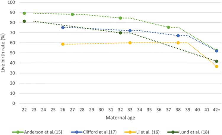
Fig. 1.1
Trend lines demonstrate that as maternal age increases, live birth rate decreases, with a sharp decline at ~40 years. Ages are medians of ranges given in the literature
Some studies suggest that poorer prognosis for live birth after the age of 35 is directly linked to diminished ovarian reserve (DOR). In a study of patients with DOR undergoing IVF, Levi et al. [19] not only found higher rates of RPL in older patients (>40 compared to 35–40 and <35) but also found higher rates of recurrent miscarriage in patients with elevated FSH levels reflecting DOR in each of the age groups. They further reported that higher rates of aneuploidy were found in embryos of patients with RPL compared with those who did not have RPL [19]. This is consistent with other studies indicating that abnormal FSH and estradiol levels are associated with RPL and aneuploidy [20, 21]. Furthermore, infertility or subfertility could also play a role in the high rates of miscarriage at increased ages, but this will be discussed in more detail further on [22].
Aneuploidy is a significant cause of fetal loss and is directly linked to advanced maternal age (AMA ) . It accounts for approximately 50 % of first trimester abortions, 30 % of second trimester abortions, and 3 % of stillborn births [14]. Trisomy is the most common cause of aneuploidy by far, followed by other polysomies and structural anomalies inherited from parental anomalies.
The rate of aneuploidy in the products of conception from couples with RPL compared to the general population is debatable. Table 1.1 summarizes previous studies reports. Generally, we can conclude that fetal aneuploid y is positively associated with AMA [25, 26] and unexplained RPL [15], while it is negatively associated with the number of previous miscarriages [24, 27, 28].
Table 1.1
The rate of aneuploidy in RPL patients compared to the general population
|
Type of study |
Number of cases |
% aneuploidy in RPL |
Rate of aneuploidy compared to the general population |
Caveats |
|
|
Carp et al. [20] |
Retrospective analysis |
126 |
29 % |
Lower |
Only used in patients with three or more miscarriages |
|
Li et al. [16] |
Retrospective, observational analysis |
105 |
32.4 % |
Similar |
|
|
Ogasawara et al. [24] |
Retrospective analysis |
234 |
51.3 % |
Similar |
Abnormal karyotypes were less common as number of miscarriages increased |
|
Marquard et al. [25] |
Retrospective cohort study |
50 |
78 % (>35 years old) |
Higher |
Patients with AMA were used |
|
Stephenson et al. [26] |
Prospective cohort study |
197 |
40 % (>35 years old), 64 % (<35 years old) |
Similar |
|
|
Stern et al. [27] |
Retrospective analysis |
94 |
57 % |
Similar |
Abnormal karyotypes were less common as number of miscarriages increased |
|
Sullivan et al. [28] |
Retrospective analysis |
122 |
25.4 % |
Lower |
Poor placentation and implantation failure are also associated with AMA. Higher rates of perinatal complications including preterm birth, stillbirth, and infertility as well as increased comorbidities including diabetes, obesity, and hypertension have been reported [29]. The increased complication rates of AMA could potentially lead to RPL in women who postpone their reproductive lives.
Karyotyping the Products of Conception
Karyotyping the products of conception (POC ) after the second miscarriage may provide reassurance to the couple as unknown etiologies tend to provoke more stress. Suigura-Ogasawara et al. [30] found that of the 70 % of couples with unexplained RPL, 41 % had abnormal karyotype in their subsequent pregnancy (see Fig. 11.1 in Chap. 11). This not only gives couples a reason for their miscarriage but it also increases the probability that their recurrent loss was due to chance alone, providing them reassurance to a live birth in their future pregnancy. Additionally, it may be more cost effective, with an average savings of $524 per couple, to analyze the POC before performing a standard work-up after two losses, especially in women >35 years old [31].
The Numbers Matter
The number of previous miscarriages is an important prognostic factor. Demonstrated in Fig. 1.2, as the number of miscarriages increases, the prognosis worsens [16–18, 30]. Li et al. [16] found that live birth rate was 64 % in couples with two miscarriages, but as low as 43.2 % in women with six or more miscarriages. Additionally, Lund et al. [18] found a 71.9 % success rate after 5 years in women with three previous losses versus 50.2 % success after six or more losses. Using a 5-year follow-up instead of risk per pregnancy is a beneficial estimate of success for patients because it approximates the overall outcome of having a child [18].
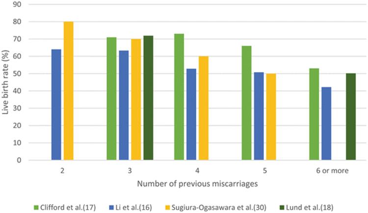
Fig. 1.2
Our figure shows that across studies, the live birth rate decreases as the number of previous miscarriages decreases
Maternal age must also be taken into account when developing a prognosis based on the number of previous miscarriages. The prospect for a live birth is dually affected as the quantity of miscarriages increases in couples with RPL because subsequent pregnancies occur at a later maternal age. Brigham et al. found that in 20-year-old women, the live birth rate after two miscarriages was 92 %, and 85 % after 5 miscarriages. These figures decreased to 77 % in 35-year-old women with two previous losses, and 62 % after five losses. At age 45, the numbers were much lower with a 60 % success rate after two losses, and 42 % after 5 losses [13].
Although the prognosis decreases as the number of miscarriages increases, it is important to reiterate that even women in their early forties with 5 or more losses still achieve a live birth rate of anywhere from 42 to 53 % [13]. Besides advising them about their worse prognosis, couples with five or more losses may warrant a different evaluation. As of now, all couples are evaluated in the RPL clinic equally, whether they are after two miscarriages or six. There is no literature that addresses this issue, but we suggest that further studies are needed for this subgroup to determine the most effective diagnostic evaluation and treatment available.
Two vs. Three
There are few implications in changing the definition of RPL from three or more to two or more losses. A small difference (30 vs. 33 %) in the index pregnancy of two as opposed to three pregnancy losses strongly supports the evaluation after two losses in order to provide the best outcome to couples [32].
Jaslow et al. [33] found no statistically significant differences in diagnostic factors identified in 1020 women with two losses versus three or more. Additionally, according to Bashiri et al. [4], there are no statistically significant differences in outcome for patients with primary RPL who had two versus three pregnancy losses. The study found there were higher levels of TSH in women with three versus two pregnancy losses (16.3 vs. 2.6 %, p = 0.033), and subsequent spontaneous pregnancy occurred more frequently in women with three pregnancy losses (91.7 vs. 77.4 %, p = 0.011). Low molecular weight heparin (LMWH) therapy was also used more in women with three or more losses (40.3 vs. 18.6 %, p = 0.016), and there were higher rates of chronic disease, unemployment, and consanguinity. Brigham et al. [13] also found no statistical differences between couples with 2 or more or 3 or more idiopathic miscarriages. Furthermore, Bhattacharya et al. [34] found that there was no statistical difference between two, three, and even four losses in estimating future pregnancy outcome in 143,595 pregnancies adjusted for maternal age, year of pregnancy, and smoking history.
The change in definition of RPL has led to earlier evaluation of couples with RPL in order to seek out an etiology. This shift raises two important considerations. First, some reviews evaluating the epidemiology of RPL struggle with the inclusion of patients with two losses because of the higher likelihood that the RPL is due to chance. As the number of miscarriages increases in a couple, the likelihood of the couple having an underlying cause increases and as a result, successful treatment reported in trials also increases. The second consideration involves a cost-benefit analysis, although this is beyond the scope of this review.
Primary vs. Secondary RPL
Primary RPL is defined as pregnancy loss with no previous live births, while secondary RPL refers to women with pregnancy loss and at least one live birth [35]. It has been suggested that secondary RPL couples make up 40–61 % of all people with RPL [35, 36]. Although some studies have found differences in couples with primary versus secondary RPL, the implications for the two groups remain to be seen.
Christiansen et al. [5] suggested that primary RPL may involve an innate immunological process after compiling data that found higher rates of thrombophilia, NK cell activity, and the effectiveness of allogenic lymphocyte immunization in primary RPL. Conversely, while secondary RPL may be linked more strongly to adaptive immunity, suggesting there are higher rates of antipaternal antibodies, HLA-DR3, and effectiveness of treatment with IVIg in secondary compared to primary RPL. The effectiveness of immunomodulating treatment will be discussed in a later chapter. However, these findings are currently more motivating for research opportunities.
Alternatively, Bashiri et al. [35] determined that there were no statistically significant prognostic differences in couples with primary versus secondary RPL in terms of live births (75.9 and 70.9 %, p = 0.262, respectively). However, higher pregnancy complications were observed in women with primary RPL such as preterm delivery, fetal growth restriction, and gestational diabetes mellitus after adjustment for age and gravidity. All diagnostic laboratory results were comparable in primary and secondary RPL patients, except for more cases of elevated prolactin in maternal blood. Most studies have agreed that there is no statistical difference [13, 16, 17, 37], and therefore, patients with primary and secondary RPL can be advised and evaluated in the same manner, although women with RPL should be monitored closely for obstetric complications.
While there is no difference in the evaluation of couples with primary versus secondary RPL, we often encounter unique circumstances in our clinic. In our area we have two special communities, the Ultra-Orthodox Jewish and the Bedouins. Both communities are characterized by families having 5–10 or more children. Couples are referred to the RPL clinic with secondary RPL after having 3, 4, and even 5 children. We desire to provide them with the counsel that they seek, but we struggle to prioritize these couples due to the limited time and resources in our publicly funded clinic, with a long queue of primary RPL couples. So far, our policy has been to perform a full patient history, discuss their prognosis, and advise them to continue to attempt to conceive. Then, if they insist on a further evaluation, we provide them the full evaluation. The logic behind this approach is that there is a possibility of acquiring pathology such as antiphospholipid syndrome, hypothyroidism, and uncontrolled diabetes.
Infertility and Superfertility
Infertility, defined as the inability to conceive after 1 year of regular intercourse without the use of contraceptives, has a negative impact on an RPL couple’s prospects of having a live birth [38]. Li et al. [16] found that those with RPL and a history of infertility had a lower live birth rate than those without (50.6 and 61.3 %). Additionally, women with infertility have reported a higher rate of fetal loss, with an odds ratio of 3.92 [38]. Studies supporting these results suggest that many infertile women are unknowingly having repeated early miscarriages [39]. Infertile women also continue attempting conception at an older age, which may contribute to their increased risk [22].
On the other hand, superfertility has also been associated with RPL. It is described as a monthly fecundity rate of >60 % (normal: ~20 %) [40], and the pathophysiology is attributed to dysregulation of the endometrium that allows implantation after the optimal window. Superfertile women conceive very easily but have complicated pregnancies and RPL [41] due to poorer placentation and more apparent pregnancy loss. Although it has not been described as a known etiology, superfertility is a new frontier of RPL that will be discussed later in this book.
Gestational Age
Gestational age is an important part of the couple’s history, as timing of previous losses could point to different etiologies. The majority of preclinical miscarriages are due to aneuploidy (70 %) [42] while thrombophilia and cervical incompetence are more causes of second trimester loss [43]. Still, no etiology is restricted to a certain gestational age, and every couple with RPL deserves a complete work-up.
Additionally, knowledge of the gestational age in the current pregnancy will provide reassurance as it advances. Detection of the fetal heartbeat confers the most reassurance for a live birth, since pregnancy loss occurs in only 2–6 % of women without RPL after fetal heartbeat detection [45]. This important finding should be visualized at 6 weeks gestation the latest [44]. It is thought that a couple with RPL has a three to five times higher chance of miscarriage after seeing a fetal heartbeat than the general population [14], as couples with RPL lose their pregnancy after a detected fetal heartbeat in 10.2–32 % of cases [45]. However, even if a fetal heartbeat is not determined around 6 weeks gestation, a repeat scan is indicated after 7 or more days before diagnosing an abortion [44].
RPL Etiology
Many entities have been examined to determine the source of RPL, but few have been significant enough to warrant investigation in all couples. Figure 1.3 demonstrates the proportions in which the etiologies contribute to RPL—parental chromosomal aberrations, uterine anomalies , endocrine abnormalities, autoimmune disorders, and thrombophilias. The next few sections discuss the known etiologies and the prognosis for patients who fall into these categories.

Fig. 1.3
Etiology of RPL
Parental Chromosomal Aberrations
Parental chromosomal anomalies account for 2–4 % of RPL in couples [32]. However, in unpublished data, Bashiri et al. have found a higher rate of chromosomal rearrangements at 11 % in an analysis of their patient database karyotyping approximately 500 couples. Translocations are the most common aberrations, followed by inversions, insertions, and mosaicism [32]. In light of its frequency and causality, the ASRM recommends chromosomal analysis of both partners during their initial evaluation [3]. Factors that have been associated with a higher likelihood of chromosomal aberrations include RPL in first-degree relatives and early age of onset of RPL [46]. The prognosis for couples with chromosomal abnormalities is difficult to express, with cumulative live birth rates ranging from 55 to 83 % for natural conception [3, 47, 48].
Couples with an abnormal karyotype can be advised to continue attempting pregnancy or they may be offered preimplantation genetic diagnosis (PGD). Continual attempts to conceive improve the chances of eventually having a child. Although the likelihood of miscarriage is higher for carriers, the prognosis for a live child is similar to those without aberrations [46].
Preimplantation genetic testing encompasses both screening and diagnostic measures and is currently the only intervention available to prevent pregnancy loss due to aneuploidy and chromosomal aberrations. PGD is a diagnostic tool for parents with known genetic anomalies. It has been found to reduce the rate of miscarriage in parents with structural chromosomal aberrations once they have become pregnant, but its ability to provide a better outcome for live birth compared to natural conception over time is controversial [49]. ESHRE determined that patients undergoing PGD for chromosomal abnormalities had the lowest pregnancy rate of all groups undergoing PGD, at less than 30 % per transfer. This was attributed to the concomitant infertility or subfertility in these patients [50] and the method used to detect healthy embryos. However, once pregnancy was achieved, women with chromosomal abnormalities had a live birth rate of 83 % in one clinical trial [51]. It is thought that a new method using microarray to determine healthy embryos will be more effective, but committees have not yet made recommendations [49]. Preimplantation genetic screening (PGS) is used largely for detecting aneuploidy and is reserved for patients with advanced maternal age , repeated implantation failure, and unexplained recurrent miscarriage. However, 11 studies have shown no benefit of PGS in terms of live birth. Moreover, PGS was suggested to negatively impact the pregnancy rate compared to natural conception in women with unexplained RPL [49, 50, 52]. Accordingly, a report by the American College of Obstetricians and Gynecologists (ACOG) concerning PGS does not support its use for AMA, recurrent unexplained miscarriage, or recurrent implantation failures [53].
Anatomic Abnormalities
Anatomic abnormalities cause 10–15 % of all RPL cases [32]. Septated uterus is the most common congenital abnormality, accounting for approximately 55–66 % of all uterine abnormalities in women with RPL [54]. Bicornuate and unicornuate make up the remaining 33 % of congenital anomalies. Acquired malformations that may contribute to RPL include polyps, fibroids, and intrauterine adhesions [54, 55]. The prevalence of uterine anomalies is approximately 3 times higher in those with RPL compared to the general population (12.6 and 4.3 %, respectively) [3]. Sugiura-Ogasawara et al. [56] found a live birth rate of untreated women with either a septated or bicornuate uterus of 59.5 %, with a cumulative birth rate of 78 %. These findings vary from others due to the fact that the septated and bicornuate uterus were combined in the study and usually treatment for a bicornuate uterus is not offered. For those with a septated uterus who wish to undergo treatment or continue to have pregnancy loss after diagnosis, hysteroscopic septectomy is offered although no randomized control trials have been performed to evaluate the effectiveness of treatment [57]. Nevertheless, Grimbizis et al. [58] performed a review of 9 retrospective and observational studies and found a cumulative live birth rate of 83.2 %, while another review of 18 retrospective trials have found an overall live birth rate of 45 % after hysteroscopic septectomy [59]. These two seemingly contradictory studies may represent the outcomes in different general groups of women. While Grimbizis et al. reviewed articles with some unspecified birth histories, Nouri et al. used articles that included women with complicated histories including infertility and RPL [58, 59]. Still, hysteroscopic septectomy should be discussed and offered to patients who have a history of RPL and a septate uterus due to its association with increased birth weight and decreased preterm delivery [60]. Unicornuate has the worst prognosis, with a live birth rate of 43.7 % with no available recommended surgical intervention [61].
Intrauterine adhesions (IUA) are also associated with RPL. Women with previous miscarriages have an increased risk of IUA, with a prevalence of 19–24 %, and an odds ratio of 1.99 in women with 2 or more miscarriages compared to one previous miscarriage [62, 63]. Although no previous studies have shown a poorer prognosis for women with RPL after repeated dilation and curettage, this may be a topic worth investigating due to its potential complications since a live birth rate of 71–88 % was found in women with IUA after treatment and a miscarriage rate of 26–30 % in untreated women [63, 64].
Lastly, it remains unclear if fibroids are associated with spontaneous miscarriage, although the size and location of the fibroids are important for prognosis determination [32]. Submucosal fibroids sized 5 cm or greater are more predictive of RPL and infertility, while intramural and subserosal remain to be associated with RPL [55, 65]. Saravelos et al. [66] found that women with RPL have a higher prevalence of fibroids than women with infertility. They suggest that women with repeated second trimester losses should be evaluated for fibroids and undergo myomectomy if present. It has been shown that myomectomy increases the chance of a successful pregnancy from 57 to 93 % [56], and should be considered in women with fibroids. See Chap. 7 for a further description of uterine anomalies.
Endocrine Abnormalities
Endocrine abnormalities affect both the implantation and maintenance of an embryo, and are the source of 17–20 % of all RPL [32]. Diabetes mellitus (DM) must be evaluated in patients with RPL using HbA1C, since uncontrolled DM increases the risk for fetal loss [3]. However, once controlled, DM is no longer a risk factor for RPL. Insulin resistance has been found at higher rates in women with RPL in the early stages of their pregnancy, with insulin resistance observed in 27 % of women with RPL compared to 9.5 % in the general population [67]. Zolghadri et al. [68] found that women with RPL had a higher prevalence of abnormal results in their glucose tolerance tests (17.6 % compared to 5.4 % of women without RPL). Furthermore, they found that those with abnormal glucose tolerance had better outcomes when taking metformin than those untreated, with an abortion rate of 15 and 55 % respectively. Therefore, these studies suggest insulin resistance is an important factor in RPL that warrants screening and treatment, although committee recommendations of this nature have not been made [67–69].
Overt hypothyroidism is seen in 0.2 % of pregnancies and subclinical hypothyroidism (SCH) is seen in 2–3 % [70]. All women with RPL should have their TSH levels monitored [3]. Although lowering the upper limit of a normal TSH level in pregnancy from 5.0 to 2.5 has been discussed, no conclusion in this matter has been made [71]. Pregnant women with known hypothyroidism must be monitored closely, as the rate of fetal loss is estimated to be 31 % in untreated women compared to 4 % in well-treated women [72]. Women with RPL have been found to have higher rates of hypothyroidism (10.5 %) and SCH (19 %) [73]. Furthermore, patients with SCH have been found to have higher rates of RPL [74]. A study by Benhadi et al. [75] of almost 2500 women showed that higher TSH levels were positively correlated with higher rates of pregnancy loss. Given the evidence that SCH may be associated with adverse outcomes, the Endocrine Society recommends that women with SCH are treated with T4 regardless of their thyroid antibody titers [76].
Thyroid autoimmunity has been directly linked with RPL [77], and 22–37 % [76] of women with RPL have positive thyroid antibodies (Tg-Ab or TPOAb), with as many as 10 % being thyroid antibody positive despite euthyroidism [74]. Kaprara et al. [78] reviewed 14 studies which found higher rates of miscarriage groups with thyroid antibodies compared to those without, with statistical significance in 10 out of 14. Additionally, 9 studies detected higher rates of auto-antibodies in groups with RPL compared to those without RPL, with statistical significance in 6 out of 9 [78]. Interestingly, high titers have not been shown to correlate with worse outcome than lower titers [70]. Levothyroxine is given for the treatment of autoimmune hypothyroidism [75]. Treatment for euthyroid women with positive thyroid antibody titers is not recommended [76], although levothyroxine has been shown in three studies to decrease fetal loss in thyroid antibody positive women [79–81].
The actual prevalence of PCOS in patients with RPL is not known, although Hudecova et al. [82] found similar live birth and miscarriage rates in both groups in a long-term follow-up. There is no correlation between PCOS and a high aneuploidy rate [82], and ultrasonographic polycystic ovaries and abnormal luteinizing hormone levels were not predictive of future miscarriage [84]. Although PCOS is not directly linked to RPL, hyperandrogenism, obesity, and hyperinsulinism are common sequelae in PCOS and have also been found in higher rates in women with RPL [81, 82].
Luteal phase defects have been studied for RPL and their association with endometrial dysfunction. However the lack of diagnostic criteria made the effects of varying luteal phase hormones on pregnancy impossible to quantify [83]. Therefore, according to the 2012 ASRM guidelines [3], progesterone treatment for luteal phase defects is not recommended. However, a recent systematic review by Carp made a compelling argument for treatment with dydrogesterone, a progestogen, in a review of 13 studies demonstrating a 13 % absolute reduction in the miscarriage rate among the treated vs. the untreated group [85].
Hyperprolactinemia is associated with RPL, and it may be involved in the pathogenesis of reproductive failure in patients with APS, PCOS, and hypothyroidism [3, 79, 86]. Hirahura et al. [86] found that treatment with bromocriptine was very effective for women with elevated prolactin levels, leading to a live birth rate of 85.7 %. On the other side, Li et al. [87] found that women with prolactin concentrations in the lower end of the physiological range had an increased risk of miscarriage when adjusted for all other factors.
Thrombophilias
Hereditary thrombophilias (HT ) elicit a prothrombotic state that has been associated with RPL. The most common HT is Factor V Leiden [88]. It is found in ~5 % of the general population of Caucasians, 1 % Africans, and is almost absent in Asians [88]. Other thrombophilias include prothrombin gene mutation, protein C deficiency, protein S deficiency, and antithrombin III deficiency. Certain thrombophilias produce a higher risk environment than others, and routine screening for HT is not mandatory in all patients with RPL. The association between Factor V Leiden mutation and pregnancy loss is thought to be as low as 4.2 % [89]. ACOG’s most recent practice bulletin [90] divides HT into high-risk and low-risk groups, recommending prophylactic treatment based on the genetic mutation and history of venous thromboembolism (VTE). Histories of VTE or high-risk thrombophilia were considered as indications for prophylactic therapy with LMWH or unfractionated heparin [90]. In spite of this, it has been found that as many as 40 % of physicians screen patients without recommendations [91]. In clinical practice, HT is hard to ignore. Early studies have supported screening [91], and discounting a possible cause of RPL, however small it may be, in couples by foregoing a genetic test or prophylactic treatment is difficult to do when the stakes are so high. See chapter on hereditary thrombophilia for further discussion of therapeutic options.
Acquired thrombophilia, mostly attributed to antiphospholipid syndrome (APS ) , is seen in 5–20 % of patients with RPL [33]. Testing for APS is indicated after 3 or more unexplained miscarriages before week 10, one unexplained miscarriage after 10 weeks gestation, or a history of preeclampsia or placental insufficiency that led to delivery before 34 weeks gestation. Anticardiolipin, lupus anticoagulant, anti-β2-glycoprotein I, and antiphosphatidylserine are the only tested antiphospholipid antibodies, and they must be found positive twice at a 12-week interval or greater [92]. The inclusion of other antiphospholipid antibodies has increased false positive results, leading to vastly greater percentages describing the incidence of APS in past studies [31].
Antiphospholipid antibodies induce thrombosis, inhibit differentiation of the trophoblast, and provoke dysregulation of the maternal immune system [3]. Inflammation may also participate in producing these effects on the trophoblast. Fetal loss rate, e.g., pregnancy loss after 20 weeks, was reported to be 50–90 % without treatment [93]. A Cochrane review of randomized control trials has shown that low-dose aspirin with heparin leads to a live birth rate of 70–80 % [94]. Although these results are encouraging, this subgroup is still at an increased risk for miscarriage. An observational study by Bouvier et al. [95] found that women with RPL and obstetric APS undergoing treatment continued to have increased rates of fetal loss as well as neonatal and obstetric complications, producing lower rates of live births compared to women without APS. Therefore, a desired successful and uncomplicated pregnancy after the diagnosis and treatment for APS is not always accomplished.
Lifestyle and Habits
Smoking, alcohol, and caffeine are three of the most discussed environmental factors associated with spontaneous single abortion. However, no direct correlation between these factors and RPL was reported. All three factors have been shown to be dose-dependent causes of single abortions, and obtaining a full patient history including questions about these factors may uncover a possible causation [3, 33].
Obesity
Obesity is defined as a BMI of ≥30 kg/m [96]. It has been a topic of discussion in the field of RPL due to its increasing prevalence and association with PCOS, type II diabetes mellitus, and hormone abnormalities. Differing results have been found concerning the association of obesity with RPL. Metwally et al. [97] found that while there was an association between obesity and miscarriage, the evidence was weak in 16 articles published including both spontaneous and assisted conception. They criticized the data for using varying definitions of obesity and advanced maternal age. Boots et al. [98] performed a similar review of the literature, including spontaneous conceptions only. They found stronger evidence of recurrent early miscarriage in obese patients with an odds ratio of 3.51. Furthermore, Lashen et al. [99] found an increased rate of recurrent miscarriage in women with high BMI compared to those without, with odds ratio of 4.68 in a study of 4932 women. Therefore, it is likely that obesity directly affects RPL, but more studies are needed to confirm these results.
Unexplained RPL
Although it is commonly recognized that unexplained RPL (URPL ) occurs in about 50 % of all RPL [32], as seen in Fig. 11.1 in Chap. 11, Sugiura-Ogasawara et al. [30] found an abnormal karyotype in the POC of 41.1 % of URPL patients, and therefore suggest that true unexplained RPL may only be present in 24.5 % of the total RPL population. Saravelos et al. [10] has shown that women with URPL have a similar risk of miscarriage compared to the general population (14–26 and 15–25 %). However, they do not suggest that all URPL is due to chance alone, but that this group should be separated into two subgroups. Those who fall under type I have RPL that is actually due to chance alone, while those in type II have truly unexplained RPL. Younger women with higher incidence of clinically diagnosed pregnancy losses and normal karyotypes in the POC are classified as type II, while women who do not meet these criteria are type I [10]. This has interesting implications for counseling these two groups, but is still not widely accepted.
A variety of therapies have been attempted on women with URPL including immunotherapy, low-dose aspirin, and psychological support [32, 100]. Interestingly, psychological support alone has offered promising results. Clifford et al. [17] found better outcomes in couples with URPL who received supportive care during early pregnancy compared to those who did not (26 and 51 % respectively). Other studies have supported these results [13, 101]; however, additional studies should be conducted due to the lack of randomization. It has been shown that women with RPL experience increased levels of anxiety. In fact, Mevorach-Zussmanet et al. [102] found that variables which compose a woman’s reproductive status are directly associated with her psychological health. They suggest that the anxiety is a long-term condition, often not resolved by TLC therapy, and may produce poor outcome in subsequent pregnancies. As was discussed earlier, PGS is not a standard of care, as the chance of having a live birth in URPL is good. It is typically only in cases in infertility, AMA, and primary RPL as a last resort.
When May I Get Pregnant Again, and Is It Ok to Try Now?
Many patients with RPL wish to know if it will be possible for them to achieve conception, and if they should wait a certain amount of time to decrease their risk of another miscarriage. Psychological stress can become an extremely important factor in counseling women who feel their “biological clock is ticking.” Kaandorp et al. [103] found a median time to pregnancy of 21–23 weeks in women with unexplained RPL, which is comparable to the average time to pregnancy for women in the general population, and can be reassuring for patients who wish to become pregnant soon after their last miscarriage.
Additionally, inter-pregnancy intervals (IPIs) have been a source of speculation for prognosis. Questions have been raised over whether psychological stress and physical recovery should delay conception after a miscarriage. However, since the World Health Organization [104] suggested at least 6 months between miscarriage and conception, studies have shown that IPIs are equivalent in prognosis [105, 106]. Furthermore, some studies have suggested that a shorter IPI of less than 6 months carries a better prognosis [107]. Bentolila et al. [106] showed that shorter IPI (<6 months) was associated with a better pregnancy outcome, although the group with an IPI of more than 6 months had equivalent outcomes when adjusted for maternal age and fertility problems. Therefore, patients who desire to become pregnant soon after miscarriage may be reassured that they are not at an increased risk of adverse effects compared to those who delay conception.
Conclusion
Each couple with RPL has a different prognosis according to their individual history and the results of their diagnostic work-up. The prognosis can be dynamic from the first meeting until right before pregnancy. Therefore, although it is difficult to create an exact percentage for each couple’s prognosis, advising them on their potential for a live birth can help make important therapeutic and reproductive decisions. Maternal age provides the best indication for prognosis, with the lowest rates of live birth seen in women over 40 years old. Higher incidence of previous miscarriages worsens prognosis and decreases the likelihood that the miscarriages are due to chance alone. While primary and secondary RPL have similar live birth rates, women with primary RPL should be monitored more closely for obstetric complications during the pregnancy. Concomitant infertility also contributes to a poor outcome. All of these factors affect the prognosis of the couple aside from etiology. Each etiology confers its own rate of live birth and varies based on whether it is treated or untreated. Some therapy—diabetes control for example—provides near normal live birth rates, while others, like PGD for parental chromosomal aberrations, are more controversial. In conclusion, each couple should be carefully evaluated and advised with emotional support in order to provide the best possible outcome.
References
1.
Jauniaux E, Farquharson RG, Christiansen OB, Exalto NE, (ESHRE). Evidence-based guidelines for the investigation and medical treatment of recurrent miscarriage. Hum Reprod. 2006;21:2216–22.PubMed
2.
Practice Committee of the American Society for Reproductive Medicine. Definitions of infertility and recurrent pregnancy loss. Fertil Steril. 2013;99(1):63. doi:10.1016/j.fertnstert.2012.09.023. http://dx.doi.org.
3.
Practice Committee of the American Society for Reproductive Medicine. Evaluation and treatment of recurrent pregnancy loss: a committee opinion. Fertil Steril. 2012;98(5):1103–11. doi:10.1016/j.fertnstert.2012.06.048. http://dx.doi.org.
4.
Bashiri A, Ratzon R, Amar S, Serjienko R, Mazor M, Shoham-Vardi I. Two vs. three or more primary recurrent pregnancy losses- are there any differences in epidemiologic characteristics and index pregnancy outcome? J Perinat Med. 2012;40:365–71.PubMed
5.
Christiansen OB. Epidemiology of recurrent pregnancy loss. In: Carp H, editor. Recurrent pregnancy loss: causes, controversies and treatment. 1st ed. UK: Informa healthcare; 2007. p. p1–14.
6.
Royal College of Obstetricians and Gynaecologists (RCOG). The investigation and treatment of couples with recurrent first-trimester and second-trimester miscarriage. Green-top guideline; no. 17. London (UK): RCOG. 2011.
7.
Christiansen OB. Recurrent miscarriage is a useful and valid clinical concept. Acta Obstet Gynecol Scand. 2014;93:852–7. doi:10.1111/aogs.12456.PubMed
8.
Chard T. Frequency of implantation and early pregnancy loss in natural cycles. Baillieres Clin Obstet Gynaecol. 1991;5(1):179–89.PubMed
9.
Farquharson RG, Jauniaux E, Exalto N. Updated and revised nomenclature for description of early pregnancy events. Hum Reprod. 2005;20(11):3008–11.PubMed
10.
Saravelos S, Li T. Unexplained recurrent miscarriage: how can we explain it? Hum Reprod. 2012;27:1882–6.PubMed
11.
Kolte A, van Oppenraaj R, Quenby S, Farquharson R, Stephenson M, Goddin M, Christiansen O. (ESHRE). Non-visualized pregnancy losses are prognostically important for unexplained recurrent miscarriage. Hum Reprod. 2014;29(5):931–7.PubMed
12.
Zeadna A, et al. A comparison of biochemical pregnancy rates between women who underwent IVF and fertile controls who conceived spontaneously. Hum Reprod. 2015;30(4):783–8.PubMed
13.
Brigham S, Colon C, Farquharson G. A longitudinal study of pregnancy outcome following idiopathic recurrent miscarriage. Hum Reprod. 1999;14:2868–71.PubMed
14.
Fritz M. Recurrent early losses. In: Fritz M, Speroff L, editors. Clinical gynecologic endocrinology and infertility. 8th ed. Philadelphia, PA: Lippincott Williams & Wilkins; 2011. p. 1191–220.
15.
Nybo Andersen A, Wohlfahrt J, Christens P, Olsen J, Melbye M. Maternal age and fetal loss: population based register linkage study. BMJ. 2000;320:1708–12.PubMed
16.
Li TC, Iqbal T, Anstie B, Gillham J, Amer S, Wood K, Laird S. An analysis of the pattern of pregnancy loss in women with recurrent miscarriage. Fertil Steril. 2002;78(5):1100–6.PubMed
17.
Clifford K, Rai R, Regan L. Future pregnancy outcome in unexplained recurrent first trimester miscarriage. Hum Reprod. 1997;12(2):387–9.PubMed
18.
Lund M, Kamper-Jorgensen M, Nielson HS, Lidegaard O, Nybo Anderson A, Christiansen O. Prognosis for live birth in women with recurrent miscarriage: what is the best measure of success? Obstet Gynecol. 2012;119(1):37–43. doi:10.1097/AOG.0b013e31823c0413.PubMed
19.
Levi A, Raynault M, Bergh P, Drews M, Miller B, Scott R. Reproductive outcome in patients with diminished ovarian reserve. Fertil Steril. 2001;76(4):666–9.PubMed
20.
Hofmann GE, Khoury J, Thie J. Recurrent pregnancy loss and diminished ovarian reserve. Fertil Steril. 2000;74:1192–5.PubMed
21.
Nasseri A, Mukherjee T, Grifo J, Noyes N, Krey L, Copperman A. Elevated day 3 follicle stimulating hormone and/or estradiol may predict fetal aneuploidy. Fertil Steril. 1999;71:715–8.PubMed
22.
Cano F, Simon C, Remohi J, Pellicer A. Effect of ageing on the female reproductive system: evidence for a role of uterine senescence in the decline in female fecundity. Fertil Steril. 1995;64:584–9.PubMed
23.
Carp H, Toder V, Aviram A, Daniely M, Mashiach S, Barkai G. Karyotype of the abortus in recurrent miscarriage. Fertil Steril. 2001;75(4):678–82.PubMed
24.
Ogasawara M, Aoki K, Okada S, Suzumori K. Embryonic karyotype of abortuses in relation to the number of previous miscarriages. Fertil Steril. 2000;73:300–4.PubMed
25.
Marquard K, Westphal L, Milki A, Lathi R. Etiology of recurrent pregnancy loss in women over the age of 35 years. Fertil Steril. 2010;94(4):1473–7. doi:10.1016/j.fertnstert.2009.06.041.PubMed
26.
Stephenson MD. Frequency of factors associated with habitual abortion in 197 couples. Fertil Steril. 1996;66:24–9.PubMed
27.
Stern J, Dorfman A, Gutierez-Najar M. Frequency of abnormal karyotype among abortuses from women with and without a history of recurrent spontaneous abortion. Fertil Steril. 1996;65:250–3.PubMed
28.
Sullivan A, Silver R, LaCoursiere D, Porter T, Branch D. Recurrent fetal aneuploidy and recurrent miscarriage. Obstet Gynecol. 2004;104(4):784–8.PubMed
29.
Nelson S, Telfer E, Anderson R. The ageing ovary and uterus: new biological insights. Hum Reprod Update. 2013;19(1):67–83. doi:10.1093/humupd/dms043.PubMedPubMedCentral
30.
Sugiura-Ogasawara M, Ozaki Y, Suzumori N. Management of recurrent miscarriage. J Obstet Gynaecol. 2014;40(5):1174–9. doi:10.1111/jog.12388.
31.
Foyouzi N, Cedars M, Huddleston H. Cost-effectiveness of cytogenetic evaluation of products of conception in the patient with a second pregnancy loss. Fertil Steril. 2012;98(1):151–5. doi:10.1016/j.fertnstert.2012.04.007.PubMed
32.
Ford H, Schust D. Recurrent pregnancy loss: etiology, diagnosis, and therapy. Rev Obstet Gynecol. 2009;2(2):76–83.PubMedPubMedCentral
33.
Jaslow CR, Carney JL, Kutteh WH. Diagnostic factors identified in 1020 women with two versus three or more recurrent pregnancy losses. Fertil Steril. 2010;93(4):1234–43.PubMed
34.
Bhattacharya S. Townsend, Bhattacharya S. Recurrent miscarriage: are three miscarriages one too many? analysis of a Scottish population-based database of 151,021 pregnancies. Eur J Obstet Gynecol Reprod Biol. 2010;150(1):24–7. doi:10.1016/j.ejogrb.2010.02.015.PubMed
35.
Bashiri A, Shapira E, Ratzon R, Shoham-Vardi I, Sejienko R, Mazor M. Primary vs. secondary recurrent pregnancy loss- epidemiological characteristics, etiology, and next pregnancy outcome. J Perinat Med. 2012;40(2012):389–96.PubMed
36.
Jivraj S, Anstie B, Cheong YC, Fairlie FM, Laird SM, Li TC. Obstetric and neonatal outcome in women with a history of recurrent miscarriage: a cohort study. Hum Reprod. 2001;16:102–6.PubMed
37.
Morikawa M, Yamada H, Kato EH, Shimada S, Sakuragi N, Fujimoto S, Minakami H. Live birth rate varies with gestational history and etiology in women experiencing recurrent spontaneous abortion. Eur J Obstet Gynecol Reprod Biol. 2003;109(1):21–6.PubMed
38.
Whitley E, Doyle P, Roman E, De Stavola B. The effect of reproductive history on future pregnancy outcomes. Hum Reprod. 1999;14(11):2863–7.PubMed
39.
Hakim R, Gray R, Zacur H. Infertility and early pregnancy loss. Am J Obstet Gynecol. 1995;172:1510–7.PubMed
40.
Teklenburg G, Salker M, Heijnen C, Macklon NS, Brosens JJ. The molecular basis of recurrent pregnancy loss: impaired natural embryo selection. Mol Hum Reprod. 2010;12:886–95. doi:10.1093/molehr/gaq079.
41.
Wilcox A, Baird D, Weinberg C. Time of implantation of the conceptus and loss of pregnancy. N Engl J Med. 1999;340(23):1796–9.PubMed
42.
Ohno M, Maeda T, Matsunobu A. A cytogenetic study of spontaneous abortions with direct analysis of chorionic villi. Obstet Gynecol. 1991;77(3):394–8.PubMed
43.
Drakeley A, Quenby S, Farquharson R. Mid-trimester loss—appraisal of a screening protocol. Hum Reprod. 1998;13(7):1975–80.PubMed
44.
Appiah A, Johns J. Endocrine and ultrasonic surveillance of pregnancies in patients with recurrent miscarriage. In: Christiansen O, editor. Recurrent pregnancy loss. 1st ed. Wiley-Blackwell: Hoboken, NJ; 2014. p. 103–14.
45.
Li TC, Spring PG, Bygrave C, Laird SM, Serle E, Spuijbroek M, Adekanmi O. The value of biochemical and ultrasound measurements in predicting pregnancy outcome in women with a history of recurrent miscarriage. Hum Reprod. 1998;13(12):3525–9.PubMed
46.
Franssen MT, Korevaar JC, van der Veen F, Leschot NJ, Bossuyt PM, Goddijn M. Reproductive outcome after chromosome analysis in couples with two or more miscarriages: index [corrected]-control study. BMJ. 2006;332(7544):759–63.PubMedPubMedCentral
47.
Sugiura-Ogasawara M, Ozaki Y, Sato T, Suzumori N, Suzumori K. Poor prognosis of recurrent aborters with either maternal or paternal reciprocal translocations. Fertil Steril. 2004;81:367–73.PubMed
48.
Carp H, Guetta E, Dorf H, Soriano D, Barkai G, Schiff E. Embryonic karyotype in recurrent miscarriage with parental karyotypic aberrations. Fertil Steril. 2006;85(2):446–50.PubMed
49.
Brezina P, Kutteh W. Clinical applications of preimplantation genetic testing. BMJ. 2015;350:g7611. doi:10.1136/bmj.g7611.PubMed
50.
Harper J, Wilton L, Traeger-Synodinos J, Goossens V, Moutou C, SenGupta S, et al. The ESHRE PGD Consortium: 10 years of data collection. Hum Reprod Update. 2012;18:234–47.PubMed
51.
Twisk M, Mastenbroek S, van Wely M, Heineman MJ, Van der Veen F, Repping S. Preimplantation genetic screening for abnormal number of chromosomes (aneuploidies) in in vitro fertilisation or intracytoplasmic sperm injection. Cochrane Database Syst Rev. 2006;1.
52.
Rubio C, Simon C, Vidal F, Rodrigo L, Pehlivan T, Remohi J, et al. Chromosomal abnormalities and embryo development in recurrent miscarriage couples. Hum Reprod. 2003;18:182–8.PubMed
53.
Preimplantation Genetic Screening for Aneuploidy. ACOG Committee Opinion No.430. American College of Obstetricians and Gynecologists. Obstet Gynecol. 2009;113:766–7.
54.
Salim R, Regan L, Woelfer B, Backos M, Jurkovic D. A comparative study of the morphology of congenital uterine anomalies in women with and without a history of recurrent first trimester miscarriage. Hum Reprod. 2003;18(1):162–6.PubMed
55.
Bajekal N, Li TC. Fibroids, infertility and pregnancy wastage. Hum Reprod Update. 2000;6(6):614–20.PubMed
56.
Sugiura-Ogasawara M, Ozaki Y, Kitaori T, Kumagai K, Suzuki S. Midline uterine defect size is correlated with miscarriage of euploid embryos in recurrent cases. Fertil Steril. 2010;93(6):1983–8. doi:10.1016/j.fertnstert.2008.12.097.PubMed
57.
Kowalik CR, Goddijn M, Emanuel MH, Bongers MY, Spinder T, de Kruif JH, Mol BW, Heineman MJ. Metroplasty versus expectant management for women with recurrent miscarriage and a septate uterus. Cochrane Database Syst Rev. 2011;6:CD008576. doi:10.1002/14651858.CD008576.pub3.PubMed
58.
Grimbizis GF, Camus M, Tarlatzis BC, Bontis JN, Devroey P. Clinical implications of uterine malformations and hysteroscopic treatment results. Hum Reprod Update. 2001;7(2):161–74.PubMed
59.
Nouri K, Ott J, Huber JC, Fischer EM, Stögbauer L, Tempfer CB. Reproductive outcome after hysteroscopic septoplasty in patients with septate uterus—a retrospective cohort study and systematic review of the literature. Reprod Biol Endocrinol. 2010;8:52. doi:10.1186/1477-7827-8-52.PubMedPubMedCentral
60.
Freud A, Harlev A, Weintraub AY, Ohana E, Sheiner E. Reproductive outcomes following uterine septum resection. J Matern Fetal Neonatal Med. 2014;1–4.
61.
Raga F, Bauset C, Remohi J, Bonilla-Musoles F, Simón C, Pellicer A. Reproductive impact of congenital Müllerian anomalies. Hum Reprod. 1997;12(10):2277–81.PubMed
62.
Yu D, Wong YM, Cheong Y, Xia E, Li TC. Asherman syndrome—one century later. Fertil Steril. 2008;89(4):759–79. doi:10.1016/j.fertnstert.2008.02.096.PubMed
63.
Hooker A, Lemmers M, Thurkow A, Heymans M, Opmeer B, Brölmann H, Mol B, Huirne J. Systematic review and meta-analysis of intrauterine adhesions after miscarriage: prevalence, risk factors and long-term reproductive outcome. Hum Reprod Update. 2014;20(2):262–78. doi:10.1093/humupd/dmt045.PubMed
64.
Pabuçcu R, Atay V, Orhon E, Urman B, Ergün A. Hysteroscopic treatment of intrauterine adhesions is safe and effective in the restoration of normal menstruation and fertility. Fertil Steril. 1997;68(6):1141–3.PubMed
65.
Casini ML, Rossi F, Agostini R, Unfer V. Effects of the position of fibroids on fertility. Gynecol Endocrinol. 2006;22(2):106–9.PubMed
66.
Saravelos S, Yan J, Rehmani H, Li TC. The prevalence and impact of fibroids and their treatment on the outcome of pregnancy in women with recurrent miscarriage. Hum Reprod. 2011;26(12):3274–9. doi:10.1093/humrep/der293.PubMed
67.
Craig L, Ke R, Kutteh W. Increased prevalence of insulin resistance in women with a history of recurrent pregnancy loss. Fertil Steril. 2002;78(3):487–90.PubMed
68.
Zolghadri J, Tavana Z, Kazerooni T, Soveid M, Taghieh M. Relationship between abnormal glucose tolerance test and history of previous recurrent miscarriages, and beneficial effect of metformin in these patients: a prospective clinical study. Fertil Steril. 2008;90(3):727–30.PubMed
69.
Tian L, Shen H, Lu Q, Norman RJ, Wang J. Insulin resistance increases the risk of spontaneous abortion after assisted reproduction technology treatment. J Clin Endocrinol Metab. 2007;92(4):1430–3.PubMed
70.
Casey B, Dashe J, Wells C, McIntire D, Byrd W, Leveno K, Cunningham F. Subclinical hypothyroidism and pregnancy outcomes. Obstet Gynecol. 2005;105:239–45.PubMed
71.
Ke RW. Endocrine basis for recurrent pregnancy loss. Obstet Gynecol Clin North Am. 2014;41(1):103–12. doi:10.1016/j.ogc.2013.10.003.PubMed
72.
Abalovich M, Gutierrez S, Alcaraz G, Maccallini G, Garcia A, Levalle O. Overt and subclinical hypothyroidism complicating pregnancy. Thyroid. 2002;12:63–8.PubMed
73.
Bernardi L, Cohen R, Stephenson M. Impact of subclinical hypothyroidism in women with recurrent early pregnancy loss. Fertil Steril. 2013;100(5):1326–31. doi:10.1016/j.fertnstert.2013.07.1975.PubMed
74.
Negro R, Mestman J. Thyroid disease in pregnancy. Best Pract Res Clin Endocrinol Metab. 2011;25(6):927–43. doi:10.1016/j.beem.2011.07.010.PubMed
75.
Benhadi N, Wiersinga WM, Reitsma JB, et al. Higher maternal TSH levels in pregnancy are associated with increased risk for miscarriage, fetal or neonatal death. Eur J Endocrinol. 2009;160(6):985–91.PubMed
76.
De Groot L, Abalovich M, Alexander EK, Amino N, Barbour L, Cobin RH, Eastman CJ, Lazarus JH, Luton D, Mandel SJ, Mestman J, Rovet J, Sullivan S. Management of thyroid dysfunction during pregnancy and postpartum: an Endocrine Society clinical practice guideline. J Clin Endocrinol Metab. 2012;97(8):2543–65. doi:10.1210/jc.2011-2803.PubMed
77.
Abramson J, Stagnaro-Green A. Thyroid antibodies and fetal loss: an evolving story. Thyroid. 2001;11(1):57–63.PubMed
78.
Kaprara A, Krassas GE. Thyroid autoimmunity and miscarriage. Hormones. 2008;7(4):294–302.PubMed
79.
Negro R, Mangieri T, Coppola L, Presicce G, Casavola EC, Gismondi R, Locorotondo G, Caroli P, Pezzarossa A, Dazzi D, Hassan H. Levothyroxine treatment in thyroid peroxidase antibody-positive women undergoing assisted reproduction technologies: a prospective study. Hum Reprod. 2005;20(6):1529–33.PubMed
80.
Negro R, Formoso G, Coppola L, Presicce G, Mangieri T, Pezzarossa A, Dazzi D. Euthyroid women with autoimmune disease undergoing assisted reproduction technologies: the role of autoimmunity and thyroid function. J Endocrinol Invest. 2007;30(1):3–8.PubMed
81.
Lincoln SR, Ke RW, Kutteh WH. Screening for hypothyroidism in infertile women. J Reprod Med. 1999;44(5):455–7.PubMed
82.
Hudecova M, Holte J, Olovsson M, Sundström PI. Long-term follow-up of patients with polycystic ovary syndrome: reproductive outcome and ovarian reserve. Hum Reprod. 2009;24(5):1176–83. doi:10.1093/humrep/den482.PubMed
83.
Weghofer A, Munne S, Chen S, Barad D, Gleicher N. Lack of association between polycystic ovary syndrome and embryonic aneuploidy. Fertil Steril. 2007;88(4):900–5.PubMed
84.
Cocksedge K, Li TC, Saravelos SH, Metwally M. A reappraisal of the role of polycystic ovary syndrome in recurrent miscarriage. Reprod Biomed Online. 2008;17(1):151–60.PubMed
85.
Carp H. Gynecol Endocrinol. 2015;31(6):422–30.PubMed
86.
Hirahara F, Andoh N, Sawai K, et al. Hyperprolactinemic recurrent miscarriage and results of randomized bromocriptine treatment trials. Fertil Steril. 1998;70:246–52.PubMed
87.
Li W, Ma N, Laird SM, Ledger WL, Li TC. The relationship between serum prolactin concentration and pregnancy outcome in women with unexplained recurrent miscarriage. J Obstet Gynaecol. 2013;33(3):285–8. doi:10.3109/01443615.2012.759916.PubMed
88.
De Stefano V, Chiusolo P, Paciaroni K, Leone G. Epidemiology of factor V Leiden: clinical implications. Semin Thromb Hemost. 1998;24(4):367–79.PubMed
89.
Rodger MA, Betancourt MT, Clark P, et al. The association of factor V Leiden and prothrombin gene mutation and placenta-mediated pregnancy complications: a systematic review and meta-analysis of prospective cohort studies. PLoS Med. 2010;7(6), e1000292. doi:10.1371/journal.pmed.1000292.PubMedPubMedCentral
90.
James A. Practice bulletin no. 123: thromboembolism in pregnancy. Obstet Gynecol. 2011;118(3):718–29.PubMed
91.
Davenport W, Kutteh W. Inherited thrombophilias and adverse pregnancy outcomes a review of screening patterns and recommendations. Obstet Gynecol Clin N Am. 2014;41:133–44. doi:10.1016/j.ogc.2013.10.005. http://dx.doi.org.
92.
Miyakis S, Lockship M, Atsumi T, Branch D, Brey R, Cervera R, Derksen R, De Groot P, Koike T, Meroni P, Reber G, Shoenfeld Y, Tincani A, Vlachoyiannopoulos P, Krilis S. International consensus statement on an update of the classification criteria for definite antiphospholipid syndrome (APS). J Thromb Haemost. 2006;4:295–306. doi:10.1111/j.1538-7836.2006.01753.x.PubMed
93.
Kwak-Kim J, Agcaoili MSL, Aleta L, Liao A, Ota K, Dambaeva S, Beaman K, Kim JW, Gilman-Sachs A. Management of women with recurrent pregnancy losses and antiphospholipid antibody syndrome. Am J Reprod Immunol. 2013;69:596–607. doi:10.1111/aji.12114.PubMed
94.
Empson M, Lassere M, Craig JC, Scott JR. Recurrent pregnancy loss with antiphospholipid antibody: a systematic review of therapeutic trials. Obstet Gynecol. 2002;99:135–44.PubMed
95.
Bouvier S, Cochery-Nouvellon E, Lavigne-Lissaide G, Mercier E, Marchetti T, Balducchi JP, Mares P, Gris JC. Comparative incidence of pregnancy outcomes in treated obstetric antiphospholipid syndrome: the NOH_APS observational study. Blood. 2014;123(3):404–13.PubMed
96.
World Health Organization. Obesity: preventing and managing the global epidemic. Report of a WHO consultation on obesity. Geneva, Switzerland: World Health Organization; 1999.
97.
Metwally M, Ong K, Ledger W, Li TC. Does high body mass index increase the risk of miscarriage after spontaneous and assisted conception? A meta-analysis of the evidence. Fertil Steril. 2008;90(3):714–26. doi:10.1016/j.fertnstert.2007.07.1290.PubMed
98.
Boots C, Stephenson M. Does obesity increase the risk of miscarriage in spontaneous conception: a systematic review. Semin Reprod Med. 2011;29(6):507–13. doi:10.1055/s-0031-1293204.PubMed
99.
Lashen H, Fear K, Sturdee DW. Obesity is associated with increased risk of first trimester and recurrent miscarriage: matched case-control study. Hum Reprod. 2004;19:1644–6.PubMed
100.
Stephenson M, Kutteh W. Evaluation and management of recurrent early pregnancy loss. Clin Obstet Gynecol. 2007;50(1):132–45.PubMed
101.
Stray-Pederson B, Stray-Pederson S. Etiologic factors and subsequent reproductive performance in 195 couples with a prior history of habitual abortion. Am J Obstet Gynecol. 1984;148:140–6.
102.
Mevorach-Zussman N, Bolotin A, Shalev H, Bilenko N, Mazor M, Bashiri A. Anxiety and deterioration of quality of life factors associated with recurrent miscarriage in an observational study. J Perinat Med. 2012;40(5):495–501.PubMed
103.
Kaandrop S, van Mens T, Middeldorp S, Hutton BA, Hof M, van der Post J, van den Veen F, Goddijn H. Time to conception and time to live birth in women with unexplained recurrent miscarriage. Hum Reprod. 2014;29(6):1146–52.
104.
World Health Organization. Report of a WHO technical consultation on birth spacing. 2005. www.who.int/making_pregnancy_safer/documents/birth_spacing.pdf.
105.
Sholapurkar SL. Is there an ideal interpregnancy interval after a live birth, miscarriage or other adverse pregnancy outcomes? J Obstet Gynaecol. 2010;30(2):107–10.PubMed
106.
Bentolila Y, Ratzon R, Shoham-Vardi I, Serjienko R, Mazor M, Bashiri A. Effect of interpregnancy interval on outcomes of pregnancy after recurrent pregnancy loss. J Matern Fetal Neonatal Med. 2013;26(14):1459–64. doi:10.3109/14767058.2013.784264.PubMed
107.
Love ER, Bhattacharya S, Smith NC, Bhattacharya S. Effect of interpregnancy interval on outcomes of pregnancy after miscarriage: retrospective analysis of hospital episode statistics in Scotland. BMJ. 2010;341:c3967.PubMedPubMedCentral