
Asher Bashiri1 , David Gilad2, David Yohai3 and Tullio Ghi4
(1)
Maternity C and Recurrent Pregnancy Loss Clinic, Department of Obstetrics and Gynecology, Soroka University Medical Center, Faculty of Health Sciences, Ben-Gurion University of the Negev, Be’er Sheva, Israel
(2)
Department of Physiology and cell Biology, Joyce and Irwing Goldman Medical school, Ben-Gurion University, Be’er Sheva, Israel
(3)
Department of Obstetrics and Gynecology, Soroka Medical Center, Ben-Gurion University of the Negev, Be’er Sheva, Israel
(4)
Department of Obstetrics and Gynecology, University of Parma, Parma, Italy
Asher BashiriDirector
Email: abashiri@bgu.ac.il
Keywords
Mullerian anomaliesCongenital uterine malformationsAcquired uterine malformationSeptated uterusMyomasUterine polypUterine synechia3D ultrasound
Introduction
The overall incidence of uterine malformations in the general population is hard to determine with accuracy due to a wide range of epidemiologic data reported in the literature. It is reasonable to estimate that uterine malformations are found in approximately 0.1–4 % of the general population and in approximately 15 % of patients with RPL [1]. Some reports describe a prevalence as high as 25 % of the general population [2]. As for the incidence of the specific types of anomalies, it appears that septate and arcuate uterus represent 55 % of congenital uterine malformations [1] with the former being the most common congenital uterine anomaly encountered in clinical practice [2]. Women suffering from Müllerian anomalies (congenital malformations of the uterus and Fallopian tube) face reproductive challenges in pregnancy maintenance as well as in conception. The associated clinical implications of uterine anomalies include an increased risk of spontaneous abortion, malpresentation, placental abruption, intrauterine growth restriction, prematurity, operative delivery, retained placenta, and fetal mortality [2].
Embryology
During embryogenesis, between 5 and 8 weeks of gestation, the development of the male and female genital systems is sex indifferent, with the presence of both the mesonephric (Wolffian) and paramesonephric (Müllerian) ducts. In a normal female fetus the absence of a Y chromosome does not allow for the expression of the testis-determining factor (TDF) gene, also known as sex determining region Y (SRY), located in the Y chromosome of a male fetus. Therefore, in a female fetus a developing testis does not form and will not liberate anti-Müllerian hormone (AMH), also known as Müllerian inhibitory factor (MIF), as occurs in the male fetus. This process promotes the degeneration of the mesonephric ducts, and bidirectional development of the Müllerian ducts along the lateral aspects of the gonads into the uterus, uterine cervix, fallopian tubes, and upper two thirds of the vagina forming the anatomy of a female reproductive system.
Between 8 and 20 weeks gestation, the Müllerian duct will progressively undergo elongation, fusion, canalization, and septal resorption in a cephalic direction. Fusion of the paramesonephric ducts occurs primarily in their caudal portions forming the uterovaginal primordium. Their nonfused cranial directions will give rise to the developing Fallopian tubes with most cranial ends forming the ostium. Once fusion is complete a median septum is formed from the now apposed walls of the two fused paramesonephric ducts. In order to form single uterine and vaginal cavities, this septum must degenerate. Abnormalities of the formation of the cranial paramesonephric ducts will give rise to anomalies of the uterine tubes, and malformation of the caudal portions may result in a myriad of possible congenital uterine anomalies collectively referred to as Müllerian anomalies, ranging from complete absence or formation of a rudimentary uterus (e.g., complete agenesis of the uterus, Müllerian aplasia, uterine hypoplasia), unilateral aplasia of the paramesonephric ducts (e.g., uterus unicornis, uterus bicornis unicollis), and partial to complete retention of the apposed walls (forming a uterus subseptus unicollis and uterus bicornis septus, respectively), with complete failure of unification forming a double uterus (uterus didelphys) that may be associated with a single or correspondingly double vagina. Defective fusion is considered to be the most common cause of congenital uterine anomalies.
The Bcl-2 gene mediates the regression of the uterine septum caused by apoptosis, and persistence of the septum has been suggested to be a result of impaired Bcl-2 gene activity. There are two suggested theories regarding the process of this regression; the classic theory suggests a unidirectional regression of the septum, from the caudal to the cranial aspect of the uterovaginal canal; the second theory suggests a bidirectional regression in which the regression occurs simultaneously in the caudal and cranial directions [3, 4].
The urinary and genital systems both arise from a common ridge of mesoderm developing along the dorsal body wall, and both rely on normal development of the mesonephric system. Hence, abnormal differentiation of the mesonephric or paramesonephric ducts may also be associated with anomalies of the kidneys including renal agenesis and renal ectopy [2, 5]. In their original study, Buttram and Gibbons [6] reported that 31 % of patients with Müllerian anomalies had coincident urinary anomalies. Moreover, unilateral abnormalities of the paramesonephric ducts are more frequently associated with renal defects. In a study specifically looking at urinary tract anomalies associated with unicornuate uterus, 40.5 % had an accompanying renal anomaly; of them, the most frequent was renal agenesis contralateral to the unicornuate uterus [7]. Thus, imaging study of the kidneys should be undertaken when a Müllerian anomaly is found, specifically an obstructive Müllerian anomaly.
Paramesonephric duct abnormalities may also rarely alter the normal anatomical location of the ovaries [8]. In patients with a unilateral rudimentary horn uterus or in patients with complete Müllerian agenesis, an ectopic location or a complete absence of the gonad on the affected side may also be found. Such occurrences may be observed in women with a normal uterus. The reported frequency of altered gonad location is as high as 20 % when the uterus is absent and 42 % in cases of unicornuate uterus. The ovaries in those patients may be found in the upper abdomen, at the level of the pelvic brim or in the inguinal canal [9, 10].
Prevalence and Inheritance
Estimating the exact incidence of Müllerian anomalies in the general population is challenging, as most of these women may not have an adverse reproductive outcome and will escape clinical detection. Overall congenital anomalies are estimated to occur in about 0.1–4 % of the general female population [2]. In a study of 679 women with normal reproductive outcomes evaluated with laparoscopy or laparotomy prior to tubal ligation, the incidence of congenital uterine anomalies was 3.2 % [11].
Interestingly, among women with adverse reproductive outcomes, the prevalence of uterine anomalies is higher. Congenital uterine anomalies were present in 12.6 % of patients with RPL. The prevalence of uterine anomalies among women with recurrent first trimester miscarriage ranged from 5 to 10 %, and reached 25 % in patients with recurrent second trimester pregnancy loss [2].
Congenital uterine malformations were found to be 21 times more frequent among infertile women than among those with normal fertility [12].
In a meta-analysis of 22 studies including unselected fertile patients referred for uterine morphologic assessment at the time of hysteroscopic tubal occlusion, or abdominal sterilization, or cesarean delivery, the prevalence of a Müllerian malformation was 1 in 594 [12]. These data clearly demonstrate a significant variation in the reported prevalence of congenital uterine anomalies amongst women with and without adverse reproductive outcomes.
A large case–control study conducted by Sugiura-Ogasawara et al. included 1676 patients with two or more consecutive pregnancy losses found major uterine anomalies in 3.2 % of the study population by hysterosalpingography and laparotomy/laparoscopy [13].
Jaslow and Kutteh [14] studied the effect of prior birth or miscarriage on the prevalence of both acquired and congenital uterine anomalies in women with RPL. The study population consisted of 875 women who suffered from two or more consecutive pregnancy losses. Uterine anomalies (both congenital and acquired) were diagnosed in 169 of the women (19.3 %). Women with primary RPL were more likely to have a structural uterine anomaly compared to women with secondary RPL. Congenital anomalies were more prevalent in the primary RPL group (9 % vs. 4.6 %) with septate uterus being significantly more common in the primary RPL group compared to the secondary RPL group (6.5 % vs. 3.2 %). Interestingly, no significant difference in the prevalence of acquired anomalies was found between the primary and secondary RPL groups. The prevalence of uterine anomalies among the 169 women was as follows: septate uterus had the highest occurrence rate (4.9 %), followed by bicornuate uterus with 0.8 %, unicornuate uterus 0.7 %, T-shaped uterus 0.3 %, and didelphic uterus 0.2 %. In summary, this important study suggests that uterine anomalies are more prevalent in cases of primary RPL compared to secondary RPL. Furthermore, not only is septate uterus the most common congenital uterine anomaly, it is also the most common uterine anomaly associated with primary RPL.
Although the incidence of the various subtypes of uterine anomalies varies across the studies, the septate uterus is consistently reported as the most common uterine anomaly encountered, while Mayer-Rokitansky-Küster-Hauser syndrome (MRKH), also known as Müllerian Agenesis, seems the rarest [15].
Familial cohorts of these disorders are uncommonly reported and the inheritance of congenital uterine anomalies remains unclear [16]. Among the genetic conditions characterized by Müllerian malformations, the hand-foot-genital syndrome presenting with bilateral great toe, thumb hypoplasia, and various grades of incomplete fusion of the Müllerian duct must be acknowledged. Additionally, 7.7 % of women with congenital uterine anomalies were found to have abnormal karyotypes. Müllerian anomalies are considered to be multifactorial and polygenic, and since many women with Müllerian anomalies do not present clinical signs, familial studies involving Müllerian defects are challenging [2].
Classification of Uterine Anomalies
Several classifications of uterine malformation were suggested over the years. In 1979, Buttram and Gibbons [6] were the first to propose a classification that was based on the failure of normal degree of development. In 1988, the American Fertility Society [17] introduced a modified version of this classification that remains the most widely accepted classification. More recently, in 2004 and 2005, two classification systems were proposed by different research groups—the embryological clinical classification system of genito-urinary malformations, by Acién et al. [18]; and the vagina, cervix, uterus, adnexae, and associated malformations system based on the tumor, nodes, metastases (TNM) system in oncology, by Oppelt et al. [19]. Both systems had limitations regarding effective categorization, clinical usefulness, and simplicity, leading some experts to strive for a comprehensive updated classification system. Finally, in 2013, a new classification by the ESHRE/ESGE (European Society of Human Reproduction and Embryology/European Society of Gynaecological Endoscopy) was introduced [20] (Table 7.1). The new classification system is based on four major leading concepts:
Table 7.1
ESHRE 2013 Müllerian anomaly classification system
|
Class |
Subclass |
Main characteristics |
Image |
|
Class U0 = normal uterus |
Straight curved line but with an internal indentation at the fundal midline not exceeding 50 % of the uterine wall thickness Uterine deformity defined by the proportion of the uterine anomaly landmarks |
|
|
|
Class U1 = dysmorphic uterus |
Normal uterine outline but with an abnormal shape of the uterine cavity excluding septa |
||
|
Class U1a/T-shaped uterus |
Narrow uterine cavity due to thickened lateral walls with a correlation 2/3 uterine corpus and 1/3 cervix |
|
|
|
Class U1b/uterus infantilis |
Narrow uterine cavity without lateral wall thickening and an inverse correlation of 1/3 uterine body and 2/3 cervix |
|
|
|
Class U1c or others |
All minor deformities of the uterine cavity including those with an inner indentation at the fundal midline level of <50 % of the uterine wall thickness |
||
|
Class U2 = septate uterus |
Normal fusion and abnormal absorption of the midline septum |
||
|
Class U2a/partial septate uterus |
Existence of a septum dividing partly the uterine cavity above the level of the internal cervical os |
|
|
|
Class U2b/complete septate uterus |
Existence of a septum fully dividing the uterine cavity up to the level of the internal cervical os. These patients could have or not cervical (e.g., bicervical septate uterus) and/or vaginal defects |
|
|
|
Class U3 = bicorporeal uterus |
All cases of fusion defects—an abnormal fundal outline Characterized by the presence of an external indentation at the fundal midline exceeding 50 % of the uterine wall thickness. This indentation could divide partly or completely the uterine corpus including in some cases the cervix and/or vagina. It is also associated with an inner indentation at the midline level that divides the cavity as happens also in the case of septate uterus |
||
|
Class U3a/partial bicorporeal uterus |
External fundal indentation partly dividing the uterine corpus above the level of the cervix |
|
|
|
Class U3b/complete bicorporeal uterus |
External fundal indentation completely dividing the uterine corpus up to the level of the cervix |
|
|
|
Class U3c/bicorporeal septate uterus |
Presence of an absorption defect in addition to the main fusion defect. The width of the midline fundal indentation exceeds by 150 % the uterine wall thickness. Could have or not co-existent cervical (e.g., double cervix/formerly didelphys uterus) and/or vaginal defects (e.g., obstructing or not vaginal septum) |
|
|
|
Class U4 = hemi-uterus |
All cases of unilateral formed uterus, defined as the unilateral uterine development; the contralateral part could be either incompletely formed or absent. It is a formation defect |
||
|
Class U4a/hemi-uterus with a rudimentary (functional) cavity |
The presence of a communicating or non-communicating functional contralateral horn |
|
|
|
Class U4b/hemi-uterus without rudimentary (functional) cavity |
Characterized either by the presence of non-functional contralateral uterine horn or by aplasia of the contralateral part. The presence of a functional cavity in the contralateral part is the only clinically important factor for complications, such as hemato-cavity or ectopic pregnancy in the rudimentary horn or hemato-cavity |
|
|
|
Class U5 = aplastic uterus |
All cases of uterine aplasia. Absence of any fully or unilaterally developed uterine cavity. In some cases there could be bi- or unilateral rudimentary horns with cavity, while in others there could be uterine remnants without cavity |
||
|
Class U5a/aplastic uterus with rudimentary cavity |
Presence of bi- or unilateral functional horn |
|
|
|
Class U5b/aplastic uterus without rudimentary (functional) cavity |
Presence of uterine remnants or by full uterine aplasia. The presence of a horn with cavity is clinically important and it is used as a criterion for subclassification because it is combined with health problems (cyclic pain and/or hemato-cavity) necessitating treatment |
|
|
|
Class U6 |
Incorporates unclassified cases. Cases of infrequent anomalies, subtle changes, or combined pathologies that could not be allocated correctly to one of the six groups |
||
1.
2.
3.
4.
Uterine Anomalies Diagnostic Modalities and Techniques
Several diagnostic modalities including both invasive and noninvasive techniques are available for the diagnosis of anatomical anomalies of the uterus. In RPL patients, imaging studies play an important role during the initial work-up.
3D Ultrasonography
A noninvasive method , currently available in most clinics and considered to be the preferred diagnostic modality. It is a relatively quick imaging method that allows the evaluation of the external contours of the uterus (Fig. 7.1) with MRI comparable results. Examples of TVS findings in various congenital uterine anomalies are presented in Figs. 7.2, 7.3, 7.4, and 7.5. The ability of 3D ultrasonography to visualize both the uterine cavity and the myometrium, as well its ability to differentiate subseptate from bicornuate uteri, makes it an accurate modality for the detection of uterine anomalies [22]. Szkodziak et al. [23] compared the performance of hysterosalpingography (HSG) and 3D transvaginal sonography (TVS) in diagnosing uterine anomalies. In 22 cases out of 155 the diagnosis of arcuate, septate, and bicornuate uterus was possible only after the use of 3D TVS. Importantly, in five patients the HSG exam could not be completed due to severe pain and lack of cooperation; a 3D TVS was performed and found all five cases to have normal uterus. The authors concluded that 3D TVS can accurately demonstrate uterine anomalies. Salim et al. [24] examined the reproducibility of the diagnosis of congenital uterine anomalies and the repeatability of the measurements of uterine cavity dimensions using 3D TVS. Two independent observers evaluated the data. Eighty-three 3D TVS volumes were examined and both investigators diagnosed 27 uteri as normal, 33 as arcuate, 19 as subseptate, and 3 as unicornuate; only a single uterine anomaly was classified by one as arcuate and by the other as subseptate (kappa 0.97). They concluded that 3D TVS is a reproducible method in diagnosing congenital uterine anomalies (Fig. 7.1).

Fig. 7.1
Multiplanar imaging of a normal uterus at volume ultrasound: the volume rendering box is as narrow as possible in the sagittal plane (panel b) and adjusted on the uterine corpus in the coronal plane (panel a). A rendered image of the normal uterus on the coronal plane is displayed in panel d. [Reprinted from Ghi, Tullio et al., Accuracy of three-dimensional ultrasound in diagnosis and classification of congenital uterine anomalies. Fertil Steril. 2009;92(2):808–13. With permission from Elsevier]

Fig. 7.2
Examples of three-dimensional transvaginal sonography findings in congenital anomalies of the uterus. (a) Arcuate uterus, (b) Bicornuate uterus, (c) Septate uterus, (d) Subseptate uterus, (e) Pregnancy in septate uterus. (a–d) [Reprinted from Ghi, Tullio et al., Accuracy of three-dimensional ultrasound in diagnosis and classification of congenital uterine anomalies. Fertil Steril. 2009;92(2):808–13. With permission from Elsevier]. (e) [Courtesy of Tullio Ghi, MD, PhD]

Fig. 7.3
Three-dimensional surface-rendered ultrasound images showing different types of uterine malformation using the American Fertility Society classification: (a) normal uterus; (b) unicornuate uterus; (c) didelphic uterus; (d) complete bicornuate uterus; (e) partial bicornuate uterus; (f) septate uterus with two cervices; (g) partial septate/subseptate uterus; (h) arcuate uterus; (i) uterus with DES drug-related malformations. [Reprinted from Bermejo, C., Martinez Ten, P., Cantarero, R., Diaz, D., Perez Pedregosa, J., Barron, E. Ruiz Lopez, L. Three-dimensional ultrasound in the diagnosis of Mullerian duct anomalies and concordance with magnetic resonance imaging. Ultrasound Obstet Gynecol, 2010;35(5), 593–601. With permission from John Wiley & Sons, Inc.]

Fig. 7.4
To distinguish bicornuate uteri from septate uteri with three-dimensional ultrasound we used the formula proposed by Troiano and McCarthy: a line was traced joining both horns of the uterine cavity. If this line crossed the fundus or was ≤5 mm from it, the uterus was considered bicornuate (a and b); if it was >5 mm from the fundus it was considered septate, regardless of whether the fundus was dome-shaped (c), smooth or discretely notched. [Reprinted from Bermejo, C., Martinez Ten, P., Cantarero, R., Diaz, D., Perez Pedregosa, J., Barron, E. Ruiz Lopez, L. Three-dimensional ultrasound in the diagnosis of Mullerian duct anomalies and concordance with magnetic resonance imaging. Ultrasound Obstet Gynecol, 2010;35(5), 593–601. With permission from John Wiley & Sons, Inc.]
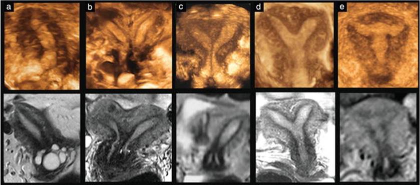
Fig. 7.5
Comparison of three-dimensional ultrasound and magnetic resonance imaging in cases of uterine malformation; the two imaging modalities are extremely similar. Images, according to the American Fertility Society classification, show: (a) unicornuate uterus (Type IId); (b) bicornuate bicollis uterus (Type IVb); (c) septate uterus with two cervices (Type Va); (d) partial septate uterus (Type Vb); (e) uterus with diethylstilbestrol (DES) drug-related malformations (Type VII). [Reprinted from Bermejo, C., Martinez Ten, P., Cantarero, R., Diaz, D., Perez Pedregosa, J., Barron, E. Ruiz Lopez, L. Three-dimensional ultrasound in the diagnosis of Mullerian duct anomalies and concordance with magnetic resonance imaging. Ultrasound Obstet Gynecol, 2010;35(5), 593–601. With permission from John Wiley & Sons, Inc.]
Ghi et al. [25] studied the accuracy of 3D ultrasound in the diagnosis of congenital uterine anomalies among a group of women with RPL.
Ultrasound scan was performed using a machine equipped with a multi-frequency volume endovaginal probe. The insonation technique was standardized according to the following criteria: probe frequency set at 9 mHz, a midsagittal view of the uterus filling 75 % of the screen, three-dimensional (3D) box size including the uterus from fundus to the cervix, sweep angle of 90°, and sweep velocity adjusted to maximum quality. As shown in Fig. 7.1, the volume reconstruction technique was standardized according to the following criteria: the volume rendering box was as narrow as possible in the sagittal plane and adjusted on the uterine corpus in the coronal plane, cut plane scrolled in anterior-posterior fashion with slice thickness set at 1 cm, transparency low (<50 %), and volume rendering by a mix of surface and maximum mode. The analysis of uterine morphology was performed in a standardized reformatted section with the uterus in the coronal view using the interstitial portions of fallopian tubes as reference points. Specific ultrasound diagnosis of uterine anomalies was based on the classification system originally proposed by the American Fertility Society and subsequently modified according to 3D ultrasound landmarks [10] (Table 7.2).
Table 7.2
Classification of congenital uterine anomalies according to volume transvaginal ultrasound
|
Uterine morphology |
Fundal contour |
External contour |
|
Normal |
Straight or convex |
Uniformly convex or with indentation <10 mm |
|
Arcuate |
Concave fundal indentation with central point of indentation at obtuse angle |
Uniformly convex or with indentation <10 mm |
|
Subseptate |
Presence of septum, which does not extend to cervix, with central point of septum at an acute angle |
Uniformly convex or with indentation <10 mm |
|
Septate |
Presence of uterine septum that completely divides cavity from fundus to cervix |
Uniformly convex or with indentation <10 mm |
|
Bicornuate |
Two well-formed uterine cornua |
Fundal indentation >10 mm dividing the two cornua |
|
Unicornuate with or without rudimentary horn |
Single well-formed uterine cavity with a single interstitial portion of Fallopian tube and concave fundal contour |
– |
Reprinted from Ghi, Tullio et al., Accuracy of three-dimensional ultrasound in diagnosis and classification of congenital uterine anomalies. Fertil Steril. 2009;92(2):808–13. With permission from Elsevier
Women with negative ultrasound findings subsequently underwent office hysteroscopy; a combined laparoscopic-hysteroscopic assessment was performed in cases of suspected Müllerian anomaly. A specific Müllerian malformation was sonographically diagnosed in 54 of the 284 women (19 %) included in the study group. All negative ultrasound findings were confirmed at office hysteroscopy. Among the women with abnormal ultrasound findings, the presence of a Müllerian anomaly was endoscopically confirmed in all. Concordance between ultrasound and endoscopy around the type of anomaly was verified in 52 of the 54 (96.3 %) cases, including all cases with a septate uterus and two out of three with bicornuate uterus. This important study concluded that volume TVS appears to be extremely accurate for the diagnosis and classification of congenital uterine anomalies and should conveniently become the first recommended step in the assessment of the uterine cavity in patients with a history of recurrent miscarriage. Three-dimensional ultrasound enables the clinician to comprehensively assess uterine morphology, thus alleviating the need for invasive tests.
Hysterosalpingography (HSG) is a radiographic procedure performed in order to mainly examine the patency of the Fallopian tubes and the morphology of the uterine cavity. It is usually indicated in the early stages of an infertility work-up [26]. The radio-opaque contrast medium fills the cavity, allowing the accurate identification of filling defects due to Müllerian malformations. However, this technique cannot accurately differentiate a septate uterus from a bicornuate uterus [22]. It is also unable to determine the myometrial thickness above the defect or the size of the defect itself. Therefore, the major limitation of this exam lies in its inability to evaluate the external uterine contour [5]. Another disadvantage is the exposure to ionizing radiation in typically young women.
Magnetic Resonance Imaging (MRI)
An expensive, powerful but noninvasive and accurate technique. It has displayed promising results in the diagnosis and categorization of uterine malformations with an accuracy of up to 100 % in the evaluation of Müllerian anomalies [5]. In a study by Bermejo et al. [27], a high degree of concordance between 3D TVS and MRI was reported for the diagnosis of uterine malformations. The structural relationship between the uterine cavity and fundus was equally well visualized with both techniques. Currently MRI is indicated as a complementary imaging modality to 3D ultrasound only in cases of complex abnormalities that involve in addition to the uterus both the cervix and the vagina [22].
Diagnostic Hysteroscopy
Hysteroscopy has become the gold standard for the evaluation of the uterine cavity and is a reliable and safe method in an office setting [22]. This technique allows visualization of the inner part of the cervix and uterus, offering direct vision of the uterine cavity and its internal structures and allows guided biopsies to be obtained if necessary [28]. However, it is also an invasive method that may cause patient discomfort. Hysteroscopy alone is unable to differentiate a septate uterus from a bicornuate uterus [22]. In a retrospective analysis performed by Valli et al. [29], 344 women with RPL and 922 controls were referred for diagnostic hysteroscopy. There was a significantly higher rate of major and minor uterine anomalies (septate and unicornuate uterus) in the RPL group compared to the control group (32 % vs. 6 %, p < 0.001). There was no significant difference in uterine adhesions between the two groups. Another retrospective analysis by Weiss et al. [30] compared the prevalence of uterine anomalies between women referred to hysteroscopy for RPL after two or more consecutive miscarriages. There was no significant difference in uterine abnormality rates between the 67 patients with 2 RPLs and the 98 patients with 3 or more RPLs (32 % vs. 28 %, respectively).
Diagnostic Laparoscopy
This modality gives the surgeon the ability to assess the outer surface of the uterus as well as other pelvic structures. Nonetheless, it is more expensive and invasive [5] compared to the previously reviewed modalities. Currently, diagnostic laparoscopy is generally reserved for women in whom interventional therapy is likely to be undertaken and rarely used for uterine anatomic evaluation purposes [22]. As shown by some, the high accuracy of 3D TVS or MRI allows a noninvasive diagnosis and characterization of uterine anomalies without the need for diagnostic laparoscopy [28].
Sonohysterography
A transvaginal sonogram used in combination with a saline contrast medium injected into the uterine cavity. This is a simple and quick procedure with minimal discomfort to the patient and is now being increasingly used for a routine evaluation of the uterine cavity [22]. Goldberg et al. [26] performed transvaginal sonohysterography on 40 consecutive patients with infertility or RPL previously diagnosed with uterine abnormalities by HSG. The study found that sonohysterography was more accurate than HSG and provided more information about uterine abnormalities. It also provides additional information on the relative proportion of the intracavitary and intramyometrial components of submucous myomas, as well as extracavitary myomas and adhesions [26].
RPL in Different Types of Müllerian Duct Anomalies and Treatment Options
Uterine anomalies have been associated with adverse pregnancy outcomes including spontaneous abortion, recurrent miscarriage, malpresentation, placental abruption, IUGR, prematurity, operative delivery, retained placenta, and fetal mortality [2, 31]. However, it is difficult to assess reproductive outcome precisely, because the majority of studies do not have a control group. In the following section we discuss the treatment of the uterine malformations among women with RPL.
Septated Uterus (ESHRE Classification Class U2)
Septated uterus is the most common Müllerian anomaly, accounting for about 55 % of all Müllerian duct anomalies [5]. It is also the most common major uterine anomaly in women with RPL [32], with a reported prevalence of 15–26 %.
The etiology of RPL in a septated uterus was originally attributed to the fibrous and vascular nature of the septum, despite the lack of histologic data [33, 34]. However, thanks to the use of MRI and histology it is now clear that the septum is composed primarily of smooth muscle and not fibrous tissue [5].
The increased risk of pregnancy loss is most probably related to the decreased connective tissue of the septum that may result in poor decidualization and reduced implantation rate, while increased muscular tissue may result in increased contractility of the tissue. In addition to the inherent deficiencies of the composition of the septum, the overlying endometrium has been shown to be defective [35]. Studies employing electron microscopy reported that the septal endometrium was found to be irregular in morphology, with a decreased sensitivity to preovulatory hormonal changes [36]. Morphologic narrowing of the cavity by the septum, causing a reduction in endometrial capacity, is also believed to play a role in the pathophysiology of adverse reproductive outcome [37].
Finally, inadequate vascularization within the septum and altered relationships between the endometrial, myometrial vessels and myometrial nerves are also considered to be associated with RPL [33, 34]. If this is true, the likelihood of miscarriage caused by septal implantation should increase with the severity of the disruption of uterine morphology [24].
Adverse pregnancy outcome seems to be increased in women with septate uterus as shown in several studies with fetal survival between 6 and 28 % [2]. Ghi et al. [38] reported on pregnancy outcome in women with incidental diagnosis of septate uterus at first trimester scan. They found that in 24 patients diagnosed at a median gestational age of 8.2 weeks, the cumulative pregnancy progression rate was 33.35 % due to the occurrence of early (≤13 weeks) or late (14–22 weeks) miscarriages in 13 and 2 cases, respectively.
Septate uterus is amenable to surgical correction. Hysteroscopic septectomy is the treatment of choice [22]. This procedure is considered to be simple and safe and is reported to increase the live birth rate in patients affected by RPL. It is important to emphasize that although hysteroscopic septectomy is easy and safe to perform, it is critical to demonstrate the external uterine contour by 3D ultrasound before the procedure to rule out a bicornuate uterus for which a different therapeutic approach should be considered (see below).
In addition to some reports on the improvement of infertility and fecundity (38.6 % vs. 20.4 %) after septectomy treatment [39], other studies suggested that the treatment improved birth rate in RPL patients [1, 39].
Fedele et al. [40] reported good results after septectomy in 102 patients with RPL and infertility who had a complete or partial septum. The cumulative pregnancy rate and birth rate after 36 months were 89 and 75 %, respectively, in the septated uterus and 80 and 67 % in the subseptated group.
Homer et al. [32] published a meta-analysis on pregnancy outcomes before and after septectomy and showed a marked improvement after surgery. However, this meta-analysis includes nonrandomized observational methodology. Grimbizis et al. [1] published a nonrandomized study that included patients with previous delivery and live birth rate of only 5 %. After septectomy, the subsequent term delivery rate was around 75 % and the live birth rate was around 85 %.
On the other hand, some authors claim that surgical therapy for septate uterus is not necessary. For example, Homer et al. [32] in their review state that septated uterus is not an indication for surgical intervention. Heinonen [41] reported on 67 patients with a uterine septum and a longitudinal vaginal septum in whom pregnancy outcome was favorable without surgical intervention.
In our opinion, resection of the septum is highly recommended for those with infertility or with RPL to optimize the uterine cavity and minimize adverse obstetrical outcomes once pregnancy is achieved [2].
The procedure includes several methods for removal of the uterine septum using a hysteroscope or resectoscope , including mechanical scissors, electrosurgery with knife electrode or vaporization by bipolar electrodes, yag laser, and mechanical morcellators. The most acceptable procedure is the use of a hysteroscope with mechanical scissors. The septum should be cut from its middle portion where its vasculature is usually most scarce. The preferable timing for the procedure is during the follicular period of the menstrual cycle and should be performed by an experienced surgeon. Complications of the procedure include bleeding, fluid overload, uterine perforation, formation of intrauterine adhesions, and uterine rupture in a subsequent pregnancy. Future deliveries following the procedure do not mandate a cesarean section [42].
It remains uncertain if prophylactic removal of incidentally discovered uterine septa detected prior to childbearing would be indicated in order to improve fertility and pregnancy outcomes [43].
In cases with a cervical septum , the resection of the septum is controversial in regard to the risk of cervical os incompetence once pregnancy has been achieved. At present, a cervical septum should be resected when surgically feasible since inadequate evidence exists for the risk of cervical os incompetence [2].
Unicornis Uterus (ESHRE Classification Class U4, Hemi-uterus)
Unicornis uterus is associated with the worst reproductive outcome, with 30 % of pregnancies resulting in miscarriage [22]. Furthermore this malformation is also associated with intrauterine growth restriction (IUGR), malpresentation, preterm labor, cesarean section, and cervical incompetence [44]. In one recent review of 175 patients and 468 pregnancies, 24.3 % ended in first trimester miscarriage and 9.75 % in second trimester miscarriage with an overall live birth rate of 49.9 % [45].
The presence of a rudimentary uterine horn containing functional endometrium is associated with endometriosis, hematometra, hematosalpinx, pelvic pain, and acute abdomen secondary to ruptured rudimentary horn containing ectopic pregnancy. In those cases, the data support laparoscopic removal in order to prevent these complications [46, 47].
Uterus Arcuatus (ESHRE Classification Class U1c)
Classification of arcuate uterus has been challenging, because it remains unclear whether this variant should be classified as a true anomaly or as an anatomic variant of normal [5]. By definition, this uterus has an intrauterine indentation less than 1 cm [22]. It has an estimated prevalence of 20 % in the general population [1]. Data regarding the reproductive outcomes of patients with an arcuate uterus are extremely limited and widely disparate. In small studies, both poor and good obstetric outcomes have been reported, although an arcuate configuration is generally thought to be compatible with normal term gestation, with a quoted live birth rate of 85 %. In a study of 38 fertile women with live newborns and a history of RPL, uterine malformations were observed in 7.5 % of the cases. The frequency of arcuate uterus was higher than the 4.6 % found in 131 fertile women with live newborns and no history of RPL [48]. However, after excluding all possible extrauterine factors for infertility, surgical hysteroscopy may be considered in selected patients with RPL, particularly those with a prominent or broad configuration of the fundal myometrium. This type of uterine malformation must be considered as part of the differential diagnosis of a partial septate uterus. Some experts consider arcuate uterus as a subtype of a partial septate uterus, even though the natural history and clinical manifestations of arcuate uterus malformation are relatively benign [1]. Even though the pathophysiology of pregnancy loss in women with this malformation remains uncertain, one can view arcuate uterus as a minor alteration of the uterine cavity shape but with no major external change of the contour. This is supported by a long-term study that found a term delivery rate of almost 80 % with a live birth rate of 82.7 % and no adverse impact on reproduction [49].
Uterine Didelphi (ESHRE Classification Class U3c)
This type of uterus, also referred to as a double uterus, may result from complete failure of the fusion of the two Müllerian ducts. This results in a separate and narrower uterus developing from each duct. Each uterus may have its own cervix or both uteri may share a single one. This type of uterus is associated with a double vagina (each referred to as “hemivagina”) in 67 % of cases, separated by a thin wall [22]. However, this anomaly is relatively rare. Nahum [12] found uterine didelphi in 11 % of all congenital uterine anomalies. Heinonen [50] studied the clinical implications and long-term follow-up of 49 cases of didelphic uterus. Complications such as an obstructed hemivagina and renal agenesis were found, as well as ovarian neoplasm [9 %]. As for pregnancies, 94 % of the women had at least one pregnancy and the miscarriage rate was 21 %. Ectopic pregnancy occurred in 2 % of the cases, prenatal mortality in 5.3 %, and prematurity in 24 %. The study concluded that women with this type of uterus do not have a notably impaired fertility. Grimbizis et al. [1] instead reported a term delivery rate of approximately 45 % in women with didelphic uteri.
While the nonobstructive type is usually asymptomatic, the didelphic uterus with a hemivaginal obstruction could become symptomatic at the time of menarche and present with dysmenorrhea. Other complications that may be associated are endometriosis and pelvic adhesions, possibly secondary to retrograde menstrual flow in patients with an obstruction [5].
Bicornuate Uterus (ESHRE Classification Classes U3a and U3b)
A bicornuate uterus results from a partial fusion of the Müllerian ducts. Bicornuate uteri may be classified into two subtypes, depending on the level of the caudal extension of the fundal indentation: bicornuate unicollis if at the level of the internal cervical os, bicornuate bicollis if at the level of the external cervical os. The horns of this uterus are not fully developed and are thus smaller than those of a didelphic uterus [22]. In a series of 67 pregnancies among 261 patients with untreated bicornuate uterus, Grimbizis et al. [1] found poor pregnancy outcomes, with a mean miscarriage rate of 36 %. The mean preterm delivery rate was 23 % and the mean live birth rate was 55.2 %. They concluded that the pregnancy outcome was significantly poorer (p < 0.001) than that of women with a normal uterus. Usually this type of malformation is not a candidate for surgical correction, but a Strassman metroplasty with a wedge resection of the medial aspect of each uterine horn followed by the unification of the two cavities may be considered in women with RPL [2].
Diethylstilbestrol Exposure
Diethylstilbestrol (DES) is an orally active synthetic estrogen that was used between 1948 and 1971 in the prevention of pregnancy loss in women suffering from RPL. When administered to pregnant women, it has been shown to increase the risk of malformations and tumors in the genitalia of the offspring. The use of this medication in pregnancy was discontinued due to its teratologic effects. When hysterosalpingography was performed on 267 DES-exposed women, 69 % of them presented a congenital malformation of the uterus. The most common abnormality was a T-shaped uterine cavity (ESHRE classification class U1a). How DES affects the uterine development is not clear. A ) relationship between in utero DES exposure and the occurrence of cervical incompetence was also noted.
Treatment of Uterine Malformations
Raga et al. [49], in their review, summarized the current management of the different types of uterine malformations. For septate and arcuate uterus, hysteroscopic metroplasty is the treatment of choice. The other anomalies, which are less frequently encountered, may require more complicated abdominal or combined procedures or may even not have a surgical solution. The strategy of management must be based either on the obstetric history of the patient or on the prognosis of the malformation itself. Most clinicians do not recommend the Strassman procedure, which was associated with a high percentage of complications. The main concept in the treatment of uterine malformations is close monitoring and follow-up in high-risk pregnancy clinic, bed rest, and progesterone treatment for the prevention of preterm delivery.
Cerclage is indicated in cases with cervical os incompetence diagnosed by medical history or shortened cervical length by ultrasound. Cervical os incompetence is difficult to diagnose and is not a common cause of RPL even in patients with profound structural uterine abnormality. However, cerclage has recently been offered as a treatment for women with RPL and a uterine anomaly other than a septate uterus. Seidman et al. [51] compared the survival rate of fetuses in 86 women with congenital uterine anomalies and 106 women with normal anatomy. The incidence of cervical os incompetence proven by HSG was 23 % in the two groups. Sixty-seven out of 86 and 29 out of 106 were managed with cervical cerclage. The obstetric outcome was stratified by cervical incompetence and obstetric history. The viable live birth rate was significantly higher in the malformed group with cerclage (88 %) compared with the malformed group without cerclage (47 %). No statistically significant differences in live birth rate were found in the normal uterus with cerclage even when only those women with a history of RPL were considered. The indications for cerclage are still controversial. The CIPRACT study found that therapeutic cerclage with bed rest reduced neonatal morbidity and preterm delivery rates in women with risk factors (including DES exposure and uterine anomaly) and/or cervical os incompetence and cervical length of <27 mm before 27 weeks gestational age [52].
Yassaee and Mostafaee [53] studied 40 patients with uterine anomalies, 26 were treated with cerclage and 14 without cerclage. In patients with bicornuate uterus and cervical cerclage, term delivery rate was 76.2 % compared to 27.35 % in the group without cerclage (p < 0.05). Among patients with uterus arcuatus, preterm delivery rate was not statistically different between those with or without cerclage.
Cervical os incompetence is still considered an infrequent cause of pregnancy loss in patients with major structural anomalies of the uterus. We recommend to consider prophylactic cerclage for those patients with uterine anomalies such as bicornuate uterus and unicornis uterus with a history of RPL or preterm delivery.
Acquired Uterine Structural Malformations
Myomas
A uterine myoma , also known as fibroid, is a benign solid tumor made of fibrous tissue of the uterus. The myomas’ size and number may vary; they are usually slow-growing and asymptomatic. Myomas are associated with abnormal uterine bleeding including heavy menstrual bleeding, infertility, RPL, and complaints related to the effect of an enlarged uterus on the adjacent structures in the pelvis [54]. RPL is usually the result of lesions that distort the endometrial cavity, being close to the endometrium. The presence of submucous myomas may deform the uterine cavity; the overlying endometrium is usually thin and therefore inadequate for normal implantation. Myomas are common in woman of reproductive age with a prevalence as high as 70–80 % in woman aged 50 years [55]. The location of the myomas may affect the reproductive outcome and function in women, and the removal of the myoma prior to conception may have a positive influence on pregnancy rate [56]. In a meta-analysis by Pritts et al. [56] women with submucous myomas had significantly higher spontaneous miscarriage rates (RR 1.68, 95 % CI 1.37–2.05, p = 0.022). The mechanism by which submucous myomas affect pregnancy outcomes is unknown. Histologic testing did show glandular atrophy of the endometrium overlying the myomas and opposite to the myomas. The atrophy has been suggested to impair the implantation and nourishment of the developing embryo [57, 58]. Other suggested mechanisms are impaired transport of gametes, altered uterine contractility, and other negative effects such as enlarging or deforming the endometrial cavity and obstructing tubal ostia [59, 60]. Myomas may also cause implantation failure by physically altering the uterine shape, preventing discharge of intrauterine blood or clots, and by altering the normal endometrial development [60]. Due to the increasing use of US, there has been a rise in the diagnosis of uterine leiomyomas in women with unexplained infertility [61].
Regarding the recommended treatment, another interesting finding of the study performed by Casini et al. [62] was that there is an important role for the removal of fibroids before conception and pregnancy. However, it is still undetermined who are the patients that may benefit the most from the invasive surgical approach. In a study by Pritts et al. [56] the relative risk of spontaneous pregnancy following submucous myomectomy was 0.77, compared to the control subjects who did not undergo myomectomy. This confirms the important role of uterine fibroids in infertility as well as the importance of fibroid removal before conception, to improve both the chances of fertilization and pregnancy maintenance. Patients who had submucous fibroids larger than 2 cm distorting their uterine cavities had a higher pregnancy rate following hysteroscopic resection [22]. It seems that subserosal myomas have little, if any, effect on reproductive outcome, especially if they are up to 5–7 cm in diameter. Intramural myomas that do not invade the endometrium may be considered as well to be relatively harmless to reproduction, as long as they are smaller than 4–5 cm in diameter [60]. Submucosal as well as some intramural myomas that compress the uterine cavity significantly reduce pregnancy rates, and surgical removal should be considered before assisted reproductive techniques are used [63].
The association between submucous myomectomy and pregnancy loss is less evident as there is insufficient data regarding this association. Moreover, an inherited bias exists due to the increased risk of first trimester loss. The available evidence is suggestive of benefit. Clearly more data are required, but this evidence suggests that, at least in selected patients, submucous myomectomy may reduce the risk of spontaneous miscarriage [61]. Therefore, one can conclude that the two parameters influencing better outcomes of a future pregnancy are the location and size of the myomas. Hysteroscopic myomectomy is the gold standard for the treatment of submucous myomas. For other myomas, abdominal or laparoscopic myomectomy by a trained surgeon is the best alternative [60]. It is now recommended that most of the intramural and subserosal uterine myomas should be treated with laparoscopic myomectomy in women who desire to preserve their uterus. The post-laparoscopy pregnancy rates are estimated to be 50–60 % [64]. Another less invasive surgical approach is laparoscopic-assisted myomectomy (LAM), which became a safe and efficient alternative to both laparoscopic myomectomy and myomectomy by laparotomy for patients with numerous large or deep intramural myomas [22].
Uterine Polyps
Polyps are growths, endometrial masses attached to the inner lining of the uterus. They can be differentiated from fibroids by the fact that while fibroids are mainly composed of muscle tissue, polyps are made of endometrial tissue. Polyps consist of benign hyperplastic endometrial growth. It is suggested that polyps with intracavity extensions may act like foreign bodies within the uterine cavity [22]. Another suggestion is that they play a role in inducing chronic inflammatory changes in the endometrium, causing the area to be less receptive for pregnancy implantation and support. However, the association between endometrial polyps and RPL is yet to be proven. Several more hypotheses have arisen more recently. A case–control study by Rackow et al. [65] evaluated the effect of hysteroscopically identified endometrial polyps on the endometrium using known molecular markers of endometrial receptivity. Marked decrease in the mRNA levels of the HOXA10 and HOXA11 was observed in a uterus with endometrial polyps. These molecular markers are thought to impair implantation. These findings offer a possible molecular mechanism to support the clinical findings of lower pregnancy rates in women with endometrial polyps.
Even though the association between endometrial polyps and pregnancy loss is yet to be proven, polyps are more common in patients with recurrent spontaneous miscarriage [29]. The current treatment approach to infertile women, particularly those undergoing in vitro fertilization and embryo transfer, is to perform hysteroscopic polypectomy when intrauterine filling defects are diagnosed [66]. Perez-Medina et al. [67] attempted to determine whether hysteroscopic polypectomy before intrauterine insemination (IUI) resulted in better pregnancy outcomes. Two hundred and fifteen infertile women scheduled to undergo IUI participated in the study and it was reported that, compared to women who did not undergo the procedure, hysteroscopic polypectomy improved the likelihood of conception, with a relative risk of 2.1 (95 % CI 1.5–2.9).
It seems that hysteroscopic polypectomy enhances fertility [68]. Therefore, one should consider performing polypectomy in women with uterine polyps and RPL, especially when no other etiology has been found.
Intrauterine Adhesions
Intrauterine adhesions , also known as Asherman syndrome, occur most often due to exaggerated postpartum or postmiscarriage dilatation and curettage. Other causes include genital tuberculosis, previous uterine surgery [69], and endometritis [22]. The adhesions may result in infertility and/or RPL [70]; although this is not a common cause it may lead to secondary infertility in these patients. These intrauterine scars can interfere with the normal implantation process, therefore being responsible for pregnancy loss. The type and extent of intrauterine adhesions vary. They are expected to be found more often in women with RPL since vacuum aspiration, which is one of the leading causes of uterine adhesions, is a common procedure in cases of early pregnancy loss. In a study performed by Ventolini et al. [71], among 23 patients with an otherwise unexplained history of three or more first or second trimester miscarriages and no live births, hysteroscopy showed that 5 (21.8 %) of the women had intrauterine adhesions. Classifying the adhesions can be done according to the amount of the involved uterine cavity. Minimal adhesions are defined by the involvement of less than one-fourth of the uterine cavity with thin and filmy adhesions. If affecting one-fourth of the uterine cavity with no agglutination of the walls, ostial areas and partial occlusion of the upper fundus, it is defined as moderate adhesions. Severe adhesions involve more than three-fourths of the uterine cavity, with agglutination of the walls or thick bands and occlusion of the ostial areas and upper uterine cavity [69].
It is agreed that adhesions should be hysteroscopically resected. In a retrospective case report series published by Pabuccu et al. [69], 40 women with RPL or infertility underwent hysteroscopic adhesiolysis. The majority of women included in the study had a history of vigorous curettage and two had a history of genital tuberculosis. Women with previous uterine surgery were excluded from the study. All women with RPL conceived after surgery and 71 % of pregnancies resulted in term or viable preterm infants. Out of eight patients with mild adhesions, seven conceived after surgery, as did three out of four patients with moderate adhesions.
Hysteroscopic synechiae resection seems to be indicated when the adhesions are classified as moderate to severe or the access to the tubal ostia is blocked [69]. When no other cause for RPL is found, and mild adhesions are present, one should consider performing adhesiolysis as well. It should be noted that uterine adhesions caused by genital tuberculosis are usually cohesive and have a tendency to recur, therefore conferring a poor prognosis. The reproductive outcome correlates with the severity of the initially diagnosed adhesions.
Summary
Congenital Müllerian and acquired uterine structural abnormalities are important in the pathophysiology, diagnosis, and therapy of RPL. Therefore, an anatomical work-up is recommended for RPL patients according to ASRM guidelines. The initial diagnostic modality is 2D followed by 3D transvaginal ultrasonography. These modalities will provide the diagnosis in more than 95 % of the cases. The findings should be classified using the newer and simplified ESHRE classification. Following diagnosis, appropriate interventions should be considered when indicated, like septectomy for a septate uterus or adhesiolysis for intrauterine adhesions. Other (non-septate) Müllerian anomalies require close monitoring and a few of them may benefit if cervical cerclage is performed. For acquired malformation, adhesiolysis as well as synechiae and resection of uterine myomas are recommended in the setting of RPL.
References
1.
Grimbizis GF, Camus M, Tarlatzis BC, Bontis JN, Devroey P. Clinical implications of uterine malformations and hysteroscopic treatment results. Hum Reprod Update. 2001;7(2):161–74.PubMedCrossRef
2.
Reichman DE, Laufer MR. Congenital uterine anomalies affecting reproduction Best practice and research clinical obstetrics and gynaecology, volume 24. Amsterdam: Elsevier; 2010. p. 193–208.
3.
Muller P, Musset R, Netter A, Solal R, Vinourd JC, Gillet JY. State of the upper urinary tract in patients with uterine malformations. Study of 133 cases. Presse Med. 1967;75(26):1331–6.PubMed
4.
Lee DM, Osathanondh R, Yeh J. Localization of Bcl-2 in the human fetal müllerian tract. Fertil Steril. 1998;70(1):135–40.PubMedCrossRef
5.
Troiano RN, McCarthy SM. Mullerian duct anomalies: imaging and clinical issues. Radiology. 2004;233:19–34.PubMedCrossRef
6.
Buttram VC, Gibbons WE. Müllerian anomalies: a proposed classification. (An analysis of 144 cases). Fertil Steril. 1979;32(1):40–6.PubMed
7.
Fedele L, Bianchi S, Agnoli B, Tozzi L, Vignali M. Urinary tract anomalies associated with unicornuate uterus. J Urol. 1996;155(3):847–8.PubMedCrossRef
8.
Narang HK, Warke HS, Mayadeo NM. Mullerian anomaly with ovary at deep inguinal ring: a rare case finding. J Obstet Gynaecol. 2013;33(3):317–8.PubMedCrossRef
9.
Ombelet W, Verswijvel G, de Jonge E. Ectopic ovary and unicornuate uterus. N Engl J Med. 2003;348(7):667–8.PubMedCrossRef
10.
Dabirashrafi H, Mohammad K, Moghadami-Tabrizi N. Ovarian malposition in women with uterine anomalies. Obstet Gynecol. 1994;83(2):293–4.PubMed
11.
Simón C, Martinez L, Pardo F, Tortajada M, Pellicer A. Müllerian defects in women with normal reproductive outcome. Fertil Steril. 1991;56(6):1192–3.PubMed
12.
Nahum GG. Uterine anomalies. How common are they, and what is their distribution among subtypes? J Reprod Med. 1998;43(10):877–87.PubMed
13.
Sugiura-Ogasawara M, Ozaki Y, Kitaori T, Kumagai K, Suzuki S. Midline uterine defect size is correlated with miscarriage of euploid embryos in recurrent cases. Fertil Steril. 2010;93(6):1983–8.PubMedCrossRef
14.
Jaslow CR, Kutteh WH. Effect of prior birth and miscarriage frequency on the prevalence of acquired and congenital uterine anomalies in women with recurrent miscarriage: a cross-sectional study. Fertil Steril. 2013;99(7):1916–22. e1.PubMedCrossRef
15.
Shulman LP. Müllerian anomalies. Clin Obstet Gynecol. 2008;51(2):214–22.PubMedCrossRef
16.
Battin J, Lacombe D, Leng JJ. Familial occurrence of hereditary renal adysplasia with müllerian anomalies. Clin Genet. 1993;43(1):23–4.PubMedCrossRef
17.
The American Fertility Society classifications of adnexal adhesions, distal tubal occlusion, tubal occlusion secondary to tubal ligation, tubal pregnancies, mullerian anomalies and intrauterine adhesions. Fertil Steril. 1988;49(6):944–55.
18.
Acién P, Acién M, Sánchez-Ferrer M. Complex malformations of the female genital tract. New types and revision of classification. Hum Reprod. 2004;19(10):2377–84.PubMedCrossRef
19.
Oppelt P, Renner SP, Brucker S, Strissel PL, Strick R, Oppelt PG, et al. The VCUAM (Vagina Cervix Uterus Adnex-associated Malformation) classification: a new classification for genital malformations. Fertil Steril. 2005;84(5):1493–7.PubMedCrossRef
20.
Grimbizis GF, Gordts S, Di Spiezio Sardo A, Brucker S, De Angelis C, Gergolet M, et al. The ESHRE-ESGE consensus on the classification of female genital tract congenital anomalies. Gynecol Surg. 2013;10(3):199–212.PubMedPubMedCentralCrossRef
21.
Di Spiezio Sardo A, Campo R, Gordts S, Spinelli M, Cosimato C, Tanos V, et al. The comprehensiveness of the ESHRE/ESGE classification of female genital tract congenital anomalies: a systematic review of cases not classified by the AFS system. Hum Reprod. 2015;30(5):1046–58.PubMedPubMedCentralCrossRef
22.
Carp HJ. Recurrent pregnancy loss- causes, controversies and treatment. 1st ed. London: Informa UK Ltd, Taylor & Francis e-Library; 2008. 305.
23.
Szkodziak P, Wozniak S, Czuczwar P, Paszkowski T, Milart P, Wozniakowska E, et al. Usefulness of three dimensional transvaginal ultrasonography and hysterosalpingography in diagnosing uterine anomalies. Ginekol Pol. 2014;85(5):354–9.PubMed
24.
Salim R, Regan L, Woelfer B, Backos M, Jurkovic D. A comparative study of the morphology of congenital uterine anomalies in women with and without a history of recurrent first trimester miscarriage. Hum Reprod. 2003;18(1):162–6.PubMedCrossRef
25.
Ghi T, Casadio P, Kuleva M, Perrone AM, Savelli L, Giunchi S, et al. Accuracy of three-dimensional ultrasound in diagnosis and classification of congenital uterine anomalies. Fertil Steril. 2009;92(2):808–13.
26.
Goldberg JM, Falcone T, Attaran M. Sonohysterographic evaluation of uterine abnormalities noted on hysterosalpingography. Hum Reprod. 1997;12(10):2151–3.PubMedCrossRef
27.
Bermejo C, Ten Martinez P, Cantarero R, Diaz D, Perez Pedregosa J, Barron E, et al. Three-dimensional ultrasound in the diagnosis of Mullerian duct anomalies and concordance with magnetic resonance imaging. Ultrasound Obstet Gynecol. 2010;35(5):593–601.PubMedCrossRef
28.
Dueholm M, Lundorf E, Hansen ES, Ledertoug S, Olesen F. Evaluation of the uterine cavity with magnetic resonance imaging, transvaginal sonography, hysterosonographic examination, and diagnostic hysteroscopy. Fertil Steril. 2001;76:350–7.
29.
Valli E, Zupi E, Marconi D, Vaquero E, Giovannini P, Lazzarin N, et al. Hysteroscopic findings in 344 women with recurrent spontaneous abortion. J Am Assoc Gynecol Laparosc. 2001;8(3):398–401.PubMedCrossRef
30.
Weiss A, Shalev E, Romano S. Hysteroscopy may be justified after two miscarriages. Hum Reprod. 2005;20:2628–31.
31.
Hua M, Odibo AO, Longman RE, Macones GA, Roehl KA, Cahill AG. Congenital uterine anomalies and adverse pregnancy outcomes. Am J Obstet Gynecol. 2011;205(6):558.e1–5.
32.
Homer HA, Li TC, Cooke ID. The septate uterus: a review of management and reproductive outcome. Fertil Steril. 2000;73(1):1–14.PubMedCrossRef
33.
Fayez JA. Comparison between abdominal and hysteroscopic metroplasty. Obstet Gynecol. 1986;68(3):399–403.PubMedCrossRef
34.
Fedele L, Bianchi S. Hysteroscopic metroplasty for septate uterus. Obstet Gynecol Clin North Am. 1995;22(3):473–89.PubMed
35.
Candiani GB, Fedele L, Zamberletti D, De Virgiliis D, Carinelli S. Endometrial patterns in malformed uteri. Acta Eur Fertil. 1983;14(5):311–8.PubMed
36.
Fedele L, Bianchi S, Marchini M, Franchi D, Tozzi L, Dorta M. Ultrastructural aspects of endometrium in infertile women with septate uterus. Fertil Steril. 1996;65(4):750–2.PubMed
37.
Patton PE, Novy MJ. Reproductive potential of the anomalous uterus. In: Seminars in reproductive endocrinology, Vol. 6, No. 2, 1988. p. 217–33. http://ohsu.pure.elsevier.com/en/publications/reproductive-potential-of-the-anomalous-uterus(55ded2b8-5dba-45ef-99b9-a3bed82fc108).html.
38.
Ghi T, De Musso F, Maroni E, Youssef A, Savelli L, Farina A, et al. The pregnancy outcome in women with incidental diagnosis of septate uterus at first trimester scan. Hum Reprod. 2012;27(9):2671–5.PubMedCrossRef
39.
Mollo A, De Franciscis P, Colacurci N, Cobellis L, Perino A, Venezia R, et al. Hysteroscopic resection of the septum improves the pregnancy rate of women with unexplained infertility: a prospective controlled trial. Fertil Steril. 2009;91:2628–31.
40.
Fedele L, Arcaini L, Parazzini F, Vercellini P, Di Nola G. Reproductive prognosis after hysteroscopic metroplasty in 102 women: life-table analysis. Fertil Steril. 1993;59(4):768–72.PubMed
41.
Heinonen PK. Complete septate uterus with longitudinal vaginal septum. Fertil Steril. 2006;85(3):700–5.PubMedCrossRef
42.
Valle RF, Ekpo GE. Hysteroscopic metroplasty for the septate uterus: review and meta-analysis. J Minim Invasive Gynecol. 2013;20(1):22–42.PubMedCrossRef
43.
Pabuçcu R, Gomel V. Reproductive outcome after hysteroscopic metroplasty in women with septate uterus and otherwise unexplained infertility. Fertil Steril. 2004;81(6):1675–8.PubMedCrossRef
44.
Cohen AW, Chhibber G. Obstetric complications of congenital anomalies of the paramesonephric ducts.
45.
Reichman D, Laufer MR, Robinson BK. Pregnancy outcomes in unicornuate uteri: a review. Fertil Steril. 2009;91(5):1886–94.PubMedCrossRef
46.
Canis M, Wattiez A, Pouly JL, Mage G, Manhes H, Bruhat MA. Laparoscopic management of unicornuate uterus with rudimentary horn and unilateral extensive endometriosis: case report. Hum Reprod. 1990;5(7):819–20.PubMed
47.
Nezhat F, Nezhat C, Bess O, Nezhat CH. Laparoscopic amputation of a noncommunicating rudimentary horn after a hysteroscopic diagnosis: a case study. Surg Laparosc Endosc. 1994;4(2):155–6.PubMed
48.
Acien P. Incidence of Mullerian defects in fertile and infertile women. Hum Reprod. 1997;12(7):1372–6.PubMedCrossRef
49.
Raga F, Bauset C, Remohi J, Bonilla-Musoles F, Simon C, Pellicer A. Reproductive impact of congenital Mullerian anomalies. Hum Reprod. 1997;12(10):2277–81.PubMedCrossRef
50.
Heinonen PK. Clinical implications of the didelphic uterus: long-term follow-up of 49 cases. Eur J Obstetr Gynecol Reprod Biol. 2000;91:183–90.
51.
Seidman DS, Ben-Rafael Z, Bider D, Recabi K, Mashiach S. The role of cervical cerclage in the management of uterine anomalies. Surg Gynecol Obstet. 1991;173(5):384–6.PubMed
52.
Althuisius S, Dekker G, Hummel P, Bekedam D, Kuik D, van Geijn H. Cervical Incompetence Prevention Randomized Cerclage Trial (CIPRACT): effect of therapeutic cerclage with bed rest vs. bed rest only on cervical length. Ultrasound Obstet Gynecol. 2002;20(2):163–7.PubMedCrossRef
53.
Yassaee F, Mostafaee L. The role of cervical cerclage in pregnancy outcome in women with uterine anomaly. J Reprod Infertil. 2011;12(4):277–9.PubMedPubMedCentral
54.
AAGL practice report: practice guidelines for the diagnosis and management of submucous leiomyomas. J Minim Invasive Gynecol 2012;19:152–71.
55.
Baird DD, Dunson DB, Hill MC, Cousins D, Schectman JM. High cumulative incidence of uterine leiomyoma in black and white women: ultrasound evidence. Am J Obstet Gynecol. 2003;188(1):100–7.PubMedCrossRef
56.
Pritts EA, Parker WH, Olive DL. Fibroids and infertility: an updated systematic review of the evidence. Fertil Steril. 2009;91(4):1215–23.PubMedCrossRef
57.
Deligdish L, Loewenthal M. Endometrial changes associated with myomata of the uterus. J Clin Pathol. 1970;23(8):676–80.PubMedPubMedCentralCrossRef
58.
Maguire M, Segars JH. Benign uterine disease: leiomyomata and benign polyps. The endometrium: molecular, cellular and clinical perspectives. 2nd ed. London: Informa Health Care; 2008.
59.
Farhi J, Ashkenazi J, Feldberg D, Dicker D, Orvieto R, Ben Rafael Z. Effect of uterine leiomyomata on the results of in-vitro fertilization treatment. Hum Reprod. 1995;10(10):2576–8.PubMed
60.
Kolankaya A, Arici A. Myomas and assisted reproductive technologies: when and how to act? Obstet Gynecol Clin North Am. 2006;33(1):145–52.PubMedCrossRef
61.
Klatsky PC, Tran ND, Caughey AB, Fujimoto VY. Fibroids and reproductive outcomes: a systematic literature review from conception to delivery. Am J Obstet Gynecol. 2008;198(4):357–66.PubMedCrossRef
62.
Casini ML, Rossi F, Agostini R, Unfer V. Effects of the position of fibroids on fertility. Gynecol Endocrinol. 2006;22:106–9.
63.
Carranza-Mamane B, Havelock J, Hemmings R, Reproductive E, Infertility C, Cheung A, et al. The management of uterine fibroids in women with otherwise unexplained infertility. J Obstetr Gynecol. 2015;37(3):277–88.
64.
Goldberg J, Pereira L. Pregnancy outcomes following treatment for fibroids: uterine fibroid embolization versus laparoscopic myomectomy. Curr Opin Obstet Gynecol. 2006;18(4):402–6.PubMedCrossRef
65.
Rackow BW, Jorgensen E, Taylor HS. Endometrial polyps affect uterine receptivity. Fertil Steril. 2011;95(8):2690–2.PubMedPubMedCentralCrossRef
66.
Silberstein T, Saphier O, van Voorhis BJ, Plosker SM. Endometrial polyps in reproductive-age fertile and infertile women. Isr Med Assoc J. 2006;8(3):192–5.PubMed
67.
Pérez-Medina T, Bajo-Arenas J, Salazar F, Redondo T, Sanfrutos L, Alvarez P, et al. Endometrial polyps and their implication in the pregnancy rates of patients undergoing intrauterine insemination: a prospective, randomized study. Hum Reprod. 2005;20(6):1632–5.PubMedCrossRef
68.
Varasteh NN, Neuwirth RS, Levin B, Keltz MD. Pregnancy rates after hysteroscopic polypectomy and myomectomy in infertile women. Obstet Gynecol. 1999;94(2):168–71.PubMedCrossRef
69.
Pabuçcu R, Atay V, Orhon E, Urman B, Ergün A. Hysteroscopic treatment of intrauterine adhesions is safe and effective in the restoration of normal menstruation and fertility. Fertil Steril. 1997;68(6):1141–3.PubMedCrossRef
70.
ASHERMAN JG. Traumatic intra-uterine adhesions. J Obstet Gynaecol Br Emp. 1950;57(6):892–6.PubMedCrossRef
71.
Ventolini G, Zhang M, Gruber J. Hysteroscopy in the evaluation of patients with recurrent pregnancy loss: a cohort study in a primary care population. Surg Endosc. 2004;18(12):1782–4.PubMedCrossRef


