
Chandu Vemuri
Sandra L. Wong
Presentation
A 50-year-old man with a history of hypertension presents for evaluation of an enlarging scrotal mass, thought to be an inguinal hernia. On examination, he has a large left scrotal mass that is firm, nontender, and not reducible. He is also noted to have firmness throughout the entire abdomen but has no abdominal pain. He reports some fatigue, early satiety, and a 15-lb unintentional weight loss over the past year, though he has increasing abdominal girth. Otherwise, he is active and continues to work full time.
Differential Diagnosis
Patients with retroperitoneal sarcoma typically present with nonspecific symptoms and will often have an abdominal mass on physical examination. Some symptoms are specific to mass effect on nearby structures. Because of the location, diagnosis is often delayed, and the majority of patients have tumors that are larger than 10 cm in size at time of initial evaluation. Retroperitoneal masses are usually malignant and about one-third of these masses are sarcomas. The differential diagnosis includes germ cell tumors, lymphoma, and other tumors of retroperitoneal solid organs, including endocrine cancers and renal cell carcinoma.
Workup
Occasionally, the mass is appreciated on cross-sectional imaging performed for other reasons, but otherwise a CT scan of the abdomen and the pelvis with oral and IV contrast is the preferred initial radiographic evaluation. Complete workup involves a detailed history and physical examination, including assessment of diffuse lymphadenopathy, scrotal exam, and serum laboratory studies to selectively evaluate excessive endocrine hormone production, elevated germ cell tumor markers (e.g., alpha-fetoprotein (AFP), beta-human chorionic gonadotrophin (β-hCG), and lactate dehydrogenase.
In this patient, physical examination was only remarkable as noted above. CT scan of the abdomen and pelvis was significant for a massive retroperitoneal mass extending through the left inguinal canal into the scrotum (Figure 1). The radiologic appearance was consistent with liposarcoma given the predominantly fatty appearance with linear septa. A CT-guided core needle biopsy confirmed a low-grade well-differentiated liposarcoma. CT scan of the chest did not demonstrate any evidence of pulmonary metastasis. Laboratory values were normal, including a creatinine of 0.8 mg/dL. He was referred for surgical resection.
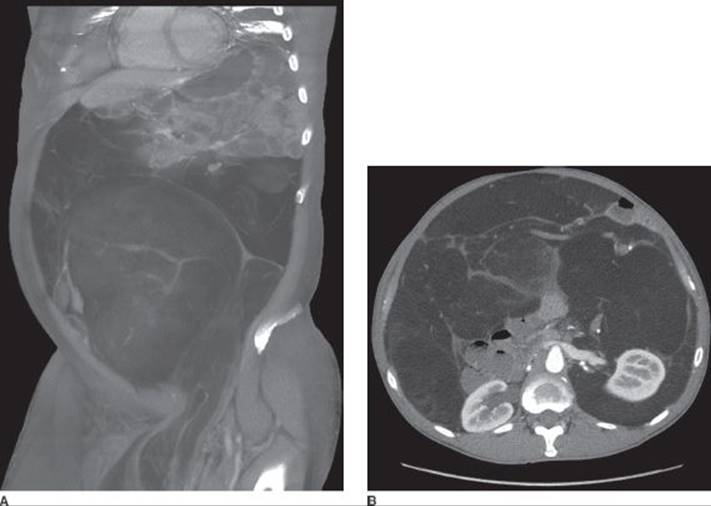
FIGURE 1 • Case presentation. A: Retroperitoneal/intra-abdominal sarcoma occupying the majority of the abdominal cavity is demonstrated on angled sagittal views from a CT scan. Note displacement of the intra-abdominal contents superiorly and extension of the tumor into the scrotum via the inguinal canal. B: Selected axial view of the liposarcoma demonstrating a homogeneous fatty appearance with linear septa. Note anterior displacement of the descending colon (arrow).
Discussion
Presenting symptoms can include vague abdominal or back pain and weight loss, as well as symptoms specific to location of the tumor such as early satiety if there is mass effect on the stomach or venous obstruction of the lower extremity from a pelvic tumor. Biopsy of suspicious masses can be performed preoperatively to establish a diagnosis. Biopsy, with a core needle biopsy approach if possible, is mandatory if there is a question about the diagnosis, if there is consideration for neoadjuvant treatment, or if the tumor is felt to be unresectable.
Imaging should be directed to preparation for surgical resection. Magnetic resonance imaging (MRI) is an alternate modality that is sufficient, but not necessary, for these patients. Workup should also include evaluation of metastatic disease, most commonly to lungs or liver. Plain chest radiographs can be used to evaluate for pulmonary metastasis, but CT should be performed if any abnormalities are seen or if the sarcoma is considered a high risk for metastasis. Positron emission tomography scans do not have a defined role in assessment of retroperitoneal sarcoma.
Diagnosis and Treatment
The cornerstone of treatment for localized disease is complete surgical resection. This patient was felt to have a resectable retroperitoneal sarcoma and this was recommended. Resection should include involved intra-abdominal/retroperitoneal structures with anastomoses and reconstruction as appropriate. Sarcomas with critical vascular involvement, peritoneal implants, involvement at the root of the mesentery, or spinal cord involvement are usually considered unresectable. Incomplete resections with microscopically positive margins lead to an increased risk of local recurrence, which represents a common form of treatment failure. Great care must be taken to ensure complete resection without capsular intrusion during the index operation. Repeat resection for recurrent disease is recommended, especially if interval surveillance is able to detect early recurrences. Unfortunately, recurrent disease often predicts further recurrences, which often become increasingly more aggressive in terms of tumor biology and more technically difficult to resect with successive operations.
Retroperitoneal sarcomas are relatively uncommon neoplasms derived from mesenchymal cells, with only an estimated 10,000 new cases of soft tissue sarcoma in the United States each year. Only 10% to 15% of these cases will be of retroperitoneal origin. More than 50 different histologic subtypes have been described, but the most commonly seen retroperitoneal sarcomas are liposarcomas or leiomyosarcomas. Prognosis is largely dictated by the ability to achieve complete surgical resection, and median survival for patients who undergo complete resection is 60 months compared to 24 months for those undergoing incomplete resection. Other factors associated with decreased survival include high histologic grade, large tumor size, and older patient age.
Multimodality treatment approaches, including systemic chemotherapy and radiation therapy, merit consideration since resection alone has unsatisfactory outcomes in high-risk tumors. There are good data to support the use of radiation in conjunction with limb-sparing procedures for extremity sarcomas. However, radiation to the retroperitoneum is complicated by dose-limiting toxicity to abdominal viscera, which are uniquely radiosensitive, and by very large treatment fields. For this reason, preoperative radiation approaches have been considered to improve rates of local control and reduce recurrences. When the sarcoma is in situ, bowel and other structures are displaced and out of the intended radiation field, allowing for more effective use of radiation. It is unlikely that radiation would change the size of the tumor or change the scope and/or extent of the planned operation. Many small series have demonstrated the safety and feasibility of radiation for selected patients with high-risk retroperitoneal sarcomas, and studies using standard external-beam radiation therapy, brachytherapy approaches, or intraoperative electron-beam radiotherapy approaches are ongoing to evaluate their efficacy in disease outcomes. The use of systemic chemotherapy for most histologic subtypes of retroperitoneal sarcoma remains controversial. Response rates are relatively low, and there is no demonstrated improvement in overall or disease-specific survival with either neoadjuvant or adjuvant chemotherapy regimens. Some commonly used cytotoxic agents include doxorubicin and ifosfamide. There is limited experience with combined chemoradiation treatments.
Surgical Approach and Special Intraoperative Considerations
Retroperitoneal sarcomas, by definition, can originate from mesenchymal tissues throughout the retroperitoneum. Hepatic sarcomas, intra-abdominal desmoid tumors (desmoids fibromatosis), and gastrointestinal stromal tumors should be considered separately since these tumors have a different presentation and management strategies. Because the location and extent of retroperitoneal sarcomas vary greatly, the surgical approach must be tailored to each patient.
A midline incision is most commonly used for access since standard laparotomy provides excellent exposure of viscera, vascular structures, and the retroperitoneum (Table 1). Upon entering the abdomen, thorough evaluation is made to ensure that the mass is resectable. Often, mobilization of intra-abdominal structures is necessary for this assessment. Once this decision has been made to proceed, further dissection should be carried out in a manner to best visualize the entirety of the mass and ensure ability to obtain vascular control if necessary during the course of dissection. Structures such as bowel, mesentery, ureters, and major vessels should not be divided until resection is known to be possible. Tumors with critical vascular involvement, peritoneal implants, involvement at the root of the mesentery, or spinal cord involvement are usually unresectable; debulking procedures are usually not considered.
TABLE 1. Resection of Retroperitoneal Sarcoma
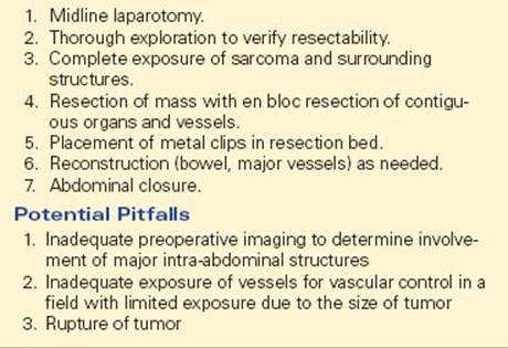
Exposure in the retroperitoneum is often limited by large tumors. With the insidious growth pattern of sarcomas, contiguous structures may have to be resected en bloc to assure complete removal of tumor. Resection of colon, small bowel, kidney, adrenal gland, and inferior vena cava (IVC) must often be considered as part of the procedure depending on the location and growth pattern of the sarcoma. En bloc nephrectomy is necessary in many cases even though the renal parenchyma is rarely invaded. Bowel resection is often necessary because of the extent of involvement of the mesentery/mesocolon.
Once the tumor is resected, metal clips should be placed in the resection bed to allow for later identification of the anatomic limits of the tumor. Necessary reconstructive procedures are performed at this time (Figure 2). Bowel anastomoses are usually performed without incident. Major vascular reconstruction techniques have been described, including primary repair or use of prosthetic graft replacements, though ligation of many vessels is generally well tolerated. Because the risk of nodal metastasis is virtually nonexistent, lymphadenectomy is unnecessary. The abdomen is closed in the standard fashion. Drains are rarely needed.
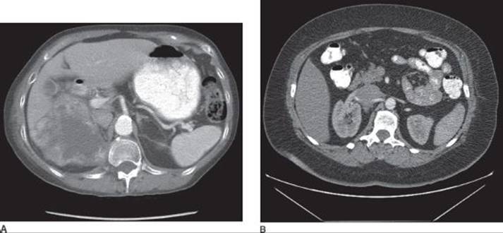
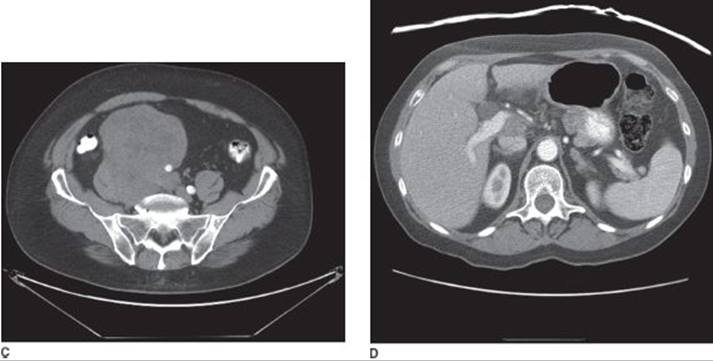
FIGURE 2 • Select cases of retroperitoneal sarcoma requiring resection of contiguous structures. A: This is a 78-year-old woman with a right-sided 10.1 × 8.7 cm retroperitoneal leiomyosarcoma involving the superior pole of the right kidney and extending to the posterior aspect of the liver. Preoperative treatment with radiation therapy led to internal necrosis of the tumor, as seen on the posttreatment/preoperative CT scan. Resection included en bloc right nephrectomy. There was no involvement of the hepatic parenchyma. B: This is a 55-year-old woman who presenting with worsening right-sided back pain. After many sessions with physical therapy and a chiropractor, she underwent evaluation with a thoracic/lumbar spine MRI. After finding a retroperitoneal mass, she underwent a CT of the abdomen and the pelvis, which demonstrated a mass involving the right renal vessels and the IVC. She underwent segmental resection of the IVC with en bloc right nephrectomy. The IVC was reconstructed using an interposition tube graft and the kidney was autotransplanted after the uninvolved kidney was dissected free from the involved portions of the renal vein and artery. C: This is a 66-year-old man with a high-grade leiomyosarcoma of the right retroperitoneum/pelvis discovered after an episode of abdominal pain. He was also noted to have right lower-extremity edema, likely due to venous compression. On CT scan, encasement of the right iliac vessels and suspected involvement of the right ureter are noted. He underwent resection of the sarcoma with en bloc resection of the right ureter, right common and external iliac artery and vein with end-to-end ureteral reconstruction (Boari flap), and vascular reconstruction of the vessels with prosthetic grafts. D: This is a 66-year-old woman with biopsy-proven leiomyosarcoma incidentally found during a laparoscopic cholecystectomy. The 3-cm mass is noted to straddle the portal vein and the IVC. Because of the extent of vascular involvement, this sarcoma was deemed unresectable. The patient was relatively asymptomatic and declined palliative treatment. She developed liver metastases 2 years after initial diagnosis.
Postoperative Management
Immediate postoperative management of these patients is dictated by the extent of resection, noting that postoperative ileus is common with long procedures or if there was extensive displacement of bowel by the sarcoma.
The long-term care of these patients requires surveillance with physical exam and imaging every 3 to 6 months for the first 2 to 3 years and then annually thereafter. Interval of surveillance may vary based on the expected risk of recurrence (dependent on completeness of surgical resection and histopathologic features) and underlying performance status of the patient. Recurrences are difficult to detect without cross-sectional imaging. Follow-up should also include chest imaging for detection of metastatic disease.
Case Conclusion
This patient underwent resection of a 13 kg, 52 × 47 × 14 cm well-differentiated liposarcoma with en bloc colon resection due to extensive involvement of the descending and sigmoid mesocolon. Because of the extent of inguinal involvement, the defect in the groin was repaired to obviate problems with an inguinal hernia. His postoperative course was uncomplicated and he noted an improvement in his prior symptoms.
TAKE HOME POINTS
· Differential diagnosis must include germ cell tumors, lymphoma, other malignancies of retroperitoneal solid organs including endocrine cancers and renal cell carcinoma.
· Preoperative imaging studies should include a CT scan of the abdomen/pelvis and chest imaging to evaluate for the presence of pulmonary metastases.
· The cornerstone of treatment is complete surgical resection. En bloc resection of contiguous structures may be necessary.
· Recurrence rates are high. Patients should be followed closely with cross-sectional imaging and clinical evaluation.
SUGGESTED READINGS
Hollenbeck ST, Grobmyer SR, Kent KC, et al. Surgical treatment and outcome of patients with primary inferior vena cava leiomyosarcoma. J Am Coll Surg. 2003;197:575–579.
Jaques DP, Coit DG, Hajdu SI, et al. Management of primary and recurrent soft tissue sarcoma of the retroperitoneum. Ann Surg. 1990;212:51–59.
Jemal A, Siegel R, Xu J, et al. Cancer statistics, 2010. CA Cancer J Clin. 2010;60:277–300.
Karakousis CP. Refinements of surgical technique in soft tissue sarcomas. J Surg Oncol. 2010;101:730–738.
Liles JS, Tzeng CW, Short JJ, et al. Retroperitoneal and intraabdominal sarcoma. Curr Probl Surg. 2009;46:445–503.
Pisters PWT, O’Sullivan B. Retroperitoneal sarcomas: combined modality treatment approaches. Curr Opin Oncol. 2002;14:400–405.
Raut CP, Pisters PW. Retroperitoneal sarcomas: combined-modality treatment approaches. J Surg Oncol. 2006; 94:81–87.