
Muneera R. Kapadia
Ann C. Lowry
Presentation
A 75-year-old man with a history of diabetes and hypertension presents with sudden onset of left-sided abdominal pain. He complains of bloody diarrhea for the last few hours and anorexia without nausea or emesis. On examination, he is mildly tachycardic with a low-grade fever, but otherwise his vitals are normal. He is mildly distended and tender in the left lower quadrant of his abdomen but does not have diffuse peritoneal signs. Rectal examination is normal but his stool is heme positive.
Differential Diagnosis
Ischemic colitis should be considered when a patient presents with sudden-onset abdominal pain with associated bloody diarrhea. The most common entities in the differential diagnosis are infectious colitides, inflammatory bowel disease, radiation enteritis, diverticulitis, and malignancy; a more complete list can be found in Table 1.
TABLE 1. Differential Diagnosis of Sudden-Onset Abdominal Pain and Bloody Diarrhea

Ischemic colitis typically develops in patients over 65 years but can occur in younger patients as well. Risk factors include cardiac or vascular comorbidities such as diabetes or hypertension. Typical presenting symptoms include cramping abdominal pain of sudden onset followed by bloody diarrhea. Some patients also experience fecal urgency as well as nausea and vomiting secondary to an ileus. The abdominal examination is usually significant for tenderness over the affected area of colon but can include peritoneal signs in more severe cases.
Workup
In the patient presenting above, serum laboratory values reveal hemoglobin of 14.2 g/dL, a white blood cell count of 15.5, and a lactic acid level of 0.8 mEq/L. A CT scan shows mild descending and sigmoid colonic thickening (Figure 1). Endoscopy demonstrates erythematous mucosa with superficial ulcerations in area of the splenic flexure (Figure 2). Stool cultures are negative.

FIGURE 1 • CT scan: The sigmoid colon is thickened (arrow)

FIGURE 2 • Endoscopic image of moderate ischemic colitis.
During the workup of ischemic colitis, blood tests and abdominal imaging are frequently obtained, but the findings are often nonspecific. The white blood cell count and the lactate level may be elevated, and metabolic acidosis may be present. Stool cultures and Clostridium difficile toxin studies should be obtained to rule out infectious colitides. Abdominal radiographs are generally not helpful unless severe ischemic colitis is present; in that case, plain films may demonstrate free air, pneumatosis, or portal venous gas. CT scans may demonstrate colonic wall thickening but are most useful for excluding other conditions. Barium enema may show the classic sign, “thumbprinting,” which represents submucosal hemorrhagic nodules. Colonoscopy with biopsies of the affected area confirms the diagnosis. Angiography is rarely indicated unless acute small intestinal mesenteric ischemia is suspected.
The diagnostic test of choice is an unprepped colonoscopy unless there is evidence of peritonitis on examination. It allows for direct visualization of mucosal changes, and biopsies can be obtained. Ischemic colon can be fragile, and colonoscopy should be performed with care to avoid overdistension of the colon. The distribution of findings depends on the vascular insult sustained, and the mucosal changes can be variable. In mild to moderate ischemic colitis, the mucosa may appear erythematous hypers and edematous with petechiae. Additionally, ulcerations and hemorrhagic nodules, which represent submucosal bleeding, may be seen. In severe ischemic colitis, the mucosa may appear gray or black indicating transmural infarction. If this is encountered, the colonoscopy should be terminated due to significant risk of perforation. If peritonitis is present on examination, a colonoscopy should be avoided and laparotomy should be performed.
Discussion
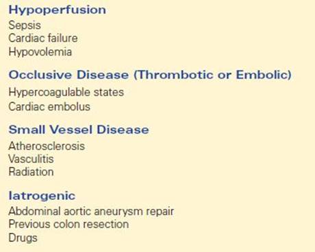
Etiologic factors are numerous (Table 2); however, in most cases, a specific cause cannot be identified. Frequently the predisposing event, such as an episode of acute illness, has resolved by the time the patient presents.
TABLE 2. Causes of Ischemic Colitis
Ischemic colitis can be divided into two categories: nongangrenous and gangrenous. Accounting for approximately 80% to 85% of cases, the nongangrenous type can be further subdivided into transient and chronic forms. Transient ischemic colitis typically involves the mucosa and submucosa and typically resolves in 1 to 2 weeks. The chronic nongangrenous form is characterized by injury to the deeper layers of the colonic wall. Healing may result in fibrosis in the colonic wall and development of a stricture. The gangrenous form with acute severe transmural injury results in perforation and/or sepsis and requires surgical resection of the compromised colon.
Typically segmental, ischemic colitis may involve any area of the colon and rectum. The splenic flexure, descending colon, and sigmoid colon are most commonly affected, which can be explained by the vascular anatomy. The blood supply to the colon and rectum occurs through the superior mesenteric artery, the inferior mesenteric artery, and the superior hemorrhoidal artery. Watershed areas, regions dependent upon collaterals between the inferior and the superior mesenteric arteries (e.g., the splenic flexure and the descending colon), are especially susceptible to ischemic injury in low flow states (Figure 3).

FIGURE 3 • Colonic bloody supply is derived from the superior mesenteric (SMA), inferior mesenteric (IMA), and superior hemorrhoidal (SHA) arteries. “Watershed” areas lie between two of the arteries and are susceptible to ischemia. Griffith’s point is located at the splenic flexure, between the SMA and IMA. Sudek’s point is located at the rectosigmoid junction, between the IMA and SHA. While these two points are particularly susceptible to ischemia, any portion of the colon and rectum can be affected by ischemic colitis.
Diagnosis and Treatment
Once the diagnosis is established, the treatment varies depending on the etiology and the severity of disease. Most patients can be managed expectantly with intravenous fluids, bowel rest, and broad-spectrum antibiotics. Restoration of adequate intravascular volume is important. If a symptomatic ileus is present, the patient may benefit from nasogastric decompression. Additionally, any specific causal agent should be addressed. If a patient has peritonitis, evidence of perforation, or deteriorates clinically with conservative medical management, laparotomy is warranted. Patients with right-sided ischemic colitis or ischemic colitis without hematochezia have been shown to need surgery more frequently and have worse outcomes. Additionally, if a patient develops a stricture or chronic colitis following ischemic colitis, colonic resection may be indicated.
Surgical Approach
In our clinical scenario from above, after 48 hours of medical management, the patient becomes acutely febrile and tachycardic. On examination his abdomen is diffusely tender. Given the patients clinical decline, emergent laparotomy is undertaken.
When operative intervention is warranted, a laparotomy should be performed through a midline incision. The abdomen is explored and the compromised segment of colon is identified. The extent of ischemic colon can be usually be easily assessed by a visual inspection. If there is doubt, the combination of intraoperative colonoscopy, palpation, doppler assessment of the colonic vessels, and intravenous fluorescein can be helpful. Once the involved region is determined, all nonviable colon should be resected. There should be brisk bleeding at the resection margins. If the remaining bowel appears viable and healthy and the patient is stable, it is reasonable to perform a primary anastomosis. If the patient is hypotensive or there is significant surrounding inflammation, a primary anastomosis should be avoided and instead an end stoma created. If there is a question about viability, the resected colonic ends are brought out as a stoma and mucous fistula. In an unstable patient, the ends may be stapled and left discontinuous, and a second look laparotomy performed after 24 to 48 hours (Table 3).
TABLE 3. Key Technical Steps and Potential Pitalls to Operative Management of Ischemic Colitis

Special Circumstance
Ischemic Colitis Following Aortic Surgery. Ischemic colitis is a well-described complication of aortic surgery. The overall incidence of ischemic colitis is about 2%; however, the incidence among patients requiring emergency surgery for ruptured aortic aneurysms is significantly higher. The underlying etiology is usually related to disruption of the inferior mesenteric artery, but additional contributory factors can include prolonged cross-clamping of the aorta and hemodynamic instability. Reimplantation of the inferior mesenteric artery may be necessary to prevent ischemic colitis if the superior mesenteric artery is stenotic and collateral bloody supply is inadequate to support the left colon. Symptoms of ischemic colitis develop in the first few days after aortic surgery and can include pain, fever, distention, and diarrhea. Diagnosis and treatment should proceed as previously discussed. If colonic resection is deemed necessary, a primary anastomosis should be avoided as an anastomotic leak risks graft contamination.
Postoperative Management
Depending on postoperative hemodynamic stability, patients may require a stay in the intensive care unit with close monitoring of urine output and volume status. A nasogastric tube is unnecessary unless the patient suffers a significant ileus. Dietary advancement should be as tolerated. If a primary anastomosis is created, patients should be monitored for evidence of an anastomotic leak. Return of bowel function should occur prior to discharge. If colonic diversion is deemed necessary at the initial operation, ileostomy or colostomy takedown can be considered at 3 to 6 months following the initial operation, depending on the patient’s overall health status.
TAKE HOME POINTS
· Sudden-onset abdominal pain followed by bloody diarrhea should prompt suspicion of ischemic colitis.
· Colonoscopy is the test of choice for diagnosis of ischemic colitis.
· Ischemic colitis most commonly involves the splenic flexure, descending and sigmoid colon.
· Most patients can be treated effectively with hydration, bowel rest, and broad-spectrum antibiotics.
· Peritonitis, evidence of perforation, or clinical deterioration should prompt immediate surgical intervention.
· Operative management should include resection of compromised colon, followed by either primary anastomosis or creation of a stoma depending on the overall status of the patient.
SUGGESTED READINGS
Brandt LJ, Boley SJ. Colonic ischemia. Surg Clin North Am. 1992;72:203–229.
Brandt LJ, Feuerstadt P, Blaszka MC. Anatomic patterns, patient characteristics, and clinical outcomes in ischemic colitis: a study of 313 cases supported by histology. Am J Gastroenterol. 2010;105:2245–2252;quiz 53.
Montoro MA, Brandt LJ, Santolaria S, et al. Clinical patterns and outcomes of ischaemic colitis: results of the Working Group for the Study of Ischaemic Colitis in Spain (CIE study). Scand J Gastroenterol. 2011;46(2):236–246. [Epub 2010.]
Mosele M, Cardin F, Inelmen EM, et al. Ischemic colitis in the elderly: predictors of the disease and prognostic factors to negative outcome. Scand J Gastroenterol. 2010;45:428–433.
Paterno F, McGillicuddy EA, Schuster KM, et al. Ischemic colitis: risk factors for eventual surgery. Am J Surg. 2010;200:646–650.
Perry RJ, Martin MJ, Eckert MJ, et al. Colonic ischemia complicating open vs endovascular abdominal aortic aneurysm repair. J Vasc Surg. 2008;48:272–277.
Scowcroft CW, Sanowski RA, Kozarek RA. Colonoscopy in ischemic colitis. Gastrointest Endosc. 1981;27:156–161.
Steele SR. Ischemic colitis complicating major vascular surgery. Surg Clin North Am. 2007;87:1099–1114, ix.
Sun MY, Maykel JA. Ischemic colitis. Clin Colon Rectal Surg. 2007;20:5–12.
West BR, Ray JE, Gathright JB. Comparison of transient ischemic colitis with that requiring surgical treatment. Surg Gynecol Obstet. 1980;151:366–368.