Angela M. Ingraham
Avery B. Nathens
Presentation
A 59-year-old male with a history of type 2 diabetes mellitus, hypertension, and a 30-pack-year smoking history underwent a left colectomy for an obstructing colon cancer. He tolerated the procedure well except for some hypotension in the operating room due to bleeding. On postoperative day 5, he was febrile to 38.7°C, was found to have a white count of 12.3, and was noted to have some erythema of the inferior aspect of the wound for which he was started on cefazolin. On postoperative day 6, he was getting out of bed when he noticed the abrupt onset of copious serosanguineous drainage from the wound.
Fascial dehiscence is the postoperative separation of the reapposed musculoaponeurotic layers of the abdomen. Failure of acute surgical wounds occurs when the load being placed across the wound exceeds the resistive capacity of the suture line and temporary matrix. Surgical wound strength increases rapidly from the first week until the fourth to sixth week postoperatively. At that time, the wound strength is between 50% and 80% of unwounded tissue. Wound strength following this initial postoperative period increases at a slower rate and never achieves the strength of unwounded tissue.
The incidence of postoperative fascial dehiscence varies depending on the study but has been reported to be between 1% and 5%. Despite the advances in antimicrobial prophylaxis, anesthesia, and suture materials, the incidence of this complication has not significantly decreased over time.
Fascial dehiscence is typically recognized within several days of the index procedure. Postoperative fascial dehiscence has been reported between postoperative days 1 and 21 with the average occurrence being on postoperative day 7. Patients often report that “something has given way” or experiencing a “ripping sensation.” In 23% to 84% of cases, serosanguineous fluid drains from the wound prior to a dehiscence. Rarely, and most commonly in late fascial dehiscence (>7 to 10 days), the fascia separates while the superficial wound layers remain intact.
Differential Diagnosis
Postoperative fascial dehiscence is easily diagnosed if evisceration is present. On the other end of the spectrum, when evisceration is not the presenting sign, there may be a delay in the recognition of a fascial dehiscence, as the superficial layers of the wound remain intact while there remains a defect in the abdominal wall. Other issues regarding the wound, such as seroma or infection, may make the identification of a postoperative fascial dehiscence more challenging.
Workup
A thorough examination of the wound, potentially including probing a draining wound, is performed. The role of advanced imaging in the diagnosis of fascial dehiscence is limited. However, a CT scan may be useful if the dehiscence is identified late in the postoperative course, a subfascial fluid collection is suspected, and operative management is not planned. The incidence of intra-abdominal infection with fascial dehiscence has been reported to be as high as 44% in some series.
When evaluating a patient for postoperative fascial dehiscence, the risk factors predisposing to the complication should also be considered. Risk factors for fascial dehiscence fall into two broad categories, those related to the patient’s comorbidities and those indicative of surgeon technique and decision making.
Patient characteristics identified as predictors of postoperative fascial dehiscence include age >65, wound infection, pulmonary disease, hemodynamic instability, presence of an ostomy within the incision, hypoproteinemia, sepsis, obesity, uremia, use of hyperalimentation, malignancy, ascites, steroid use, and hypertension. In a case control study, patients with five risk factors were reported to have an incidence of 30%, while those with eight or more comorbidities all had postoperative dehiscence.
Although patient factors are important in dehiscence, there are important technical factors to consider. The most common cause of postoperative fascial dehiscence is the suture tearing through fascia. “Bites” of tissue should reapproximate the fascia without impeding perfusion of the healing tissue. Excessive suture tension impedes blood flow causing muscle/fascial necrosis. Failure of the suture to hold occurs in the area just adjacent to the wound edge. In this area, the native tissue integrity is reduced due to proteases, which have been activated during the tissue repair process. Other causes of postoperative fascial dehiscence include a broken suture, a slipped knot, a loose stitch, and excessive travel between stitches. Lastly, whether continuous versus interrupted, closure techniques to decrease the risk of postoperative fascial dehiscence continue to be debated in the literature. A meta-analysis of 23 randomized, controlled studies found that interrupted closure is associated with a significantly decreased risk of dehiscence (Odds ratio: 0.58, P = 0.014). However, a second meta-analysis of 15 randomized studies with at least 1 year of follow-up found no difference in the risk of fascial dehiscence using continuous versus interrupted suture. Furthermore, a multicenter randomized trial comparing three parallel groups (interrupted Vicryl, continuous polydioxanone, and continuous Monoplus) found no significant difference in the incidence of fascial dehiscence.
Finally, characteristics of the incision have also been proposed as risk factors for fascial dehiscence, although this remains controversial. Retrospective data have suggested that upper abdominal incisions are at higher risk for dehiscence than those in the lower abdomen. Retrospective data have also found a higher incidence of fascial dehiscence in midline as compared to transverse incisions due to abdominal wall contractions approximating the edges of transverse incisions while separating those of midline incisions.
Diagnosis and Treatment
As fascial dehiscence can be complicated to treat, attention should be directed toward preventing fascial dehiscences and making a prompt diagnosis when appropriate. Surgical techniques that can minimize the incidence of fascial dehiscence include appropriate antibiotic coverage, improved operative technique (minimizing dead space, appropriate use of electrocautery currents in making incisions), controlling intraoperative risk factors (minimizing operative time, avoiding hypothermia), and proper closure technique (appropriate choice of suture material, utilization of drains). Proper suture placement improves bursting strength of abdominal incisions.
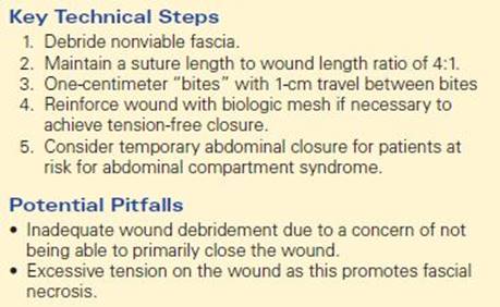
Decreased tissue strength along the border of the acute wound has prompted investigations into the identification of an ideal suture length to wound length (SL-to-WL) ratio for primary closure of midline celiotomies. An SL-to-WL ratio of 4:1 reduces the occurrence of fascial dehiscence and incisional hernia formation. This is the basis for the surgical dogma of 1-cm “bites” with progress between bites of 1 cm. The ideal SL-to-WL ratio allows the wound to be approximated with an appropriate amount of tension along the suture line. Increased tension causes the wound to fail at the suture-native tissue interface. Studies have found no difference in acute fascial wound dehiscence with mass abdominal wall closures versus layered closures. Randomized trials comparing one-layer (peritoneum not reapproximated) and two-layer closures (peritoneum reapproximated) have found no difference in the rate of fascial dehiscence of paramedian and midline incisions.
Surgical Approach
Management of dehiscence follows several surgical principles with customization of the treatment based upon the patient’s condition and the available resources. The management of fascial dehiscence must take into account the most probable cause of the complication. Primary closure is acceptable if the cause of the dehiscence was technical in nature and occurred in an otherwise healthy patient. In more complicated patients, options for closure include component release, temporary packing with an overlying plastic silo or packs, use of mesh or bioprosthesis, and skin closure only.
In less complicated cases of fascial dehiscence (i.e., without evisceration), the operative management of fascial dehiscence is dependent upon when the dehiscence occurs in the postoperative course. Dehiscence in the early postoperative period when adhesions are at a minimum potentially warrants immediate operative repair. However, if a dehiscence has occurred later in the postoperative course when new adhesions are more likely to be encountered, the risks of inadvertent enterotomies and fistulas may outweigh the development of a hernia that can be repaired at a later date. When early repair of the dehiscence is pursued, intestinal decompression via a nasogastric tube is often utilized to facilitate closure. The patient is placed under general anesthesia with adequate use of muscle relaxants to minimize abdominal wall tension. The abdomen should be explored to identify any injury related to the dehiscence. Wide debridement of compromised fascia and subcutaneous tissues is carried out. Fascial edges should be debrided back to healthy/bleeding tissue. Debridement should not be compromised due to a concern of having inadequate tissue for closure. If adequate healthy tissue is present and primary closure can be accomplished without tension, the fascia can be primarily repaired. Closure technique (use of internal and external retention sutures and running versus interrupted stitches) is primarily surgeon dependent.
When there is inadequate tissue for primary repair, a mesh closure can be considered. Since most wounds are contaminated, biologic mesh is usually used after acute wound failure. This is especially true for patients with perforation, gross spillage, or intra-abdominal abscess. However, if the fascia cannot be closed primarily, the placement of an absorbable mesh (e.g., polyglycolic acid—Vicryl or Dexon) or a bioprosthesis (Surgisis or Alloderm) is recommended. Although the use of biologic mesh may result in hernia formation, using prosthetic mesh could result in chronic mesh infection, a dreaded complication. Similar to the use of mesh in elective general surgical cases, when using mesh to treat a fascial dehiscence, an attempt should be made to place omentum between the bowel and the mesh to minimize the development of fistula.
Special Intraoperative Considerations
The intraoperative management of fascial dehiscence is heavily directed by individual patient factors. Due to excessive tension on the wound, primary closure may not be feasible in patients with significant intra-abdominal edema. Closing an abdominal wall under excess tension predisposes the patient to a repeated dehiscence, respiratory compromise, and even compartment syndrome. Therefore, in patients with anasarca or visceral edema, a temporary wound closure, such as the abdominal wound vac, should be considered.
Postoperative Management
Patients whose index surgery was complicated by fascial dehiscence will often require intensive care and will have prolonged hospital stays. It is important to note that the same comorbidities that placed the patient at risk for fascial dehiscence predispose them to developing other postsurgical complications.
After closure of a fascial dehiscence, particular attention should be paid to modifying risk factors to prevent repeat dehiscence. Patients should be educated regarding their postoperative surgical and wound care. They should be instructed to avoid straining and heavy lifting for a minimum of 6 weeks. An abdominal binder is often prescribed to reduce tension of the wound. Finally, the patient’s nutritional status should be optimized to promote healing (Table 1).
TABLE 1. Key Technical Steps and Potential Pitfalls
Case Conclusion
The patient’s wound was opened at the bedside. Examination of the wound revealed an approximately 10-cm area where the fascia had dehisced and the suture had torn through the fascia. The patient was taken back to the operating room emergently. No visceral injuries from the fascial dehiscence were identified. After debridement of some of the fascial edges, the musculoaponeurotic layer was reapproximated without tension using interrupted, figure of eight stitches with 0-Prolene suture, paying special attention to suture placement. The superficial wound was packed with moist gauze.
TAKE HOME POINTS
· The incidence of postoperative fascial dehiscence has not decreased significantly despite advances in surgical care.
· Both patient- and surgeon-dependent factors contribute to postoperative fascial dehiscence.
· Emphasis on appropriate surgical technique will decrease the modifiable risk of fascial dehiscence.
SUGGESTED READINGS
Carlson MA. Acute wound failure. Surg Clin North Am. 1997;77(3):607–636.
Cliby WA. Abdominal incision wound breakdown. Clin Obstet Gynecol. 2002;45(2):507–517.
Diener MK, Voss S, Jensen K, et al. Elective midline laparotomy closure: the INLINE systematic review and metaanalysis. Ann Surg. 2010;251(5):843–856.
Dubay DA, Franz MG. Acute wound healing: the biology of acute wound failure. Surg Clin North Am. 2003;83(3):463–481.
Graham DJ, Stevenson JT, McHenry CR. The association of intra-abdominal infection and abdominal wound dehiscence. Am Surg. 64(7):660–665.
Gupta H, Srivastava A, Menon GR, et al. Comparison of interrupted versus continuous closure in abdominal wound repair: a meta-analysis of 23 trials. Asian J Surg. 2008;31(3):104–114.
Seiler CM, Bruckner T, Diener MK, et al. Interrupted or continuous slowly absorbable sutures for closure of primary elective midline abdominal incisions: a multicenter randomized trial. Ann Surg. 2009;249(4):576–582.
van ‘t Riet M, Steyerberg EW, Nellensteyn J, et al. Metaanalysis of techniques for closure of midline abdominal incisions. Br J Surg. 2002;89(11):1350–1356.
Webster C, Neumayer L, Smout R, et al. Prognostic models of abdominal wound dehiscence after laparotomy. J Surg Res. 2003;109(2):130–137.