
Pete S. Batra
INTRODUCTION
Optic neuropathy (ON) most frequently results from blunt and penetrating trauma. Estimates suggest that traumatic ON occurs in 0.5% to 5% of all closed head injuries and up to 10% of patients with craniofacial fractures. The mechanisms of traumatic ON are likely multifactorial, with both direct and indirect mechanisms contributing to the visual loss. Direct injury, resulting from penetrating trauma from midfacial and orbital fractures, can lead to avulsion of the nerve, partial transection, orbital or hemorrhage into the optic nerve sheath, and orbital emphysema. Indirect injury results from ischemia caused by damage from the mechanical shearing of the optic nerve axons and contusion necrosis. The vascular ischemia and/or trauma induce swelling of the optic nerve within the confines of the optic canal further contributing to the death of retinal ganglion cells. Nontraumatic compressive ON can also lead to loss of vision due to a variety of pathologic processes, such as benign and malignant neoplasms of the sphenoid and sellar region, mucoceles, and Graves orbitopathy.
A variety of surgical approaches have been described for decompression of the optic nerve. Traditionally, open techniques have been employed including craniotomy, extra nasal transethmoidal, transorbital, transantral, and intranasal microscopic approaches. The introduction of rigid endoscopes, refinement of surgical instrumentation, and advent of image-guided surgery have facilitated the consideration of management of orbital and skull base pathology with minimally invasive endoscopic techniques. Indeed, endoscopic optic nerve decompression (EOND) now represents the procedure of choice to address traumatic and nontraumatic ON, given its reduction of morbidity, preservation of olfaction, superior cosmetic result, rapid recovery time, and less operative stress, especially in the patient with multisystem trauma.
HISTORY
Given that traumatic ON often occurs in patients having suffered significant blunt force trauma, the diagnosis may be often delayed as the patients are unable to provide a history due to an altered level of consciousness. This underscores the importance of maintaining a high incidence of suspicion for traumatic ON in this setting. Evaluation by an ophthalmologist is imperative in order to assess visual acuity at the earliest possible juncture. Patients with nontraumatic compressive ON may report vague ocular symptoms with complaints of blurry or “fuzzy” vision. Patients with paranasal sinus and skull base neoplasms may have associated nasal obstruction, epistaxis, headaches, proptosis, or trigeminal hypo- or anesthesia. Patients with a sphenoid mucocele may have a history of previous trauma or sinus surgery.
PHYSICAL EXAMINATION
Patients with traumatic ON require comprehensive evaluation by the trauma team. Concomitant intracranial, spinal, thoracic, and abdominal injuries must be ruled in or out. Significant blunt concussive injury or penetrating trauma may result in cerebrospinal fluid (CSF) rhinorrhea or otorrhea. Any fractures of the carotid canal at the skull base require angiography to rule out an internal carotid artery (ICA) aneurysm or cavernous–carotid fistula. Timely ophthalmologic evaluation is imperative to determine and document baseline vision. Commonly, visual acuity will be 20/400 or less in the affected eye. Detailed examination may reveal a multitude of ocular abnormalities, including visual field deficit, decrease in color vision, and an afferent papillary defect on the affected side. Funduscopic examination is essential to rule out optic nerve atrophy; further, this may rule out other etiologies of decreased vision, such as choroidal rupture, retinal detachment, or vitreous hemorrhage. Patients with nontraumatic compressive ON often have similar ocular defects and require complete neuroophthalmologic evaluation including visual field testing. Patients suspected of skull base neoplasms require comprehensive head and neck and neurologic examination. Nasal endoscopy is important to rule out exophytic masses in the middle meatus or sphenoethmoid recess (SER).
INDICATIONS
EOND should be considered in the setting of traumatic ON in patients with persistent visual loss who have failed a trial of high-dose steroids and who have evidence of a fracture of the optic canal, a hematoma of the optic nerve sheath, or a compressive hematoma at the orbital apex demonstrated on computed tomography (CT). Patients without an obvious fracture or hematoma but with suspected edema of the nerve in the bony optic canal confines may also benefit from EOND. Theoretically, this may relieve constrictive pressure from edema of the nerve in a rigid bony canal or allow for removal of an impinging bone fragment or hematoma, thus facilitating reestablishment of nerve function. Patients with a multitude of etiologies resulting in nontraumatic compressive ON may also benefit from EOND, including primary tumors of the optic nerve, such as meningiomas or gliomas, benign and malignant neoplasms of the sphenoid sinus, sellar and suprasellar tumors, fibrous dysplasia of the central skull base, mucoceles of the sphenoid sinus or sphenoethmoid (Onodi) cell, Graves orbitopathy, and benign intracranial hypertension.
CONTRAINDICATIONS
Long-standing complete optic nerve atrophy is an absolute contraindication to EOND as vision restoration is not possible in this setting. Traumatic ON presenting with injury to the nerve in the orbital portion and complete nerve transection are also contraindications to the procedure. Comatose patients should not be considered candidates for surgery until adequate visual assessment can be performed. PREOPERATIVE PLANNING
Anatomic Considerations
Intimate knowledge of the anatomy of the sphenoid sinus and optic nerve–ICA relationship is imperative prior to embarking on surgery. Embryologically, the sphenoid sinus originates from the cartilaginous nasal capsule. Through the process of ossification and resorption between the 9th and 12th years of life, it comes to occupy a central location at the cranial base. The sphenoid pneumatization may be conchal, presellar, sellar, or postsellar, with optic nerve and ICA protuberances being more prominent with increasing pneumatization. Pneumatization of the posterior ethmoid cells more posterior and superior to the sphenoid sinus results in a sphenoethmoid or Onodi cell. This is evident in 25% to 30% of cases and results in the optic nerve being closely associated with the Onodi cell, instead of the sphenoid sinus.
Multiple important structures are present on the surface of the sphenoid sinus. The opticocarotid recess (OCR) represents the pneumatization of the optic strut of the anterior clinoid process. The optic nerve courses in the optic canal just above the OCR, while the anterior bend of the ICA (C3 segment) is present just inferiorly. Dehiscence of the bone and direct septal insertions of the medial optic canal can be seen in 15% and 30% of cases, respectively. Dehiscence of the bone and direct septal insertions of the ICA canal can be seen in 20% and 40% of cases, respectively. The median distance between the ICA protuberances is 12 mm, and the median length of the OCR is 5 mm.
The optic canal is formed by the two struts of the lesser wing of the sphenoid transmitting the optic nerve and the ophthalmic artery. The nerve is a direct continuation of the brain carrying all three meningeal layers, including the pia, arachnoid, and dura. The optic nerve is divided into three segments—intraorbital, intracanalicular, and intracranial. The intracanalicular segment is most prone to injury with blunt head trauma and is most likely to benefit from EOND. The optic nerve sheath is attached to the bone in the canalicular segment of the optic canal; consequently, fractures in this area may result in a higher incidence of injury to the optic nerve. The ophthalmic artery originates from the subdural cavity and accompanies the optic nerve in the dural sheath in the optic canal. The ophthalmic artery typically enters the nerve sheath from an inferolateral direction and is typically not in the surgical field during EOND. However, 15% of patients may have the artery entering the medial aspect of the optic canal, thus making it susceptible to injury during the medial approach.
Preoperative Imaging
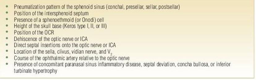
High-resolution CT imaging (1 mm or less) is an absolute requisite prior to considering EOND. It will help to delineate key anatomic relationships in the sphenoethmoid region, to identify dehiscence of the bone or presence of septations of the optic nerve and ICA, and to provide a roadmap for computer-aided surgery. Indeed, a preoperative checklist must be created prior to the surgical endeavor (Table 1.1). CT imaging will also identify fractures of the optic canal, ICA canal, or the skull base in cases of traumatic ON. Magnetic resonance (MR) imaging may be problematic in critically injured patients. However, when possible, it may demonstrate optic nerve swelling and intraorbital or optic canal hematoma. CT and MR imaging are imperative in cases of nontraumatic compressive ON. It will assist in defining the full extent of the skull base neoplasm and its relationship to the optic canal. CT imaging will help to demonstrate the site of a compressive lesion in cases of Graves orbitopathy.
TABLE 1.1 Anatomic Checklist for EOND
SURGICAL TECHNIQUE
General endotracheal anesthesia is induced with the patient in the supine position. The endotracheal tube is secured to the left side out of the surgical field. The head is secured in a doughnut, and eyes are carefully taped shut with Steri-Strips or thin pieces of tape after placement of lubricating ointment. The eyes should be palpated at the beginning of the case to assess firmness at baseline. They should remain accessible and clearly visible throughout the surgery should any orbital complication be suspected during the surgery. The nose is maximally decongested using cotton pledgets soaked in oxymetazoline. Image guidance is registered and verified at this juncture. The face is prepped and draped in the standard sterile fashion.
The procedure is started with a 0-degree endoscope. One percent lidocaine with 1:100,000 epinephrine is injected along the lateral nasal wall and the sphenopalatine foramen. In general, a transethmoid approach to the sphenoid sinus will provide the best exposure of the orbital apex and optic nerve region. A standard uncinectomy with maxillary antrostomy is performed to improve access to the middle meatus and to provide a place for blood to collect out of the surgical field. The floor of the orbit also provides a general landmark to the level of the sphenoid ostium in the SER. Total ethmoidectomy is now performed to skeletonize the orbit from the lacrimal system to the orbital apex. Great care is taken to avoid violating the lamina papyracea or periorbita as resulting herniation of orbital adipose tissue will obscure the surgeon’s vision. The superior turbinate is identified in the SER; the lower third is sharply resected to identify the sphenoid ostium. The sphenoid sinus is now opened widely to expose the optic nerve and ICA bulges. If the optic nerve courses through an Onodi cell, this should be fully dissected and the relationship between this cell and the sphenoid sinus established.
The bone at the orbital apex is now removed approximately 1 cm from the optic nerve tubercle. The bone at the orbital apex can be thick; a diamond burr drill may be required to expose the periorbita and annulus of Zinn. The bone over the medial optic canal is next addressed with a long 2- or 3-mm diamond burr drill. Concurrent suction irrigation is critical to clear bone dust and blood from the surgical field and to minimize transmission of heat to the optic nerve sheath (Fig. 1.1). The drill should be circumferentially visible when being used; this will decrease the risk of inadvertent injury to the ICA canal or the planum sphenoidale. The bone is initially blue lined with the drill and then can be subsequently removed with curettes or otologic picks. The entire optic nerve sheath, typically ranging from 10 to 15 mm, is exposed from the lateral wall of the sphenoid to the optic chiasm. The optic nerve sheath is decompressed 180 degrees along the medial and inferior aspects (Fig. 1.2).
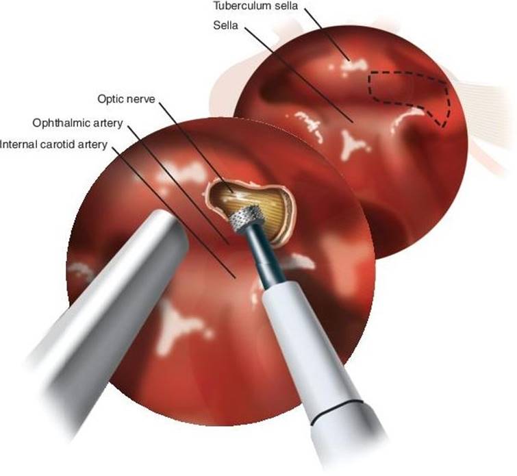
FIGURE 1.1 Endoscopic view demonstrates drilling of the medial optic canal with a diamond burr. Concurrent irrigation is critical to minimize heat transmission, and suction is imperative to clear blood and bone dust from the operative field.
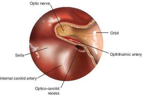
FIGURE 1.2 Endoscopic view illustrates 180-degree decompression of the medial and inferior optic nerve sheath from the orbital apex to the optic chiasm. The ophthalmic artery is visible coursing just inferior to the optic nerve.
Incision of the optic nerve sheath has been advocated by some authors to further decompress the optic nerve. This maneuver is controversial and can be potentially associated with risk of damage to the underlying optic nerve and accompanying ophthalmic artery and possible intraoperative CSF leak. It may be considered in cases with known intrasheath hematoma or severe edema of the nerve. However, routine incision of the sheath of the optic nerve is to be discouraged as proper studies demonstrating clear benefit outweighing the potential risks are not available at the present time.
POSTOPERATIVE MANAGEMENT
Patients are typically observed overnight to monitor for any associated complications, such as epistaxis, CSF rhinorrhea, or orbital issues. High-dose steroids should be continued overnight to decrease the risk of edema of the optic nerve. Serial visual acuity checks should be performed as clinically warranted. Careful ophthalmologic evaluation is obtained on postoperative day 1 to establish a baseline for future testing. Oral antibiotics and steroid taper is continued for 7 to 10 days. Gentle saline rinses are started on postoperative day 1 and continued until all mucosal healing is complete. Initial postoperative debridement is performed 5 to 7 days after surgery; this facilitates removal of any nasal crusting or early granulation tissue to ensure patency of the paranasal sinuses. The periorbita or optic nerve region is not debrided at this juncture; mucosalization of these areas will occur within 4 to 6 weeks.
COMPLICATIONS
Potential complications include adhesions in the nasal cavity and paranasal sinus, bleeding, postoperative infectious sinusitis, epiphora, and alteration in smell and/or taste. More serious complications include complete, irreversible loss of vision, CSF leak, and ICA injury. Though these serious risks are low, the expected incidence would be higher than standard endoscopic sinus surgery given the proximity of drilling close to these critical structures.
RESULTS
The optimal management of traumatic ON has been a source of considerable debate over the years, given the unclear natural history and multiple confounders in studies published to date. Multiple retrospective case series have demonstrated benefit for EOND over steroids or observation. The largest series thus far, the International Optic Nerve Trauma Study, comprised of 133 patients with traumatic ON injury was unable to demonstrate clear benefit from either steroid therapy or decompression of the optic canal when compared to observation alone. However, a treatment bias likely existed as patients in the surgery group were statistically more likely to have no light perception, relative to the steroid and observation groups. A systematic review of the literature by Cook et al. evaluated outcomes of steroids, surgery, combination, and no treatment for traumatic ON. They noted that treatment with steroids, surgery, or both was better than no treatment; furthermore, patients with moderately severe injuries had a greater recovery of vision than patients with less severe injuries. The accrued literature for traumatic ON suggests that surgery should not be considered the standard of care for patients with traumatic ON. However, careful patient selection on an individualized basis is imperative in patients with severe visual loss who have failed high-dose steroid therapy and have objective CT evidence of optic nerve lesions, that is, optic canal fracture with bony fragment impingement or hematoma.
Patients with nontraumatic ON may also benefit from EOND. Pletcher and Metson performed 10 EONDs in 7 patients with a variety of pathologic entities, including skull base neoplasms, mucoceles, and Graves disease. Mean visual acuity improved from 20/300 to 20/30 at mean follow-up of 6 months. Outcomes for nontraumatic ON will continue to evolve with growing adaptation of skull base approaches.
PEARLS
· Careful ophthalmologic evaluation is crucial in patients with traumatic and nontraumatic ON.
· High-resolution CT imaging is a requisite to define key anatomic relationships in the sphenoethmoid region and to provide a roadmap for image-guided surgery.
· Multidisciplinary coordination is important in cases of skull base neoplasms with optic nerve encroachment.
· Comprehensive paranasal sinus dissection is essential to identify salient anatomic structures including the medial orbital wall, ethmoid roof, sella, and ICA in relation to the optic nerve.
· The bone of the orbital apex and optic nerve should be drilled with a diamond burr, preferably with concurrent suction and irrigation, to optimize view of the surgical field and to minimize risk of heat trauma to the optic nerve.
· The entire optic nerve sheath is exposed from the lateral sphenoid wall to the optic chiasm and is decompressed 180 degrees along the medial and inferior aspects.
· Postoperative care should include antibiotics and steroids for 7 to 10 days, gentle saline rinses starting the day after surgery, and meticulous nasal debridement 1 week postoperatively.
PITFALLS
· The course of the ophthalmic artery should be considered prior to embarking on EOND.
· The entire drill tip should be circumferentially visible to minimize risk of injury to the skull base or ICA.
· Incision of the optic sheath is controversial and may be associated with CSF leak or trauma to the optic nerve.
INSTRUMENTS TO HAVE AVAILABLE
Endoscopic skull base set should be present for any tumor resection in this region. High-speed diamond burr drill, preferably with concurrent irrigation and suction. Bipolar cautery. Unipolar cautery should be avoided under all circumstances given potential for deeper heat penetration and risk of damage to critical orbital and intracranial structures. Image guidance should be strongly considered, especially in cases with significant anatomic alteration from extensive trauma or skull base neoplasms.
SUGGESTED READING
Cook MW, Levin LA, Joseph MP, Pinczower EF. Traumatic optic neuropathy. A meta-analysis. Arch Otolaryngol Head Neck Surg 1996;122(4):389–392.
Luxenberger W, Stammberger H, Jebeles JA, et al. Endoscopic optic nerve decompression: the Graz experience. Laryngoscope 1998;108:873–882.
Rajiniganth MG, Gupta AK, Gupta A, et al. Traumatic optic neuropathy: visual outcome following combined therapy protocol. Arch Otolaryngol Head Neck Surg 2003;129(11):1203–1206.
Pletcher SD, Sindwani R, Metson R. Endoscopic orbital and optic nerve decompression. Otolaryngol Clin North Am 2006;39:943–958.
Pletcher SD, Metson R. Endoscopic optic nerve decompression for nontraumatic optic neuropathy. Arch Otolaryngol Head Neck Surg 2007;133:780–783.
Locatelli M, Caroli M, Pluderi M, et al. Endoscopic transsphenoidal optic nerve decompression: an anatomical study. Surg Radiol Anat 2011;33:257–262.