
Ian J. Witterick
INTRODUCTION
The petrous apex is the most medial portion of the temporal bone. The temporal bone is a pyramidal structure with the base composed mostly of the bony labyrinth (semicircular canals and cochlea); the superior surface makes up a large portion of the middle cranial fossa floor. The posterior surface forms the anterolateral wall of the posterior cranial fossa where petrous apex lesions may impinge on Dorello's canal affecting the function of the abducens nerve. Inferiorly, the petrous pyramid is bounded by the jugular bulb and the inferior petrosal sinus. The internal carotid artery (ICA) canal and the transverse (petrous) portion of the ICA traverse the petrous pyramid.
The petrous apex can be divided into anterior and posterior areas by a vertical plane through the modiolus of the cochlea and the internal auditory canal. Most lesions of the petrous apex are located anterior to this plane because posteriorly, the compact bone between the internal auditory canal and the vestibular apparatus rarely pneumatizes. Approximately 10% of the population has pneumatization of the anterior portion of the petrous apex.
Traditional otologic approaches to the petrous apex include surgery through or around the labyrinth (translabyrinthine, infralabyrinthine, retrolabyrinthine, retrocochlear) or approaching it from above (middle fossa routes). Drawbacks to these otologic approaches are the following: They are technically demanding, the view and access are often narrow and restricted, there is real or potential risk of audiovestibular loss, brain retraction may be required with potential neurologic sequelae, and there is no natural drainage pathway postoperatively. As an alternative approach, lesions in the petrous apex often bulge medially into the medial sphenoid sinus and are therefore potentially amenable to endoscopic transsphenoidal approaches in selected patients.
Transsphenoidal surgical access to the petrous apex is useful for biopsy and drainage purposes. It can be particularly helpful in selected cystic lesions such as cholesterol granuloma because marsupialization, rather than complete excision of the lesion, is the goal of therapy. The transsphenoidal approach usually avoids injury to the audiovestibular apparatus and provides a drainage pathway into the sphenoid sinus if required. Some lesions approached endoscopically may also require a transpterygoid approach with or without mobilization of the ICA. Lesions arising from the central petrous apex are considered “primary” and account for approximately 40% of petrous apex disease. Secondary lesions encroach on the petrous apex from bordering regions or may be metastatic.
Cholesterol granulomas are one of the most common benign pathologic lesions of the petrous apex, followed by mucoceles and cholesteatomas. Malignant tumors of the petrous apex are uncommon. The most common primary malignant tumor is a chondrosarcoma characterized by intratumoral chondroid calcification and bone destruction on computed tomography (CT) and low to intermediate signal on T1 and high signal on T2 magnetic resonance imaging (MR) images. Neoplasms originating in other sites may also spread to the petrous apex. For example, nasopharyngeal carcinoma may reach the petrous apex by direct extension, and hematogenous metastases from elsewhere in the body are occasionally seen.
HISTORY
Slow-growing or small lesions in the petrous apex may remain asymptomatic for years. Cholesterol granuloma is typically asymptomatic and is often an incidental discovery on imaging. Fast-growing or large lesions left untreated may continue to expand and cause increasing signs and symptoms including headache, balance problems, vertigo, diplopia, progressive hearing loss, facial weakness, and lower cranial nerve neuropathies.
PHYSICAL EXAMINATION
The examination of the head and neck, including fiberoptic nasal endoscopy, is usually normal. Examination of the cranial nerves may identify subtle cranial nerve palsies; of importance is an ipsilateral lateral rectus (CN VI) palsy. Typically, the examination of the ear is normal. Palpation and imaging of the neck help to rule out metastases to the cervical lymph node in cases of malignancy.
INDICATIONS
The endonasal, endoscopic transsphenoidal approach is safe, providing a low rate of recurrence and durable marsupialization of cystic lesions and a means to biopsy solid lesions. Lesions of the petrous apex may require a biopsy to arrive at a diagnosis in order to direct future therapy. If the imaging appearance is “classic” for a common cystic disease process (e.g., cholesterol granuloma) and the patient is asymptomatic, then observation with periodic imaging is a reasonable option. If the patient is symptomatic or the nature of the lesion is unclear on imaging, then biopsy, drainage, or decompression is indicated.
The transsphenoidal approach is appropriate for lesions that create a definite impression or “mass effect” on the posterolateral wall of the sphenoid sinus (posterior to the carotid artery) (Fig. 8.1). If the optic nerve and carotid artery can be identified and kept in view, the transnasal endoscopic approach to the petrous apex is relatively straightforward. The area of the “bulge” can be opened, marsupialized, or biopsied depending on the situation. The difficulty comes when the lesion is not so obvious in the sphenoid, the sphenoid is poorly pneumatized, or the lesion does not “bulge” into the lateral sphenoid sinus and is situated posterolateral to the clival carotid artery. Endoscopic lateralization of the carotid artery has been performed in certain centers but is technically demanding and requires a great deal of endoscopic skill to perform safely. For more lateral pathology, a transpterygoid infrapetrous approach with dissection of the eustachian tube and drilling of the bone of the petrous apex inferior to the horizontal petrous ICA can be performed but again is technically demanding and applicable only to surgical teams with extensive endoscopic skull base experience (Fig. 8.2).
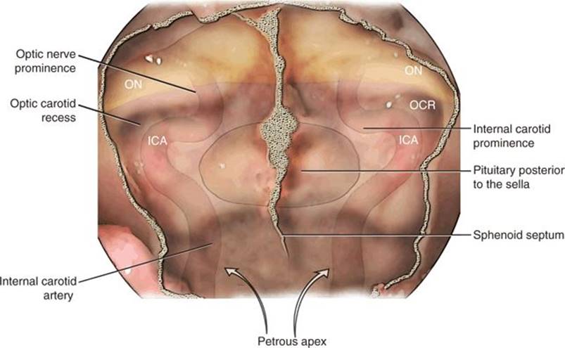
FIGURE 8.1 Endoscopic view of the anatomic features of the sphenoid sinus. The clival recess is bounded by the carotid arteries laterally and sella superiorly. The relationship of the internal carotid artery (ICA) and optic nerve (ON) is often accentuated by a well-pneumatized anterior clinoid (opticocarotid recess [OCR]). The petrous apex may be approached medially (direct access or lateral displacement of ICA) or inferior to the petrous ICA (arrows).
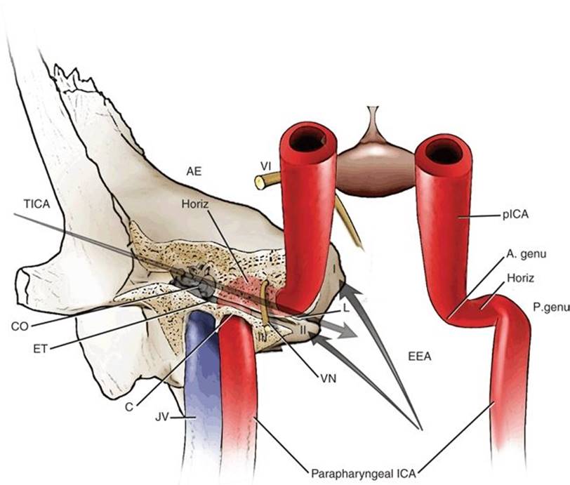
FIGURE 8.2 Schematic 3D figure to provide subjective comparison between both approaches (EEA and TICA) to petrous apex. Frontal view simulating the endoscopic endonasal approach in the operation room. Note the limits of each zone of petrous apex and the structures surrounding it, as well as the main direction provided by each approach. I, zone I of petrous apex; II, zone II; III, zone III; VI, abducens nerve; AE, arcuate eminence; CO, cochlea; JV, jugular vein, ET, eustachian tube; C, carotid canal; L, lacerum; VN, vidian nerve; P. genu, posterior genu petrous ICA; Horiz., horizontal petrous ICA; pICA, paraclival ICA; A. genu, anterior genu petrous ICA; ICA, internal carotid artery; EEA, endoscopic endonasal approach; TICA, transcanal infracochlear approach.
CONTRAINDICATIONS
Relative contraindications to an endoscopic endonasal approach include the usual patient comorbidities precluding surgery and difficult patient anatomy. If the patient has serviceable hearing, a transsphenoidal or traditional hearing sparing otologic approach should be considered (e.g., infralabyrinthine, transcanal infracochlear, or middle fossa approach). If the patient has minimal serviceable hearing and/or vestibular loss or if the direct endoscopic approach is technically difficult, then a transtemporal approach should be considered. For transtemporal operations, the temporal bone should be sufficiently pneumatized to navigate around or through the labyrinth. The middle fossa approach is technically demanding, there is no permanent drainage option, and some degree of brain retraction is required with the potential for injury to the brain. The transsphenoidal approach is better suited if continuous drainage is desirable (e.g., cholesterol granuloma).
PREOPERATIVE PLANNING
The success of any endoscopic skull base procedure depends on a thorough history, preoperative evaluation, and an informed consent conversation with the patient about the risks, benefits, and alternatives of various forms of treatment. Patients must understand the goals of surgery, which may only be diagnosis (biopsy) and the likelihood of achieving relief from symptoms. The surgical team needs to be prepared for the unexpected and have a well-developed plan in place for life-threatening complications including injury to the carotid artery.
CT and MRI are commonly used to assess petrous apex lesions. Cholesterol granuloma will typically appear on CT as an expansile lesion of the petrous apex with bony erosion and scalloping (Fig. 8.3A). Thinning of the posterior wall of the sphenoid and petrous carotid artery dehiscence may also be seen. Cholesterol granulomas have a characteristic appearance on MRI that is hyperintense on both T1-weighted and T2-weighted sequences, owing to the lipid and fluid content of these lesions (Fig. 8.3B and C). In contrast, a petrous apex cholesteatoma on MR will show a smooth, expansile mass with low signal intensity on T1-weighted imaging and high signal on T2-weighted imaging. A variety of MR pulse sequences have been described to help differentiate various petrous apex pathologies (e.g., graded recall echo).
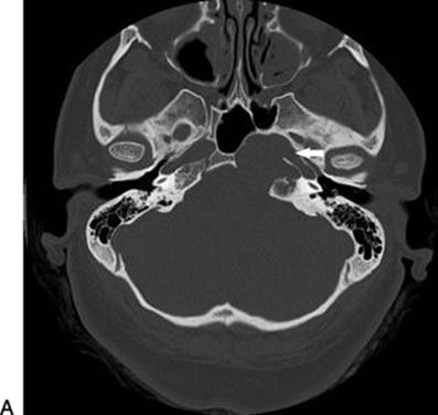
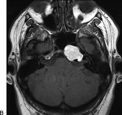
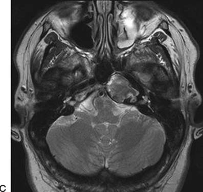
FIGURE 8.3 Axial imaging with CT and MR at similar locations of a patient with a left petrous apex lesion (biopsy proven to be a cholesterol granuloma). A. Axial CT showing an expansile lesion “bulging” into the medial sphenoid sinus. The arrow points to the left carotid canal with a dehiscent medial bony wall. B. An axial T1-weighted MR of the same patient showing a bright signal. C. An axial T2-weighted MR of the same patient showing an intermediate signal.
It is important to study the CT and MR to define the anatomy including the location of the carotid artery, optic nerve, jugular bulb, and facial nerve as well as the pneumatization of the sphenoid sinus and temporal bone (Fig. 8.4). The position and degree of bone covering/thinning of the petrous and clival portions of the carotid artery are very important. It is critical to ensure that the petrous apex pathology is not a vascular lesion such as a thrombosed aneurysm before any biopsy or drainage procedure is considered. The degree to which the pathology will be visible medial to the ICA after bone removal can be estimated on axial images by drawing a line from the pyriform aperture to the cyst (Fig. 8.4). The medial boundary is the medial edge of the pathology, and the most lateral boundary is the lacerum segment of the ICA, which cannot be retracted laterally.
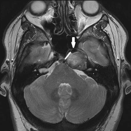
FIGURE 8.4 Axial MR showing a left petrous apex lesion pushing the carotid artery anteriorly (arrow). The artery must be identified intraoperatively and avoided during a transsphenoidal approach for management. The position of the artery narrows the corridor to access the lesion posteromedially (line).
SURGICAL TECHNIQUE (VIDEO 8.1)
The patient is placed supine on the operating room table with the head in a neutral position. The nasal mucosa is topically decongested with 1:1,000 epinephrine-soaked Neuro Patties. The image guidance system is appropriately set up, calibrated, and used to confirm important anatomy during the operation. A 0-degree 4-mm rigid nasal telescope attached to a high-definition video system is used for the majority of the operation. Angled telescopes (e.g., 30 degrees to 70 degrees) are often used to look into marsupialized cysts (e.g., cholesterol granuloma) once they have been opened.
It would be rare to need to place a nasoseptal flap for a complication such as a cerebrospinal fluid leak, but the surgeon should anticipate this ahead of time so that it can be raised initially and safeguarded in the nasopharynx or maxillary sinus. Alternatively, the posterior septal artery pedicle can be preserved on one side, allowing a viable nasoseptal flap to be raised later should it be required. A small nasoseptal flap can also be inserted through a marsupialized cholesterol granuloma as a substitute for a temporary silastic stent to keep the cyst opening patent.
A large, well-pneumatized sphenoid sinus with a lesion in the ipsilateral petrous apex can be approached through a wide unilateral sphenoidotomy. However, this usually means that the surgeon has to hold the scope and instruments as for standard sinus surgery without the ability for a second surgeon to help. Most lesions in the petrous apex are approached with an expanded endoscopic binarial technique similar to the exposure performed for endoscopic pituitary surgery (Fig. 8.5). This may require removal of a portion of one middle turbinate depending on the dimensions of the nasal cavity and the surgeon's preference. Ethmoidectomy and antrostomy are usually not required unless there is coexistent sinus disease or a transpterygoid approach is being performed. The posterior bony septum is disarticulated from the sphenoid rostrum, and the posterior 1 to 2 cm of septum is removed. The anterior wall of the sphenoid sinus is removed with Kerrison rongeurs and high-speed drill with a 3- or 4-mm coarse diamond burr. Care is taken to cauterize the posterior nasal branches of the sphenopalatine arteries coursing across the sphenoid rostrum to prevent postoperative epistaxis (on one side only if a nasoseptal flap has been raised). As with any endoscopic skull base approach, it is important to widely open the sphenoid sinuses and make them into a rectangular box so that the bony edges do not limit mobility of the instruments. The surgeon should always be aware of the location of the optic canal and carotid artery siphon during this step of the dissection.
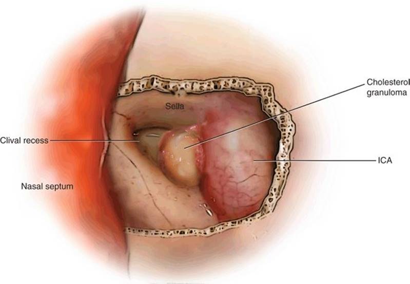
FIGURE 8.5 A bilateral sphenoidotomy with resection of the rostrum and posterior 1 cm of the nasal septum provides increased exposure for instrumentation and improves the angle of visualization.
Sphenoid septations are removed with angled Kerrison rongeurs +/− high-speed drill following the septations onto the bone covering the carotid arteries. The sphenoid sinus landmarks are identified visually and with image guidance as required. The surgeon should look for the clival recess, optic nerves, planum sphenoidale, medial and lateral opticocarotid recesses, and the indentation of the sella. An intraoperative Doppler probe may be helpful to confirm the location of the clival carotid artery if there is any doubt. It is absolutely critical that the surgeon is confident of the location of the carotid artery before any instrumentation of the petrous apex lesion is commenced.
It is helpful to remove the mucosa of the sphenoid sinus, which reduces bleeding and helps to define the bony anatomy. If the petrous apex lesion extends into the sphenoid sinus, there is often bone over the lesion, which can be evaluated by palpation with a blunt instrument such as a ball probe. If there is bone over the lesion, it must be removed, usually starting with a high-speed drill and coarse diamond burr of appropriate size for the dimensions of the area (Fig. 8.6). It is important not to rest the drill shaft on the carotid artery during the drilling process to avoid inadvertent injury or heat transfer. Drilling starts at the medial surface of the lesion running vertically and parallel to the carotid artery. When the bone is thin enough to fracture, the small pieces are removed with a small Kerrison rongeur (e.g., 1 or 2 mm). The suspected pathology and surgical access required will dictate how much bone is removed over the lesion.
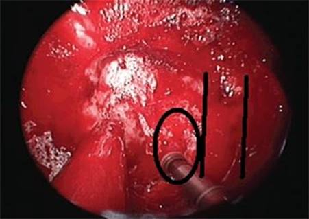
FIGURE 8.6 After identification of the wall of the cholesterol granuloma, the bone on the medial aspect of the cystic cavity is carefully thinned with a 3-mm coarse diamond drill. The opening is enlarged with Kerrison rongeurs. Circle, cholesterol granuloma; parallel lines, paraclival internal carotid artery. (Figure and legend reprinted with permission from Snyderman C, Kassam A, Carrau R, et al. Endoscopic approaches to the petrous apex. Operative Techniques in Otolaryngology 2006;17(3):168–173.)
If the lesion is suspected to be cystic (e.g., cholesterol granuloma), then as much bone as possible over the lesion is removed. A sharp sickle knife is used to open the cyst wall and drain the contents (Fig. 8.7). The exposed cyst wall is removed with angled thru-cutting forceps and the specimen sent for permanent pathology. Some surgeons use a cruciate incision and drape the cut surfaces over the bony edges. If bleeding from the capsule of the lesion is anticipated, then bipolar electrocautery can be used to outline the incision. The interior of the cyst is inspected with angled telescopes and irrigated with saline to remove any additional debris as required (Fig. 8.8). If the lesion is solid and all that is required is a biopsy or debulking, then appropriate angled instruments are used to carry out the biopsy. Intraoperative frozen section analysis is helpful to confirm that tissue representative of the lesion has been obtained and to give an initial indication as to the nature of the lesion (e.g., benign vs. malignant). If temporary or permanent drainage from the petrous apex into the sphenoid sinus is desirable, a rolled piece of thin pliable silastic sheeting is cut and tailored to the defect to sit securely through the marsupialized defect into the sphenoid sinus. Care is taken to avoid resting the cut surface on the opposite carotid artery or optic nerve to prevent pressure erosion and injury. Some surgeons have also described leaving a pediatric tracheal T tube or other small catheter to stent the area. There is no consensus on how long to leave the stent in place, but the usual time period is 3 to 6 months.
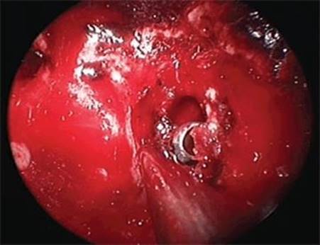
FIGURE 8.7 The contents of the cholesterol granuloma are removed with curettes, suctions, or irrigation. (Figure and legend reprinted with permission from Snyderman C, Kassam A, Carrau R, et al. Endoscopic approaches to the petrous apex. Operative Techniques in Otolaryngology 2006;17(3):168–173.)
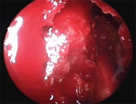
FIGURE 8.8 The final defect can be inspected with angled telescopes. (Figure and legend reprinted with permission from Snyderman C, Kassam A, Carrau R, et al. Endoscopic approaches to the petrous apex. Operative Techniques in Otolaryngology 2006;17(3):168–173.)
If the above standard approach will not allow access to the petrous apex because of poor sphenoid pneumatization or posterolateral location of the petrous apex pathology, then decompression and lateralization of the carotid artery can be considered, or alternatively, an otologic approach can be used. To decompress and lateralize the carotid artery, a wide middle meatal antrostomy is performed. The sphenopalatine artery is identified, and the bone over the posteromedial maxillary sinus is removed to expose the pterygopalatine fossa. The sphenopalatine and posterior nasal arteries are ligated and divided. The vidian canal and its contents are identified in the base of the pterygoid plates (pterygoid wedge). The bone medial and inferior to the vidian canal is removed, and the vidian artery is traced to the second genu of the carotid artery. The bone of the vertical clival carotid is thinned with a diamond burr and removed allowing several millimeters of lateral displacement of the clival carotid artery. If this is enough exposure based on the preoperative and intraoperative assessment of the petrous apex lesion, then the lesion can be managed as described above for the expanded binarial transsphenoidal approach.
Nasal packing is usually avoided unless there is excessive bleeding at the end of the operation. Absorbable packing gently placed around the operative site may help to minimize minor postoperative bleeding.
POSTOPERATIVE MANAGEMENT
Patients may be discharged the same day if the surgery is uncomplicated. The surgeon's standard postoperative regimen for routine sinus surgery can be followed. Typically, this involves 1 week of avoiding strenuous activity, nasal saline irrigations multiple times each day, and possibly a broad-spectrum antibiotic such as amoxicillin–clavulanate. Nasal debridement is performed depending on the surgeon's preference and patient tolerance. In most uncomplicated cases, gentle debridement is required every 2 to 3 weeks for approximately 6 to 8 weeks or until the cavity has healed (Fig. 8.9).
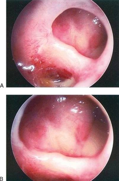
FIGURE 8.9 After several months, a well-mucosalized and widely patent opening into the petrous apex persists (A); Closer view of A (B). (Figure and legend reprinted with permission from Snyderman C, Kassam A, Carrau R, et al. Endoscopic approaches to the petrous apex. Operative Techniques in Otolaryngology 2006;17(3):168–173.)
COMPLICATIONS
The most feared complication is inadvertent injury and hemorrhage from the ipsilateral carotid artery, which is a rare but life-threatening event. Management is beyond the scope of this paper but requires the surgeon and team to have a plan in place to tamponade the bleeding (e.g., fresh muscle patch), manage intravascular volume and blood replacement, and involve interventional radiology. Avoidance is key and requires a thorough knowledge of the anatomical variations of the carotid artery, visualization intraoperatively, and careful instrumentation around the artery. Intraoperative image guidance and Doppler probe assessment help to confirm the clinical impression of the location of the artery.
Postoperative hemorrhage is uncommon unless the posterior nasal arteries are not adequately cauterized. Transient cranial nerve palsies, especially the abducens nerve or audiovestibular nerve, have been reported. Symptomatic dry eye requiring artificial tears occurs uncommonly from sacrificing the vidian nerve if a transpterygoid approach is used. Injury to the orbit and cerebrospinal fluid leaks are also uncommon with this type of surgery. Stenting of cystic lesions helps to keep the marsupialized area open, but stenosis and recurrence of symptoms can still occur months to years later requiring reoperation. Adhesions may block access for transnasal removal of the silastic stent necessitating a second operation for removal.
RESULTS
Transsphenoidal surgery for petrous apex lesions has been popular for only the last 15 to 20 years. For benign cystic lesions of the petrous apex, the endoscopic transsphenoidal approach to the medial petrous apex offers a high degree of success with a low recurrence rate, low risk to inner ear structures, and a natural drainage pathway into the sinonasal cavity.
There are multiple case reports or small case series reported in the literature. One of the largest case series of cholesterol granulomas described by Gore et al. stated the rate of clinical and/or radiologic recurrence of petrous apex cholesterol granulomas following endoscopic drainage to be in the range of 5% compared with approximately 15% for transotic approaches. Most authors prefer to leave stents from the petrous apex into the sphenoid sinus, usually made of medical grade silicone, and most leave the stents in for several months. Placing a small nasoseptal flap as a stent has been reported with good results in a small number of patients. Topical mitomycin has been used around the edges of marsupialized cystic lesions in the petrous apex with the goal of preventing postoperative stenosis. There are no good data on whether this antineoplastic agent is effective for this indication or if there are long-term sequelae associated with its use. Concerns of malignancy associated with mitomycin have limited its use.
PEARLS
· Open the sphenoid rostrum widely to maximize exposure, and permit a two-surgeon, four-hand technique.
· Optimal visualization is important throughout the procedure; maintain good hemostasis with one or more methods such as warm irrigations, topical vasoconstrictors, and topical hemostatic agents.
· Confirm the location and position of the carotid artery by multiple methods before making any incisions or taking a biopsy of lesions in the lateral sphenoid sinus.
· Have a plan of action developed and known by the surgical team in case of injury to the carotid artery.
· If the patient requires lateralization of the carotid artery, consider another approach or referral to a center with significant experience or expertise.
· If the lesion is solid, obtain a frozen section early, which may indicate pathology (e.g., lymphoma) that will not require further or more extensive surgical intervention.
PITFALLS
· Attempting to access the medial petrous apex through a unilateral or small sphenoidotomy will usually give suboptimal exposure of the lateral wall of the sphenoid sinus. This will result in poor marsupialization of a cystic lesion (and a higher chance of postoperative stenosis) and increase the chance of injury to neurovascular structures.
· Cystic lesions that have been marsupialized are much more likely to stenose without postoperative stenting.
INSTRUMENTS TO HAVE AVAILABLE
· Standard endoscopic skull base and sinus instruments
· Image guidance system
· High-speed endoscopic skull base drill
· Intraoperative Doppler probe
SUGGESTED READING
Isaacson B, Kutz JW, Roland PS. Lesions of the petrous apex: Diagnosis and management. Otolaryngol Clin North Am 2007;40:479–519.
Gore MR, Zanation AM, Ebert CS, et al. Cholesterol granuloma of the petrous apex. Otolaryngol Clin North Am 2009;44:1043–1058.
Zanation AM, Snyderman CH, Carrau RL, et al. Endoscopic endonasal surgery for petrous apex lesions. Laryngoscope 2009;119(1):19–25.