
Michael Lanuti
 INDICATIONS/CONTRAINDICATIONS
Indications
Despite the lack of large multicenter randomized controlled trials to compare oncologic outcomes of video-assisted thoracic (VATS) lobectomy to open lobectomy, comparative studies have demonstrated equivalent oncologic outcomes, shorter length of stay, and fewer postoperative complications with the VATS approach. VATS lobectomy is commonly performed for selected peripherally located T1 or T2 tumors in the absence of any significant hilar or mediastinal lymphadenopathy. Thoracoscopic resection of the left upper lobe can be the most challenging lobe by virtue of a heterogeneous branching pattern of the left pulmonary artery.
Contraindications
Although there are no absolute contraindications to VATS left upper lobectomy, the surgeon should consider the following characteristics as relative contraindications: Tumors >6 cm, centrally located tumors, bulky hilar (N1) or mediastinal (N2) lymphadenopathy, endobronchial disease within 2 cm of the origin of the left upper lobar bronchus, and patients who received induction radiation for locally advanced disease (stage IIIA). Performing VATS lobectomy on patients with previous coronary revascularization, particularly in patients who harbor a left internal mammary artery graft, can be challenging where early conversion to thoracotomy should be implemented.
 PREOPERATIVE PLANNING
All patients should undergo preoperative examination with lung function testing, PET/CT, bronchoscopy, and endobronchial ultrasound (EBUS)/mediastinoscopy for preoperative staging (unless it is a peripherally placed T1 tumor with no PET activity in the hilum or mediastinum). All anatomically enlarged mediastinal lymph nodes should be sampled to rule out N2 disease. Epidural anesthesia can be considered for VATS lobectomy but is not routinely required. Hemodynamic monitoring with a radial arterial line should be routine for VATS lobectomy given the potential for vascular injury and rapid blood loss. It is always a good idea to have a thoracotomy tray in the operating room and the availability of a sponge stick and 5-0 Prolene suture for emergency control of hemorrhage.
 SURGERY
Positioning
After successful placement of a right-sided (preferred) double-lumen endotracheal tube, the patient is placed in the maximally flexed right lateral decubitus position tilted slightly backward to prevent the hip from obstructing downward movement of the thoracoscope. The camera port is placed at the eighth or ninth interspace along the posterior axillary line, avoiding the apex of the heart. The posterior port is placed where the left lower lobe edge touches the diaphragm (in line with the scapular tip). Once intrapleural visualization is achieved, the left hilum and major fissure should be identified. Nerve blocks can be considered at this time using 0.25% to 0.5% bupivacaine under direct thoracoscopic guidance. An axillary utility incision (between 4 and 6 cm) is placed perpendicular to the anterior axillary line directly over the left superior pulmonary vein (Fig. 16.1). The soft tissue of the utility port can be separated with either a Weitlaner retractor or a wound protector (Fig. 16.2) to facilitate passage of instruments and avoid loss of pneumothorax while suctioning.
Technique
Instrumentation is important when performing thoracoscopic pulmonary resection, including the use of a 30-degree videoscope and long, curved instruments to improve retraction and dissection. High-definition video equipment improves visualization for difficult dissections. Linear staplers are used to control and divide lung parenchyma, vessels, and bronchus. After inspection of the pleural cavity (to rule out occult pleural-based metastases) and confirmation of the indication for lobectomy, the structures are divided as you encounter them during the operation from anterior to posterior. The pleura overlying the hilum is divided and the artery and superior pulmonary vein are identified. The left main pulmonary artery emerges from beneath the aortic arch and is located superior and posterolateral to the superior pulmonary vein. It is often a good time to confirm the presence of the inferior pulmonary vein to ensure no aberrant anatomy. The plane between the left main pulmonary artery and the upper lobe vein is opened, so the vein is exposed by a vessel loop coming from the anterior utility incision. Although there are numerous variations of which anatomic structures to isolate and divide in sequence, the recommended sequence is listed in Table 16.1. The left superior pulmonary vein is usually the first vascular structure to be transected with the thoracoscopic stapler introduced from the posterior port. If a lingular-sparing lobectomy is being considered, the lingular vein must be preserved along with the lingular artery. Next the first apical branch of the pulmonary artery is isolated and divided (Fig. 16.3) with a vascular stapler. This branch is a short, broad vessel that can be easily avulsed or torn if too much traction is applied when retracting the lobe. A plane between the artery and the bronchus can be created. The bifurcation of the left upper and lower lobe bronchi is identified, and the left upper lobe bronchus is transected with a thick tissue thoracoscopic stapler from the posterior port. Prior to bronchial transection, the surgeon should inflate the lung to be sure that the left mainstem or lower lobe bronchus has not been mistakenly dissected. The lobe is pushed posterior and the remaining branches on the pulmonary artery including the lingular artery are exposed and transected by a thoracoscopic stapler with a vascular load (Fig. 16.4) or energy-based devices. The fissure is finally transected with multiple applications of a linear stapler via the posterior port. The left upper lobe should be removed with a large endocatch bag to avoid port site contamination and contain any blood spilling from the resected specimen which may contain tumor cells.

Figure 16.1 The patient is placed in the maximally flexed right lateral decubitus position tilted slightly backward to prevent the hip from obstructing downward movement of the thoracoscope. The port incisions are depicted.

Figure 16.2 Utility port wound protector in place. (© SurgiSleeve 2.5 × 6 cm, Covidien, Mansfield, MA)
TABLE 16.1 The Conduct of the Operation for Left Upper Lobectomy and Best Port Approach

Mediastinal lymph node sampling in the setting of NSCLC is the minimum requirement for all pulmonary resections. Lymph nodes are sampled or removed en bloc from station 5 and 6 (as seen on Figure 16.5 between the aorta and the main pulmonary artery). The subcarinal lymph nodes (station 7) should also be sampled or removed while reflecting the lung medially and exposing the space between the left inferior pulmonary vein and the inferior border of the left mainstem bronchus. If visualized, lymph nodes from station 8 and 9 should be sampled or removed while dividing the left inferior pulmonary ligament. Station 10 nodes are in upper lobe resections and removed as part of the procedure to expose other structures. Figure 16.6 depicts completion of the lobectomy and lymph node dissection. After lymph node sampling, the divided left upper lobe bronchial stump should be tested to 20 to 30 mm Hg while inflating the lung to ensure the absence of a bronchial stump air leak. One intercostal drain is placed in through the camera incision and the lung is re-expanded.

Figure 16.3 First apical branch of the left pulmonary artery is isolated and divided with a linear vascular stapler.

Figure 16.4 After division of the left upper lobe bronchus, additional segmental pulmonary artery branches can be divided including the lingular artery.

Figure 16.5 Sampling of aortopulmonary lymph nodes.
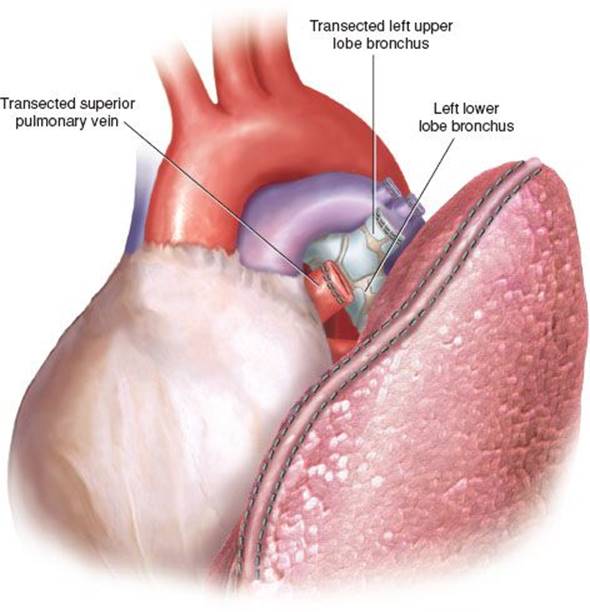
Figure 16.6 Left upper lobe bronchial and vascular stumps are visualized, after left upper lobectomy and en bloc removal of station 5 and station 6 lymph nodes.
 POSTOPERATIVE MANAGEMENT
Routine management of VATS lobectomy patients is no different than open lobectomy, except that they may not have a thoracic epidural. Most patients will need patient-controlled anesthesia for the first 24 hours with a quick transition to oral narcotics and nonsteroidal anti-inflammatory medications on postoperative day 1. Prolonged air leaks after VATS left upper lobectomy are relatively uncommon unless the major fissure is manipulated or incomplete. Routine discharge to home after removal of the chest tube can be achieved by postoperative day 2 or 3 with an uncomplicated VATS lobectomy.
 COMPLICATIONS
Life-threatening intraoperative complications are very uncommon and overall hospital mortality (0.5% to 3.6%) and conversion rates (2% to 13%) are low. One of the feared complications of VATS left upper lobectomy is vascular injury that cannot be controlled before the need of transfusion or resuscitation. This can be mitigated by good port placement and meticulous technique. The surgeon should be prepared to tamponade bleeding with a 5- to 10-mm Endo Peanut or sponge stick. Usually, conversion is best achieved by extending the anterior utility incision to an anterior thoracotomy, as opposed to a separate posterolateral thoracotomy. It is important to note that most bleeding can be controlled with a direct pressure technique where conversion can be performed in a nonemergent manner. The surgeon should also communicate with their anesthesia colleagues to prepare for blood loss and have crossmatched blood in the room upon identification of a major vascular injury. Another important pitfall is the application of excessive torque to the thoracoscopic port sites, which can be associated with intercostal nerve injury, resulting in significant and prolonged postoperative pain.
 RESULTS
In recent years, a number of large institutional studies, multi-institutional registries, and meta-analyses have demonstrated the perioperative safety and long-term oncologic efficacy of VATS lobectomy for patients with early stage NSCLC. Despite this growing body of evidence there is still skepticism within the thoracic oncology community regarding the VATS approach, as no randomized controlled trials have been completed comparing oncologic outcomes of VATS lobectomy versus open thoracotomy. Large series of VATS lobectomy have been published demonstrating reduced complications and length of stay, reduced incidence of atrial fibrillation, quicker return to work, and better compliance with adjuvant therapy. There is sufficient clinical equipoise among many thoracic surgeons that the techniques are oncologically equivalent; however, recent studies have demonstrated increased nodal upstaging with open lobectomy compared to VATS invoking a compromise to mediastinal lymph node dissection or sampling with a thoracoscopic technique.
 CONCLUSIONS
Thoracoscopic left upper lobectomy can be technically demanding. The pulmonary arterial anatomy is variable and the segmental branches are short with less room for technical error. Sampling the subcarinal lymph nodes (station 7) can be challenging but should be a routine part of routine mediastinal lymph node sampling. Although the thoracoscopic strategy may be difficult to learn, it is increasingly becoming the preferred method of anatomic lobectomy.
Recommended References and Readings
Boffa DJ, Kosinski AS, Paul S, et al. Lymph node evaluation by open or video-assisted approaches in 11,500 anatomic lung cancer resections. Ann Thorac Surg. 2012;94(2):347–353; discussion 353.
D’Amico TA. Long-term outcomes of thoracoscopic lobectomy. Thorac Surg Clin. 2008;18(3):259–262.
Licht PB, Jorgensen OD, Ladegaard L, et al. A national study of nodal upstaging after thoracoscopic versus open lobectomy for clinical stage I lung cancer. Ann Thorac Surg. 2013;96(3):943–949; discussion 949–950.
McKenna RJ Jr., Houck W, Fuller CB. Video-assisted thoracic surgery lobectomy: Experience with 1,100 cases. Ann Thorac Surg. 2006;81(2):421–425; discussion 425–426.
Onaitis MW, Petersen RP, Balderson SS, et al. Thoracoscopic lobectomy is a safe and versatile procedure: Experience with 500 consecutive patients. Ann Surg. 2006;244(3):420–425.
Paul S, Altorki NK, Sheng S, et al. Thoracoscopic lobectomy is associated with lower morbidity than open lobectomy: A propensity-matched analysis from the STS database. J Thorac Cardiovasc Surg.2010;139(2):366–378.
Swanson SJ, Herndon JE 2nd, D’Amico TA, et al. Video-assisted thoracic surgery lobectomy: Report of CALGB 39802–a prospective, multi-institution feasibility study. J Clin Oncol. 2007;25(31):4993–4997.
Yan TD, Black D, Bannon PG, et al. Systematic review and meta-analysis of randomized and nonrandomized trials on safety and efficacy of video-assisted thoracic surgery lobectomy for early-stage non-small-cell lung cancer. J Clin Oncol. 2009;27(15):2553–2562.