
Daniel G. Nicastri, Oskar Kizhner, and Todd S. Weiser
 INDICATIONS/CONTRAINDICATIONS
Indications
Thoracoscopic or video-assisted thoracic surgery (VATS) left lower lobectomy has similar indications/contraindications, preoperative planning, positioning, and technique to right-sided lower lobectomy. Left lower lobectomies account for up to 15% of all lobectomies according to a recent series. The most common indication for VATS left lower lobectomy is primary nonsmall cell lung carcinoma (NSCLC). Approximately 170,000 deaths each year are attributable to lung cancer, surpassing the next four most common cancers combined.
For early staged cancers, complete surgical resection remains the cornerstone of curative therapy for NSCLC. An anatomic resection; lobectomy, segmentectomy, or pneumonectomy; is the standard surgical approach for stage I and II NSCLC. Nonanatomic wedge resection is often reserved for those patients with suitable tumor location and significant cardiopulmonary comorbidities that preclude a formal anatomic resection. Other indications for thoracoscopic left lower lobectomy include: Pulmonary metastases not amenable to sublobar resection, benign lung tumors not amenable to wedge resection due to anatomic considerations, congenital lesions such as arteriovenous malformations and pulmonary sequestrations, and infectious or inflammatory pathologies.
Contraindications
Contraindications to VATS are growing increasingly rare for experienced thoracoscopic surgeons. However, according to a recent publication analyzing data submitted to the Society of Thoracic Surgeons General Thoracic Database, only approximately one-third of all lobectomies are being performed thoracoscopically. The following are relative contraindications to proceeding with thoracoscopic left lower lobectomy: Necessity of bronchial sleeve resection to achieve negative margins; locally advanced tumors with need for concurrent chest wall, diaphragm, or major vascular resection with reconstruction; and preoperative chemotherapy or radiation therapy. Absolute contraindications for VATS lobectomy include very large tumors requiring a sizable thoracotomy for specimen extraction, the inability of the patient to tolerate single lung ventilation, and the surgeon being uncomfortable with performing the procedure. We feel that this last contraindication has resulted in the lack of widespread adoption of this technique; thoracoscopic lobectomy is more technically demanding than that done via thoracotomy.
 PREOPERATIVE PLANNING
There are two components in assessing the patient that presents with pathology requiring pulmonary resection, especially those requiring the removal of at least a lobe or greater of pulmonary parenchyma. The first part centers on the appropriateness of the intended surgical intervention from an oncologic standpoint. Patients with biopsy-proven lung carcinoma or those with suspicious pulmonary lesions believed to be malignant should undergo thorough clinical staging. In addition to conventional CT imaging, positron emission tomography (PET) should be employed to evaluate for evidence of regional or distant metastatic disease. Concerning extrathoracic abnormalities found on PET imaging should be considered for tissue sampling to disprove stage IV disease. Surgical or endoscopic staging of the mediastinum to rule out stage III disease should be contemplated for those patients with PET abnormalities in the hilar or mediastinal lymph nodes or those with large, central tumors. When compared to NSCLCs in other lobes, left lower lobe carcinomas have a higher prevalence of lymph node drainage to the contralateral mediastinum. As a result, patients with left lower lobe lung cancer who demonstrate right paratracheal lymphadenopathy by CT or PET criteria should undergo tissue sampling to exclude regionally advanced disease. National Comprehensive Cancer Network guidelines have been established allowing for appropriate preoperative staging and management of patients with NSCLC.
The second component in the evaluation of patients presenting with pulmonary pathology requiring lobectomy involves preoperative cardiopulmonary risk assessment. Despite the tremendous advances made in surgical technique, anesthetic management, and postoperative care, the overall risk of perioperative morbidity and mortality after lobectomy remains relatively high. The ability to predict those patients that are at significant risk allows physicians to appropriately select patients for lobectomy. There have been many useful studies identifying factors associated with major morbidity and mortality in patients undergoing pulmonary lobectomy.
The scope of this chapter precludes an expansive review of this data. We suggest performing a careful history and physical examination accompanied by spirometry and measurement of diffusion capacity (DLCO) along with arterial blood gas sampling. Predicted postoperative forced expiratory volume in 1 second (FEV1) and DLCO expressed as a percentage of normal are calculated by utilizing the number of pulmonary segments to be removed. For postoperative predicted FEV1 or DLCO less than 40% predicted, quantitative pulmonary perfusion imaging is performed for a more accurate calculation. For those patients with concerning results, exercise testing can be completed to determine oxygen consumption during maximum exercise. Other adjunct tests that may be useful include cardiac stress testing and echocardiography. Nevertheless, absolute numbers should not be the sole factor in determining the best treatment for a patient; the importance of sound clinical judgment cannot be overstated.
 SURGERY
The VATS lobectomy operation consists of individual hilar ligation via three to four incisions without rib spreading. Thoracoscopic lobectomy should duplicate the oncologic principles as those attained via traditional thoracotomy. That is, resection of tumor with negative margins while performing individual vascular and bronchial division with complete hilar lymph node dissection. In addition, a mediastinal lymph node dissection, or sampling, is performed. Certain aspects in VATS lobectomies, such as avoiding rib spreading, are stressed with the goal to improve the patient’s postoperative recovery. One variant of thoracoscopic lobectomy is the “video-assisted simultaneously stapled lobectomy.” This iteration does not employ individual bronchovascular ligation and is, therefore, basically a different operation and will not be discussed in this chapter.
There are some unique aspects to a left lower lobectomy compared to a right-sided lower lobectomy. These include adjusting the camera port to the posterior axillary line to avoid the heart, sampling level 5 and 6 nodes while avoiding recurrent laryngeal and phrenic nerve injury, and understanding the relationship of the lower lobe vein to the upper lobe vein while avoiding division of a common venous trunk. Standard thoracotomy instruments are generally the ones used for VATS lobectomies: Harken’s clamp, Pearson’s scissors, suction, sponge sticks or dental pledgets, Roberts or Fraser-Kelly clamps, straight or curved Foerster ring clamps. We also utilize a suction/irrigation device and endoscopic Kittner dissectors and stapling devices.
Positioning
Positioning for VATS left lower lobectomy, as in all minimally invasive surgery, is extremely important. Once anesthetized and intubated with either a single- or double-lumen endotracheal tube, careful bronchoscopic examination of the airway is performed to evacuate all secretions and to ensure that no endobronchial tumor is present. Once that has been completed, we prefer using a bronchial blocker, passed through the end of the single-lumen tube, to obtain lung isolation. An alternative method for obtaining lung isolation is through the use of a double-lumen endotracheal tube. The patient is then placed in the right lateral decubitus position and then taped in place after table flexion. The patient’s anterior-superior iliac crest is positioned at the level of the break of the bed and the table is optimally flexed to open up the intercostal spaces. The patient’s hips are rotated slightly posteriorly to minimize the camera from hitting the hips when looking more anteriorly. An axillary roll is placed under the chest to protect the axilla. The upper arm and shoulder are abducted as far above the head as possible while being sure to prevent brachial plexus stretch (approximately 100 degrees). Prior to prepping, double-lumen tube position or location of the bronchial blocker is reconfirmed with bronchoscopy. Two video monitors are placed at the right and left sides of the head of the bed.
Technique
After prepping and draping, we begin by placing the first of three ports (Fig. 18.1). The camera port, placed in the eighth intercostal space, is aligned with the posterior axillary line. Again, no rib spreading is done. We generally use a metal thoracoscopic port for our 30-degree 10-mm thoracoscope. We almost exclusively use a 30-degree thoracoscope. It provides optimal views not afforded by a 0-degree scope, particularly during the difficult dissection around the superior hilum.
It is important to be aware of a possible elevated left hemidiaphragm and to use the lateral chest radiograph to help make adjustments of this port placement either more anterior, or more posterior, or even one rib space up on occasion. Injuries to the diaphragm can occur in an obese patient or one with an elevated hemidiaphragm if the correct approach is not taken.
Our second port is usually placed in the fourth or fifth intercostal space, either inferior or posterior to the scapular tip. This port usually serves as the lung retraction port and for placement of the suction/irrigation device. This is done under direct vision. Some muscles are divided with the cautery and we dissect directly down to the intercostal space. Care is taken not to injure the corresponding intercostal neurovascular bundle.
We then use the camera and lung retraction via the posterior port to choose the location of the final port site. This anterior port should be placed right over the hilum since this will be used as the access port. Dissection of both the hilum and the fissure will be performed through this port. This incision is initially 1 to 2 cm. It is not extended to 5 cm in length until we have decided to proceed with the VATS lobectomy. The port is usually created anterior to the latissimus dorsi in the fifth intercostal space for left lower lobectomy. A 22-gauge spinal needle may be useful to properly localize the optimal interspace in obese patients. This is an area relatively devoid of major musculature as it is located posterior to the pectoralis major muscle and anterior to the latissimus dorsi. There are usually intercalations of serratus anterior in this area. Hemostasis is very important when creating the ports as bleeding from the port sites onto the camera and onto the surgical field during the procedure is a nuisance and can significantly prolong the operation.
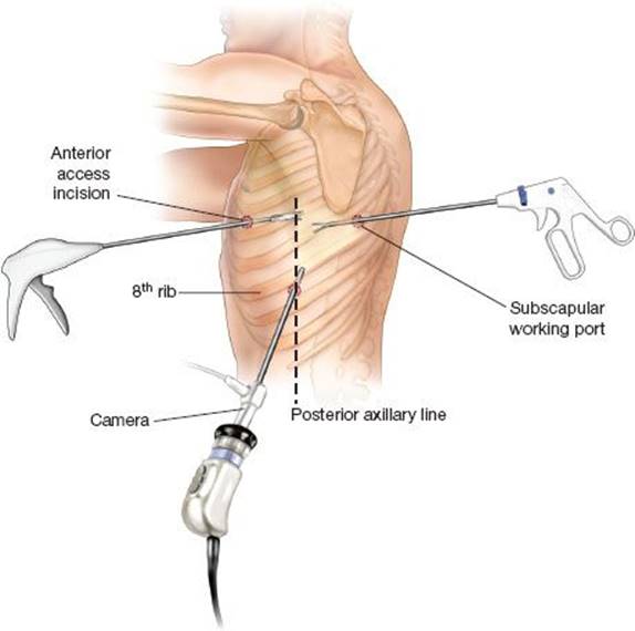
Figure 18.1 Port Placement. Camera port is placed in the eighth intercostal space in the posterior axillary line. The subscapular working port is placed in the interspace just posterior to the tip of the scapula. The anterior access port is placed at the fourth or fifth intercostal space in the anterior axillary line. The exact location is based on visualization of the anterior hilar structures.
Prior to beginning the dissection, the pleural cavity is thoroughly inspected. The pleural surfaces are explored for metastases and any adhesions are then divided. As in open surgery, total lysis of adhesions is completed to restore the normal anatomy of the left lung. This helps avoid judgment errors regarding anatomic anomalies, allows the remaining lung to occupy the pleural space after resection, and generally makes the rest of the case proceed more expeditiously. Adhesions are divided either sharply, with cautery, or with an ultrasonic cutting and coagulation device. An endoscopic Kittner dissector may be passed through the camera port to provide countertraction against the lung when dividing adhesions between the lower lobe and diaphragm. Again, full mobilization of the lung is vital to effectively complete the lower lobectomy.
Once our ports are created and any adhesions are divided, we then turn our attention toward isolating the pulmonary venous drainage of the left lower lobe. The lung is retracted toward the lateral chest wall with a ring forceps brought through the posterior port. Electrocautery is used to divide the inferior pulmonary ligament once it is under tension (Fig. 18.2A) and any level 9 lymph nodes are removed at this time. The posterior mediastinal pleura is then opened from the inferior pulmonary vein to the interlobar pulmonary artery. This is best accomplished by aiming the thoracoscope posteriorly and directing the 30-degree lens medially. The lung is grasped with a ring forceps through the anterior port and dissection is completed with instruments brought through the posterior port. Care should be placed not to dissect onto the esophagus, which lies posteriorly. The anterior mediastinal pleura is opened in a similar fashion. While working anteriorly, identification and preservation of the left phrenic nerve should occur. When necessary, the nerve is swept medially onto the pericardium using blunt dissection.
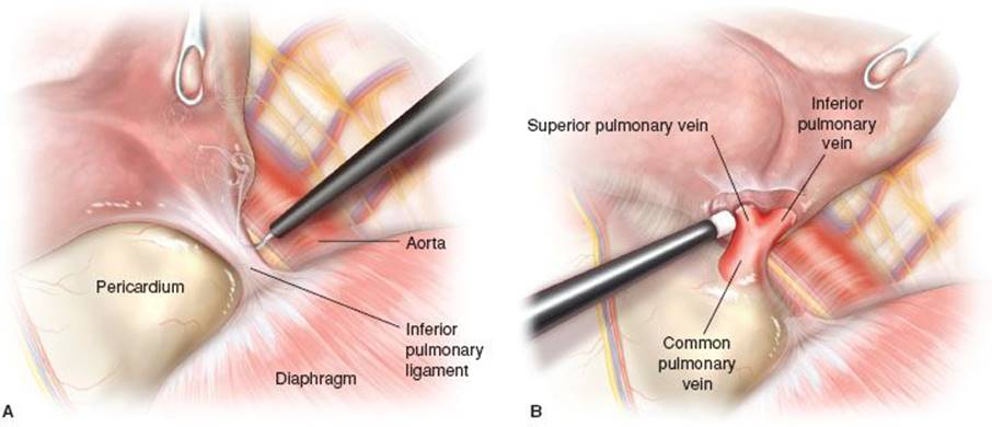
Figure 18.2 A: Division of the Inferior Pulmonary Ligament. Providing lateral traction on the lung, we take down the inferior pulmonary ligament using blunt dissection and electrocautery. B: Isolation of the Left Lower Lobe Venous Drainage. Here the ligament is taken down along with the anterior and posterior mediastinal pleura to reveal the pulmonary vein. Blunt dissection should be performed to ensure that only the inferior pulmonary vein, and not a common left pulmonary vein, is encircled. This anatomic variant occurs in approximately 14% of the population. Division of a common pulmonary vein will often result in the need for pneumonectomy.
We then prepare to isolate and encircle the inferior pulmonary vein. The inferior aspect of the vein should be well delineated by division of the pulmonary ligament. The lung is retracted posteriorly and with the use of endoscopic Kittner, the superior extent of the pulmonary vein should be identified at its junction with the pericardium. At this point, the upper lobe venous drainage should be identified. The venous drainage of the upper and lower lobes may lie in close proximity to each other. In fact, they may empty into a common venous trunk before entering the pericardium and left atrium (Fig. 18.2B). Dividing a common pulmonary vein would lead to an unintentional conversion to pneumonectomy. It should be noted that while the posterior mediastinal pleura is opened in the previous step, the lower lobe superior segmental vein is separated from the connective tissue between it and the membranous wall of the lower lobe bronchus. By opening this space, risk to the lower lobe bronchus is minimized when the vein is encircled. Once the upper lobe venous drainage is identified, the lower lobe vein is encircled with a right-angle clamp. Vascular division is not performed at this time.
The next step in performing a thoracoscopic left lower lobectomy involves subcarinal node dissection. Left-sided nodal dissection in this area is technically more difficult than when done during right-sided surgery. It is for this reason that we perform subcarinal nodal dissection with the left lobe in situ as retracting the lobe opens up the subcarinal space better than after the lobe has been removed. The lower lobe is grasped with a ring forceps through the anterior port and dissection is performed with instruments brought through the posterior port. With the lung retracted anteriorly and a Kittner used to gently displace the esophagus posteriorly; the subcarinal space can be safely dissected. Ultrasonic shears are used for coagulating and dividing bronchial vessels in this region.
Attention is placed toward completing the major fissure between the upper and lower lobes to gain access to the pulmonary arterial supply to the lower lobe (Fig. 18.3). In patients with near-complete fissures, dissection begins by carefully grasping and then sharply dividing the tissue on top of the interlobar pulmonary artery. This will give access to the perivascular plane around the artery known as the plane of Leriche. With less developed fissures, the pulmonary artery may not be clearly seen. In these cases, blunt dissection in the fissure is accomplished with caudal tension placed on the lower lobe. Often only minimal blunt work is required to identify the interlobar pulmonary artery. In cases with practically no major fissure, we recommend isolating the pulmonary artery in the posterior hilum (Fig. 18.4A). Anteromedial traction is placed on the upper and lower lobes and the mediastinal pleura has been opened. The pulmonary artery should be easily identified in this region and followed distally into the fissure.
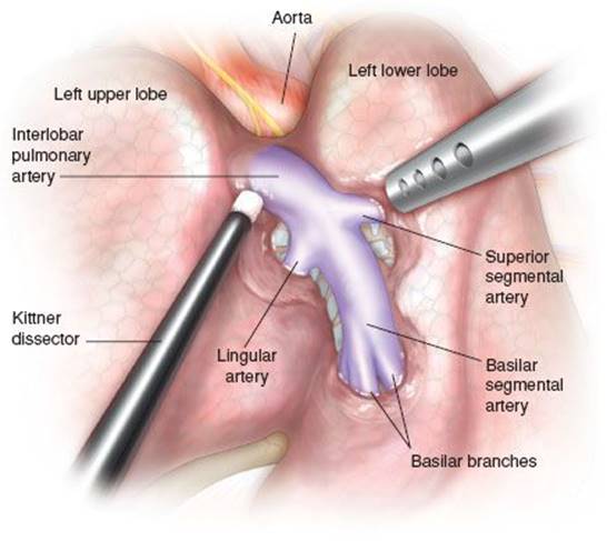
Figure 18.3 Dissection in the Major Fissure. Using an endoscopic Kittner dissector and suction catheter we bluntly dissect out the interlobar pulmonary artery and its branches. The superior segmental and basilar trunk pulmonary arterial branches should be identified. To prevent inadvertent division, the branches to the lingular segments of the upper lobe should be recognized as well.
Lung parenchyma can then be divided working from a posterior to anterior direction. As with all dissection involving the pulmonary artery, it is best to enter the plane of Leriche and to continue further dissection in this plane. With these very difficult fissures, a tunnel is developed posteriorly with the artery below and lung parenchyma above. The fissure is then completed with serial firings of the endoscopic stapler brought through the posterior incision. While working in the posterior hilum, the posterior aspect of the superior segmental artery is identified and the tissue plane between it and the lower lobe bronchus is developed. There is often a proximal posterolateral branch, which can be inadvertently injured while attempting to divide these vessels (Fig. 18.4B). We, therefore, recommend identifying this branch in the posterior hilum, so that risk of trauma to this posterior vessel is minimized during further dissection and subsequent vascular division.
Completing the fissure before division of the pulmonary arterial branches to the lower lobe should effectively prevent unintentional sacrifice of branches to the upper lobe. The lingular arterial branch arise from the distal aspect of the interlobar artery and on occasion, can arise from the basilar trunk to the lower lobe. If the major fissure is not completed, inadvertent division of the lingular blood supply can occur (Fig. 18.5A). With the fissure divided, the pulmonary arterial supply to the lower lobe and a portion of the upper lobe is clearly visualized (Fig. 18.5B). The basilar trunk and superior segmental branches are then encircled with the use of a right-angled clamp and long tonsil clamp respectively. Any resistance encountered while passing instruments around these thin-walled vessels should be carefully investigated. As described earlier, systematic dissection in the posterior hilum to free the origin of the superior segmental pulmonary arterial branch from the lower lobe bronchus should allow safe isolation of this vessel.
Attention is now placed toward division of the pulmonary arterial blood supply. It is often more facile to begin with addressing the superior segmental artery first. An attempt to pass a stapler across the basilar trunk with the superior segmental branch in place is often challenging. With the lung retracted posteriorly with a ring forceps brought through the posterior port, the camera is then moved to the access incision. An endoscopic Kittner is then passed through the camera port and then across the superior segmental artery. The use of the Kittner in this step is beneficial to those surgeons just embarking on thoracoscopic lobectomies as it provides a less obtrusive instrument to confirm the trajectory of the stapling device. The endoscopic stapler with a 30-mm vascular staple load is then brought through the camera port to divide the superior segmental artery. Care is taken not to provide undue tension on this vessel during transection. As with thoracoscopic division of all named vessels, a sponge stick or dental pledget on a clamp should be readily available to tamponade bleeding from stapler malfunction or avulsion of vascular branches.
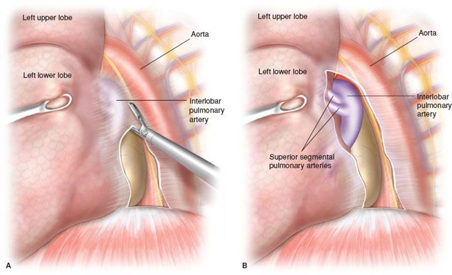
Figure 18.4 A: Posterior Fissure Dissection. Anteromedial traction is placed on the upper and lower lobes and the mediastinal pleura has been opened. The pulmonary artery should be easily identified in this region and followed distally into the fissure. Lung parenchyma can then be divided working from a posterior to anterior direction. B: Posterior Hilar View. We have taken down the pleura and lung to reveal a posterior view of the two branches of the superior segmental PA, enough length should be dissected in order for the endostapler to slide easily and prevent traction injury to the superior segmental pulmonary artery branches.
With the superior segmental artery divided, the thoracoscope is brought back through the camera port and the basilar trunk is transected with an endostapler passed through the anterior port. An alternative approach to dividing the pulmonary arterial supply to the lower lobe is to divide all branches with one firing of the vascular stapler; a 45-mm cartridge is often necessary. Care must be made to ensure effective mobilization of the pulmonary arterial supply, so that the stapler, which is brought through the access incision, passes across without difficulty (Fig. 18.5C). A silk tie can be passed around the lower lobe arterial branches, with gentle traction placed through the posterior port, to facilitate placement of the stapling device. A recent advance in stapling technology is the inclusion of a flexible introducer, which, when attached to the vascular staple load, assists passage of the cartridge across challenging vessels.
The pulmonary venous drainage of the lower lobe is then divided. The inferior pulmonary ligament has already been divided and any level 9 lymph nodes were removed. The venous drainage has already been encircled with attention placed to identifying and preserving the venous drainage of the upper lobe. Depending on the size of the pulmonary vein, either a 30- or 45-mm vascular load is selected. The lung is retracted toward the lateral chest wall with a ring forceps through the posterior port. The stapler is then brought through the access incision and passed across the inferior pulmonary vein. Slight articulation of the cartridge away from the mediastinum may be required to avoid injury to the descending thoracic aorta. We often will use an endoscopic Kittner, brought through the camera port along the thoracoscope, to keep the entirety of the pulmonary vein in the staple cartridge.
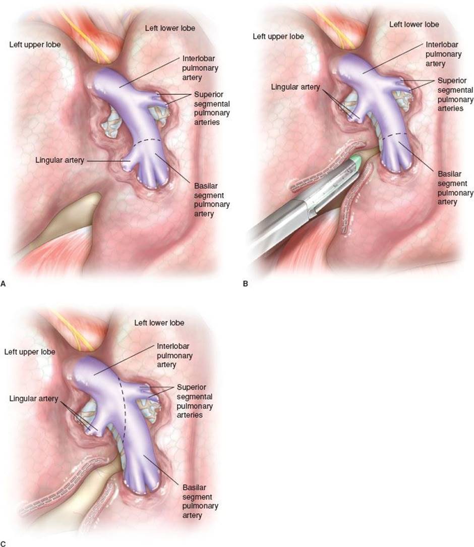
Figure 18.5 A: Inadvertent Lingular Artery Transection. The lingular artery may have an origin off of the very distal interlobar artery or can possibly arise from the basilar trunk itself. If the major fissure is not completely divided before initiating detachment of the arteries supplying the lower lobe, inadvertent lingular artery division is possible, as depicted here. B: Safe View of Lower Lobe Pulmonary Arterial Branches. The fissure has been completed. This enables safe division of the pulmonary arterial branches to the lower lobe while preserving upper lobe blood supply. C: Division of Lower Lobe Pulmonary Artery. In some cases it may be straightforward to divide the lower lobe arterial branches with one firing of the 45-mm endostapler brought through the anterior incision.
The last remaining structure to divide is the lower lobe bronchus. All lymph node tissue that has not already been removed during hilar dissection is swept up into the pulmonary parenchyma. Care is taken not to devascularize, or cause thermal injury, to the future bronchial stump. The lobe is then retracted inferiorly and laterally to fully expose the lower lobe bronchus. An endoscopic stapler with a thick tissue cartridge is then brought through the anterior port and placed across the bronchus. The stapler is then closed, but not fired, and gentle hand ventilation is given to the left lung either through the bronchial lumen of the double-lumen tube or through the single-lumen tube with the bronchial blocker deflated. Rapid inflation of the left upper lobe should be confirmed prior to lower lobe bronchial transection. The lobectomy specimen is then placed in a heavy extraction sac and brought out through the anterior port.
With the specimen removed, all that remains is completion of mediastinal lymphadenectomy. Level 9 and subcarinal lymph nodes have been removed earlier in the operation. Remaining nodal tissue in the aortopulmonary window and para-aortic region are then carefully dissected from surrounding tissue and removed through the anterior port. Attention should be placed toward identifying and preserving the left phrenic and recurrent laryngeal nerves while dissection is performed in these regions.
All areas of dissection and resection are inspected for hemostasis and the chest is then thoroughly irrigated with warm saline solution. The bronchial stump is then tested for pneumostasis under saline immersion with an airway pressure of 30 to 35 mm Hg. A 24-French chest tube is then brought through the camera port incision and positioned in the posterior hemithorax for postoperative drainage. The remaining upper lobe is again inflated under visualization to ensure proper positioning. The thoracoscope is removed and the remaining ports are closed in layers with absorbable sutures. After the patient is returned to a supine position, flexible bronchoscopy is performed to clear any remaining blood or secretions.
 POSTOPERATIVE MANAGEMENT
Postoperative management does not have any unique challenges for patients undergoing thoracoscopic left lower lobectomy compared to those after other anatomic lobectomies. In general, the patients should be extubated in the operating room and kept euvolemic postoperatively, trending toward hypovolemia. They are out of bed the same day of surgery, and ideally begin ambulating the following morning. The chest tube can be placed on water seal immediately or for patients with a sizable air leak, left on suction for 24 to 48 hours. It remains until air leaks resolve and drainage is less than approximately 350 mL over a 24-hour period. After the chest tubes are removed, the patient is discharged home on oral pain medications as needed.
 COMPLICATIONS
There is a lack of data comparing the complications of thoracoscopic left lower lobectomy versus other thoracoscopic lobectomies. Most reports document complications associated with VATS lobectomies as a whole and these compare quite favorably to lobectomy via thoracotomy. The most common perioperative complications include prolonged postoperative air leaks, atrial fibrillation, and pneumonia. Overall complications from multiple series range from 10% to 25%; although the American College of Surgeons ACOSOG Z0030 trial reported all minor and major complications up to 40%.
Given the technical approach of this textbook it is our intention to describe the potential intraoperative complications encountered during thoracoscopic left lower lobectomy. The most feared complication of VATS lobectomy is an uncontrolled vascular injury. Scant data is available regarding division of intrathoracic vessels during thoracoscopic lobectomy. Endoscopic staplers have a reported stapling failure rate of 0.1%. A sponge stick or a dental pledget on a clamp should always be readily available to tamponade bleeding from stapler malfunction or avulsion of vascular branches. In thoracoscopic resections, this single maneuver allows time for adequate control and conversion to an open thoracotomy, if needed. Minor vascular avulsion injuries during VATS resections can be adequately repaired utilizing direct suture repair of the vessel through the access incision. More significant injuries, difficulties in exposure, and cases in which patients exhibit hemodynamic compromise from vascular injuries should be converted to a thoracotomy for effective vascular control.
Having a thorough understanding of the vascular anatomy is crucial in preventing vascular injury during pulmonary lobectomy. The relatively thin walls of pulmonary arterial branches make these structures more prone to injury than their venous counterparts. We advocate that pulmonary arterial vessels are never directly grasped with any instrument. Extra care must be directed toward the isolation and division of these vessels. During VATS resections, optimization of exposure with an angled thoracoscope along with correct port placement while adhering to standard thoracic surgical principles minimizes the risk of vascular complications.
Other possible intraoperative complications during thoracoscopic left lower lobectomy include injury to the intercostal, phrenic, and recurrent laryngeal nerves; the esophagus; and the thoracic duct or one of its tributaries. Injury to the intercostal bundles can result in the “postthoracotomy” pain syndrome. It is important to avoid the intercostal nerve bundles when creating ports. Left phrenic nerve injury can occur during dissection near the anterior hilum while incising the mediastinal pleura. Inadvertent injury of the phrenic nerve leads to paralysis of the ipsilateral hemidiaphragm. Although this complication has not been adequately described in the literature for patients undergoing pulmonary lobectomy, the deleterious effects on the respiratory status of patients with chronic lung disease in the setting of lobectomy could be quite significant. The recurrent laryngeal nerve can be traumatized during mediastinal nodal dissection in the aortopulmonary and para-aortic nodal basins. Subsequent laryngeal dysfunction with aspiration could be potentially hazardous in patients undergoing lobectomy.
The esophagus can be injured while dividing the inferior pulmonary ligament or during mediastinal nodal dissection. If recognized early, it can be repaired with layered suture repair augmented with a vascularized tissue buttress. Delayed recognition with empyema and sepsis will possibly require esophageal stenting or diversion along with control of infection and hyperalimentation. The salient method of preventing this injury is through careful division of the pulmonary ligament to be observant of the underlying esophagus during nodal dissection particularly in the subcarinal space. Again, adequate exposure should prevent esophageal injury.
Thoracic duct or lymphatic tributary injury resulting in a chylothorax is rare complication for left-sided resections. Knowledge of the anatomic course of the thoracic duct may assist the surgeon in avoiding this potential complication. However, this may not be preventable due to the frequent existence of large collateral channels amongst mediastinal lymph nodes. Intraoperative realization and ligation of a significant lymphatic injury may prevent a chylothorax from occurring. Careful lymphatic ligation with electrocautery or ultrasonic shears should minimize postoperative lymphatic leaks. Postoperative chylothorax is often selflimited and resolves with dietary modifications, but reoperation with thoracic duct ligation is occasionally required for high-output leaks.
 RESULTS
To date, there have been no large, prospective randomized studies comparing thoracoscopic lobectomy with lobectomy performed via the traditional open approach. There are, however, results of several large series of available VATS lobectomies, which compare favorably with those seen by lobectomy performed via thoracotomy. The Cancer and Leukemia Group B (CALGB) reported data from a prospective, multi-institutional registry of 127 patients who underwent VATS lobectomy. In this series, the mortality was 2.7% with a median length of stay of 3 days. There have been multiple other large and prospective studies evaluating the safety of thoracoscopic lobectomy. These have either similar or lower mortality rates than the CALGB study. Conversion to thoracotomy ranges from 0% to approximately 15% and appear to decrease over time and experience.
Oncologic results should be comparable to those of other lobectomies and by thoracotomy, in theory. There are no large, randomized, prospective trials directly comparing oncologic results between thoracoscopic and thoracotomy lobectomies. There is reasonable data to conclude that the operations are at least equivocal in terms of achieving complete resection and amount of lymph nodes harvested. Long-term, actual, survival rates for patients with early staged cancers undergoing resection by VATS lobectomy are beginning to emerge. These suggest 5-year survival rates that are, at least, as good, if not better, than as those achieved via thoracotomy. Proposed explanations for this potential survival improvement in VATS lobectomy patients include: Improved preservation of the immune system, less systemic release of inflammatory mediators, and higher proportion of patients tolerating intended adjuvant chemotherapy. With particular reference to left lower lobe NSCLC, a recent retrospective study (Kudo et al., 2012) suggests that patients with left lower lobe cancers and hilar nodal disease may have worse prognosis than similar size tumors in other lobes.
 CONCLUSIONS
The VATS left lower lobectomy while similar to right lower lobectomy has some unique challenges. It is important to have proper port placement, be aware of a common pulmonary vein trunk, and be cognizant of a distal origin of the lingular artery to the upper lobe. It is imperative that lymph nodes are sampled or dissected for accurate staging as it impacts further treatment and prognosis.
VATS lobectomy is intended to accomplish the same oncologic principles as traditional lobectomy: Complete hilar dissection with individual ligation of vessels and bronchus. Thoracoscopic lobectomy has been demonstrated to be comparable with regard to safety and oncologic results, as measured by operative major morbidity and mortality, completeness of resection, extent of nodal dissection, and short-term survival, when compared to results seen with lobectomy via thoracotomy.
Recommended References and Readings
Allen M, Darling G, Pechet T, et al. Morbidity and mortality of major pulmonary resections in patients with early-stage lung cancer: Initial results of the randomized, prospective ACOSOG Z0030 trial. Ann Thorac Surg.2006;81:1013–1020.
Demmy T, James T, Swanson S, et al. Troubleshooting video-assisted thoracic surgery lobectomy. Ann Thorac Surg. 2005;79:1744–1752.
Grogan E, Jones D. VATS lobectomy is better than open thoracotomy: What is the evidence for short-term outcomes? Thorac Surg Clin. 2008;18:249–258.
Kudo Y, Saji H, Shimada Y, et al. Do tumours located in the left lower lobe have worse outcomes in lymph node-positive non-small cell lung cancer than tumours in other lobes? Eur J Cardiothorac Surg.2012;42:414–419.
Lee P, Nasar A, Port J, et al. Long-term survival after lobectomy for non-small cell lung cancer by video-assisted thoracic surgery versus thoracotomy. Ann Thorac Surg. 2013;96:951–961.
Marom E, Herndon J, Kim Y, et al. Variations in pulmonary venous drainage to the left atrium: Implications for radiofrequency ablation. Radiology. 2004;230:824–829.
Park B, Flores R. Cost comparison of robotic, video-assisted thoracic surgery and thoracotomy approaches to pulmonary lobectomy. Thorac Surg Clin. 2008;18:297–300.
Paul S, Altorki NK, Sheng S, et al. Thoracoscopic lobectomy is associated with lower morbidity than open lobectomy: A propensity-matched analysis from the STS database. J Thorac Cardiovasc Surg.2010;139:366–378.
Swanson S, Herndon J, D’Amico T, et al. Video-assisted thoracic surgery lobectomy: Report of CALGB 39802–a prospective, multi-institution feasibility study. Ann Thorac Surg. 2007;25:4993–4997.
Wright CD, Gaissert HA, Grab JD, et al. Predictors of prolonged length of stay after lobectomy for lung cancer: A Society of Thoracic Surgeons General Thoracic Surgery Database risk-adjustment model. Ann Thorac Surg.2008;85:1857–1865.