
Cameron D. Wright
 INDICATIONS/CONTRAINDICATIONS
Patients should be thoroughly staged and have no N2 disease when considered for aggressive induction therapy followed by resection for a superior sulcus lung cancer. Generally patients will undergo induction chemoradiotherapy and have restaging CT scans. Resection is performed as long as there is no progressive disease. Relief of pain during induction therapy is a favorable sign for a good therapy response.
Indications
Potentially resectable posterior-based superior sulcus tumor (Figs. 30.1 and 30.2)
Adequate cardiopulmonary function and performance status
Contraindications
Extensive involvement of the brachial plexus
N2 disease
Extensive involvement of the vertebral body (with the caveat that a very few centers of excellence offer combined superior sulcus resections in conjunction with total vertebrectomy with reasonable results)
 PREOPERATIVE PLANNING
Patients with known involvement of the subclavian vessels should have an anterior approach to their tumors to enhance the ability to perform a complete resection. Adequate recovery from induction therapy, both in terms of functional status and hematologic values should be ensured prior to surgery. Generally waiting at least 4 to 6 weeks is prudent before proceeding with surgery following induction therapy. An epidural catheter is usually placed in the induction area for postoperative pain relief.
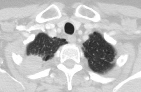
Figure 30.1 Axial CT scan of a patient with a small right superior sulcus lung cancer involving ribs 1, 2, and 3. Induction chemoradiotherapy was administered followed by a posterior approach for resection.
 SURGERY
Positioning
The patient is placed in the lateral decubitus position and carefully padded. Special attention to the arm on the involved side is made so that it is out of the way of the surgeon but not so far abducted or superiorly placed that a nerve stretch injury will occur. Sequential compression devices are placed on the calves to reduce the possibility of deep venous thrombosis. The skin preparation should include the posterior midline and the nape of the neck as the parascapular incision is carried very high onto the back.
Technique
The incision is generally almost entirely a posterior parascapular incision midway between the spine and scapula border (Fig. 30.3). The muscles that are usually transected include the latissimus dorsi, the trapezius, the major and minor rhomboids, the levator scapulae, and the serratus posterior superior (Fig. 30.4). The chest is typically entered in the fourth interspace and the limits of the invasion of the ribs ascertained. Generally the first two or three ribs are involved and require resection. The outer chest wall can be marked with a cautery mark or other marker to guide the surgeon as to where to end the anterior aspect of the chest wall resection. Several centimeters of free rib margin should be planned for. Alternatively a video camera can be inserted in the chest in the beginning and using a needle, the limits of the chest wall resection defined on the outer surface of the ribs. It is helpful to place a chest spreader under the scapula to provide good exposure to the chest wall during this phase of the operation (or use a table-mounted hook retractor).
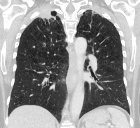
Figure 30.2 Coronal CT of the same patient demonstrating a small superior sulcus lung cancer.

Figure 30.3 Long posterior parascapular incision, which divides the posterior aspect of the latissimus dorsi and most of the trapezius muscles. The scapula is also marked as well as the spinous processes of the midline.
The chest wall resection usually begins anteriorly from the lowest involved rib taking them sequentially until the first rib is transected (Fig. 30.5). Neurovascular bundles are ligated and divided, the intercostal muscle is divided with cautery, and the rib is transected with a bone shear. The first rib bears special attention due to the brachial plexus and subclavian vessels just above it. The anterior, middle, and posterior scalene muscles must be divided from their attachments to the first and second ribs. The superior surface of the first rib must be cleared taking care to preserve the neurovascular structures (Fig. 30.6). I generally use a large Cobb periosteal elevator for this as the tip is rounded, relatively atraumatic, and the large handle allows measured force to be applied. Once the top side is free of all important structures it can be cut either with a rib shear, a Gigli saw, or by a rongeur.

Figure 30.4 The incision with the scapula retracted showing the apical chest wall. A table-mounted retractor or a chest spreader can be used for exposure. The posterior superior serratus muscle must be mobilized off the posterosuperior chest wall.

Figure 30.5 The chest wall resection is begun anteriorly by cutting the most inferior rib that requires removal. The ribs and neurovascular bundles are then taken sequentially moving superiorly until the first rib is reached.
Attention is then turned to the posterior chest wall. The paraspinal ligaments and muscles are elevated off the posterior aspect of the ribs with a combination of cautery and periosteal elevators. Again the rib resection proceeds from below to the first rib. In most cases the rib just needs to be disarticulated from the transverse process and the vertebral body. I use a Cobb elevator generally to do this but an osteotome is also a useful instrument (Fig. 30.7). Once the rib is disarticulated and loose the posterior space opens up and the neurovascular bundle can be seen. The bundle is clamped, divided, and ligated as it comes out of the foramen. Once the first rib is disarticulated the specimen is only attached by mediastinal pleura usually and by a combination of working both outside the chest wall and inside the chest these remaining attachments can be divided. This drops the upper lobe into the chest and allows the hilar dissection for the lobectomy to proceed. The upper lobectomy is done in the routine fashion and then a mediastinal lymph node dissection is done. Generally if only the first two or three ribs are removed no chest wall reconstruction needs to be done as this area is well covered by the scapula and thick chest wall muscles. If the fourth rib is removed sometimes this does have to be reconstructed to prevent scapula entrapment, which is a problem that is best avoided. If there are any apical areas of concern for close or positive margins they are marked with metal clips to aid possible postoperative radiation. The chest is then closed in the routine way with a chest drain in place.
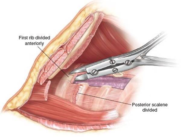
Figure 30.6 The scalene muscles must first be divided to fully expose the first rib. The superior surface of the first rib must be carefully cleared of the important structures—the subclavian vein, artery, and brachial plexus prior to cutting the rib. The first rib is very strong and hard to cut. It can be divided by rib shears, a Gigli saw, or by nibbling through it with a rongeur.

Figure 30.7 A: The posterior aspect of the ribs is then dealt with. The paraspinal muscles are mobilized off the posterior chest wall to expose the transverse processes of the vertebral bodies. A Cobb periosteal elevator or an osteotome is then used to separate the rib from the ligamentous attachments of the rib to the transverse process and vertebral body. A firm twisting motion usually achieves the desired result. B: The transverse process can be removed if needed to achieve a better margin if there is abutment or invasion of the posterior rib in this area. This is done with an osteotome.
 POSTOPERATIVE MANAGEMENT
Routine pulmonary resection postoperative care is given with emphasis on good pain control, early mobilization, and full lung expansion. The epidural is usually left in for 3 to 4 days. The chest tube is removed when drainage is less than 150 cc per day and there is no air leak. Patients are reassured that some numbness of the anterior chest wall from the chest wall resection is expected (Fig. 30.8).
 COMPLICATIONS
Patients can experience all the usual complications of pulmonary resection including bleeding, prolonged air leak, pneumonia, atrial fibrillation, deep venous thrombosis, or pulmonary embolism etc. Specific to Pancoast resections are Horner syndrome, cerebral spinal fluid leak, and brachial plexus injury. Generally Horner syndrome is permanent after Pancoast resections and reflects injury or removal of the bottom of the stellate ganglion. A cerebrospinal fluid (CSF) leak is a significant complication and almost always requires reoperation and the help of a neurosurgeon to close the leak. The dural sheaths extend to a variable extent onto the intercostal nerves and it is possible to injure the dura either at the canal level or further later along the proximal intercostal nerve. Ligation of the nerve and sheath or patching the tear usually controls the problem. Unipolar cautery should not be used around the brachial plexus-only bipolar cautery. If the T1 nerve trunk must be cut it should be cut with a knife and not the cautery. Occasionally bleeding occurs from the neural foramen during retraction or dividing posterior chest wall elements. Unipolar cautery should not be used for fear of injury to the spinal cord. Packing the foramen with oxidized cellulose should also not be performed as the swelling of the material or ongoing bleeding could compress the spinal cord. If there is significant bleeding it is best to consult with a spine surgeon to have the foramen enlarged so proper control of the vessel can be achieved. Traction injuries of the intercostal nerves are to be avoided as again injury to the spinal cord could ensue. Stretch injuries to the brachial plexus should be avoided by careful arm positioning and attention to the arm position throughout the case.

Figure 30.8 Postoperative chest radiograph of the patient from Figure 30.1 who had the first three ribs removed. Notice the complete absence of the posterior ribs adjacent to the transverse processes of the first three vertebral bodies.
 RESULTS
Rusch et al. reported the results of the Intergroup 0160 trial of induction chemoradiation followed by resection for Pancoast tumors. Induction therapy was completed by 104 patients. Of 95 patients eligible for surgery, 88 underwent thoracotomy and 83 (76%) had a complete resection. There were three deaths with induction therapy and two deaths after operation. A complete pathologic response was seen in 32 patients. The 5-year survival was 44% in all patients and 54% in patients who had a complete resection. A pathologic complete resection led to a better survival (risk ratio 2.1, p = 0.02).
 CONCLUSIONS
Modern treatment of superior sulcus tumors involves induction chemoradiotherapy followed by complete resection including the initially involved chest wall. Posteriorly based tumors are best approached by the classic Paulson posterior parascapular approach, which provides excellent exposure to the posterior chest wall including the transverse processes of the vertebrae. A good response to induction therapy and a complete resection are the most favorable prognostic factors.
Recommended References and Readings
Detterbeck FC. Changes in the treatment of Pancoast tumors. Ann Thorac Surg. 2003;75:1990–1997.
Kappers I, van Sandick JW, Burgers JA, et al. Results of combined modality treatment in patients with non-small cell lung cancer of the superior sulcus and the rationale for surgical resection. Eur J Cardiothorac Surg. 2009;36:741–746.
Kwong KF, Edelman MJ, Suntharalingam M, et al. High dose radiotherapy in trimodality treatment of Pancoast tumors results in high pathologic complete response rates and excellent long-term survival. J Thorac Cardiovasc Surg.2005;129:1250–1257.
Rusch VW, Giroux DJ, Kraut MJ, et al. Induction chemoradiation and surgical resection for superior sulcus non-small-cell carcinomas: Long-term results of Southwest Oncology Group Trial 9416. J Clin Oncol. 2007;25:313–318.
Tamura M, Hoda MA, Kleptko W. Current treatment paradigms of superior sulcus tumors. Eur J Cardiothorac Surg. 2009;36:747–753.