
Valerie W. Rusch
 INDICATIONS
The most common indication for extrapleural pneumonectomy (EPP) is the resection of malignant pleural mesothelioma (MPM). Although there has been a recent shift away from EPP in favor of pleurectomy/decortication (P/D), EPP may still be the optimal operation for a select group of MPM patients with early stage disease. EPP can also be a useful approach for the resection of other malignancies such as large intrathoracic sarcomas, or recurrent fibrous tumors of the pleura. In addition, EPP has occasionally been used to resect nonsmall cell lung carcinomas (NSCLC) metastatic to the pleura without associated extrathoracic disease.
 PREOPERATIVE PLANNING
Preoperative evaluation should determine whether the patient has potentially resectable tumor and assess cardiopulmonary reserve and overall surgical risk.
Computed tomography (CT) scan of the chest and upper abdomen is the primary means of assessing the extent of the primary tumor and identifying metastatic disease in the peritoneum or the contralateral lung and pleura. In MPM, some institutions also use magnetic resonance imaging (MRI) to determine whether the primary tumor invades the chest wall or diaphragm but in general MRI does not add significantly to CT in preoperative staging. MRI may also be useful in other diseases where there is a need to assess involvement of cardiac or neurologic (e.g., spine, spinal foramina, brachial plexus) structures.
Positron emission tomography (PET) adds to CT in staging MPM. It identifies metastatic disease not detected by CT in approximately 10% of patients. The standardized uptake value (SUV) on PET is also an independent prognostic factor for overall survival and is useful in selecting patients for treatment. We use PET-CT routinely in the initial evaluation of patients with MPM. It is also important in the extent of disease evaluation of other malignancies being considered for EPP.
Mediastinoscopy has been advocated as a routine staging procedure before EPP because the presence of mediastinal nodal metastases (N2 disease) is an adverse prognostic factor in MPM, and CT and PET are known to be inaccurate in detecting nodal disease. However, mediastinoscopy fails to identify N2 disease in some patients because the pattern of nodal metastases from MPM differs from that of lung cancer. In addition, N2 disease is only one of several important prognostic factors in MPM and does not uniformly identify patients who have a poor prognosis. At the current time, we do not routinely perform mediastinoscopy as part of the initial staging evaluation for MPM.
Endobronchial ultrasound (EBUS)-guided transbronchial needle aspiration (TBNA) has largely replaced mediastinoscopy for the detection of hilar and mediastinal nodal metastases. It may be useful in the preresection evaluation of some malignancies such as NSCLC, which would not be considered for EPP if nodal disease was present.
Laparoscopy has also been advocated as a staging maneuver before resection of MPM because it identifies transdiaphragmatic tumor extension or intra-abdominal metastases. However, routine laparoscopy is not required in patients whose imaging studies show earlier stage tumors and no intra-abdominal abnormalities.
The assessment of cardiopulmonary reserve is a pivotal part of the preoperative evaluation for EPP. Complete pulmonary function testing (PFT) should be performed, including a diffusion capacity (DLCO), especially in MPM patients who have had asbestos exposure and often, underlying interstitial lung disease. A quantitative ventilation/perfusion (V/Q) lung scan should also be done to help calculate the patient’s postoperative pulmonary function following an EPP.
Most patients being considered for EPP are older and have medical comorbidities, especially underlying cardiovascular disease. An EPP places patients at high risk for myocardial ischemia because of intraoperative blood loss and postoperative fluid shifts. Therefore, some form of stress testing should be considered preoperatively.
In summary, routine preoperative evaluation of patients being considered for EPP includes a CT scan of the chest and upper abdomen, a PET-CT scan, complete PFTs, a quantitative V/Q scan, and usually, some form of stress testing. Additional evaluation may include MRI, laparoscopy, mediastinoscopy, or EBUS is performed selectively.
 SURGERY
Preparation, Positioning, and Incision
An epidural catheter is placed for postoperative analgesia. After induction of general anesthesia, a double-lumen endotracheal tube is inserted. In addition to standard intraoperative monitoring (arterial line, pulse oximetry), a central venous catheter is inserted to help manage perioperative fluid shifts.
The patient is placed in the standard lateral decubitus position. An extended S-shaped posterolateral thoracotomy incision is made (Fig. 32.1). Extension of the incision down toward the costal margin provides exposure for diaphragmatic resection and reconstruction. Some surgeons add a second small posterior thoracotomy incision at the level of the eleventh rib to provide exposure to the costophrenic sulcus but this causes chest wall edema and pain and does not significantly improve exposure. If postoperative hemithoracic radiation is planned there is no need to excise previous chest wall incisions because the radiation treats potential tumor implants. Both the latissimus dorsi and the serratus anterior muscles are divided.
Some authors recommend using a median sternotomy rather than a thoracotomy, especially for right-sided resection. However, this approach does not provide as much exposure for resection and reconstruction of the posterior aspect of the diaphragm.
Resection Technique
The sixth rib is excised to expose the extrapleural plane. This approach is lower than for a standard pulmonary resection to facilitate exposure of the diaphragm. The intercostal muscles are carefully preserved for reclosure at the end of the operation. Blunt dissection is performed in the extrapleural plane freeing the parietal pleura from the endothoracic fascia using a sweeping motion of the hand up to apex of the chest, then down to the diaphragm, anteriorly to the pericardium, and posteriorly to the spine (Fig. 32.2). Hemostasis is obtained as the dissection is performed to prevent substantial blood loss. The most effective hemostatic tool for chest wall bleeding is the TissueLink (TissueLink Medical, Inc., Dover, NH). Once the parietal pleura is mobilized away from the chest wall, a chest retractor is inserted, and dissection continued under direct vision mobilizing the pleura circumferentially away from the mediastinum. On the left side, care is taken to identify the plane between the tumor and the adventitia of the aorta and esophagus. On the right side, dissection along the superior vena cava must be performed very gently. In some patients, there is a clean plane of dissection between the mediastinal pleura and the pericardium. In others, this plane is obliterated and the anterior mediastinal pleura has to be resected en-bloc with the pericardium later in the operation. After the pleura and lung are completely mobilized in the upper half of the chest exposing the superior and posterior aspects of the hilum, an en-bloc dissection of the subcarinal lymph nodes is performed for staging purposes and to expose the mainstem bronchus.
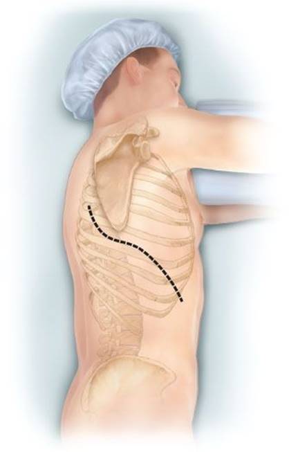
Figure 32.1 Example of the S-shaped thoracotomy incision used for EPP.
The diaphragmatic tumor is then resected. There is always a palpable “edge” posteriorly and laterally between the tumor and the normal diaphragmatic muscle or peritoneum at the level of the costophrenic sulcus. This plane is developed and the tumor mobilized along its diaphragmatic surface by blunt dissection similar to a Kocher maneuver. Once mobilized from the posterior costophrenic angle, the tumor is rotated up into the thoracotomy incision, rolling it back upon itself, and placing strong traction on the diaphragm. If the involvement of the diaphragm is extensive, it is removed entirely, peeling it away from the peritoneum. If the involvement of the diaphragm is superficial, dissection can be carried through the diaphragmatic muscle using the electrocautery. In MPM patients, every effort is made not to enter the peritoneum because of the propensity of MPM to produce tumor implants. However, entry into the abdomen at the level at the central tendon is unavoidable because of the fusion of tissue planes in that area. The peritoneum is immediately reclosed as the central tendon is removed. The diaphragm is mobilized back to the pericardium medially (Fig. 32.3). If resection of the pericardium is required, it is entered only when the tumor has been mobilized as fully as possible from all other directions because traction on the pericardium causes arrhythmias and hemodynamic instability. The hilar structures are divided in whatever sequence is technically easiest and requires the least manipulation of the large tumor mass, usually the mainstem bronchus first, then the inferior pulmonary vein, the superior pulmonary vein, and lastly the main pulmonary artery. If the pericardium is resected, it is gradually opened as this portion of the dissection is carried out. Traction sutures are placed on the pericardium to prevent it from retracting toward the opposite hemithorax and to minimize changes in the position of the heart, thereby reducing hemodynamic instability. The specimen consisting of pleura, lung, and diaphragm, with or without pericardium, is removed en-bloc. Dissection of the paratracheal lymph nodes if the operation is on the right, or of the aortopulmonary window nodes if the operation is on the left, is performed for staging purposes.

Figure 32.2 The parietal pleura is bluntly dissected away from the endothoracic fascia after opening the extrapleural plane. This is accomplished using a sweeping motion of the fingers and palm before inserting a chest retractor.

Figure 32.3 Intraoperative view of the peritoneum and remaining strands of diaphragmatic muscle after resection of the hemidiaphragm during EPP. The pericardium is visible in the lower right-hand corner of the figure and in this case did not require resection.

Figure 32.4 The completed pericardial and diaphragmatic reconstruction. If the diaphragm was detached from its costal insertion, the prosthetic material is secured laterally by sutures placed around the ribs and medially by sutures to the pericardium and edge of the contralateral hemidiaphragm.
Reconstruction of the Diaphragm and Pericardium
Reconstruction of the diaphragm is performed using 2-mm thick Gore-Tex (W.L. Gore & Associates, Flagstaff, AZ). Laterally, the prosthetic patch is secured with no. 2 Vicryl sutures around the ribs. Posteriorly, it is sutured to the crus or gently tacked with fine sutures to the wall of the esophagus. Medially it is sewn to the edge of the pericardium with O-Prolene interrupted sutures. It is extremely important to place the diaphragmatic reconstruction at the same level as the native diaphragm, namely at the tenth intercostal space posteriorly and at the eighth and ninth intercostal spaces anteriorly and laterally. Placing the reconstruction any higher than this makes it difficult to deliver adjuvant radiation safely, especially to the posterior costophrenic sulcus, and increases the risk of radiation hepatitis after right-sided and radiation gastritis after left-sided resections (Fig. 32.4). Although some surgeons recommend reconstructing the diaphragm with a latissimus dorsi reverse flap, this technique is more complex and does not provide the tensile strength and stability of a prosthetic material such as Gore-Tex.
If the pericardium is resected, it is best reconstructed with absorbable mesh to prevent cardiac herniation into the empty hemithorax, and facilitate postoperative radiation to the hemithorax by maintaining the heart in a central position (Fig. 32.5). Some surgeons prefer 1-mm thick Gore-Tex patch fenestrated for pericardial reconstruction. However, this is more difficult to size for the pericardial defect than absorbable mesh and is associated with a risk of epi- and pericarditis. A chest tube, usually a 32-French right-angle tube, is inserted and placed on the diaphragmatic reconstruction to drain the blood that inevitably oozes from the chest wall dissection. The thoracotomy incision is closed, reapproximating the intercostal muscles to prevent leakage of fluid from the pleural space.

Figure 32.5 Intraoperative view of a partially completed pericardial reconstruction using absorbable mesh. The figure is shown from the anterior aspect of the incision. The esophagus and spine are visible in the upper part of the figure.

 Postoperative Management and Complications
Fluid management is critical after EPP. Ongoing fluid shifts during surgery make hemoglobin an unreliable guide to transfusion. Transfusing the patient according to measured intraoperative blood loss is more appropriate and avoids perioperative hypotension. Gradual intravascular equilibration and hemodilution during the first 4 days postoperatively is common and usually requires additional transfusions. Monitoring of the central venous pressure during the first 24 hours postoperatively is helpful in assessing fluid management. As for any pneumonectomy, administration of intravenous crystalloid solution should be minimized.
The chest tube is placed to gravity drainage using a balanced drainage system to equilibrate the mediastinum. Leaving the chest tube in for 48 hours until drainage becomes serosanguinous avoids the accumulation of a large hemothorax. A purse-string suture should be placed around the chest tube and tied upon removal of the tube to prevent leakage of pleural fluid from the chest tube site.
Atrial fibrillation occurs in approximately one-third of patients after EPP. We routinely start antiarrhythmic medication (e.g., diltiazem, beta-blockers, and amiodarone) prophylactically on the first postoperative day and continue that for up to 6 weeks. Careful attention should be paid to the position of the mediastinum after the chest tube has been removed. Because the pleural space usually fills with fluid faster than air is resorbed from it, the mediastinum often shifts away from the operated side during the first 5 days postoperatively. Mediastinal shift can cause refractory atrial arrhythmias, which respond immediately to aspiration of air from the pleural space but are not simply controlled by medication. Prophylactic aspiration of the pleural space performed as soon as the tracheal silhouette is seen to shift even slightly on chest x-ray, will prevent these arrhythmias and also relieve the sense of dyspnea experienced by patients when the mediastinum is compressed. Aspiration of the pleural space is performed by inserting a catheter or spinal needle into the first or second intercostal space at the midclavicular line under sterile conditions with local anesthesia while the patient is sitting upright. The catheter is attached to a three-way stopcock and a 50 mL syringe. No more than 500 mL of air and/or fluid is aspirated at one time to avoid rapid shift of the mediastinum.
Respiratory insufficiency (atelectasis, retained secretions, pneumonia, and acute lung injury) is the most common complication and great attention is given to early ambulation and to the maintenance of pulmonary toilet.
Follow-up Care after Discharge from the Hospital
Early follow-up care is similar to that after any pulmonary resection. An initial postoperative visit occurs 2 to 3 weeks after discharge from the hospital for a wound check, chest x-ray, and adjustment of medications. If indicated, referral to Radiation Oncology for the planning of adjuvant hemithoracic radiotherapy is made, so that treatment commences 4 to 6 weeks postoperatively. Fatigue, skin reaction, and chest wall edema caused by radiation are common side effects and resolve about 1 month after radiotherapy. CT scan of the chest and upper abdomen is performed for radiation treatment planning, about a month after radiation, then every 4 to 6 months for the first 2 to 3 years postoperatively, then annually thereafter. In MPM, CT detects most of the sites of disease progression after EPP and adjuvant radiation, which are most frequently the peritoneum and contralateral lung or pleura. We do not utilize PET/CT routinely for follow-up because it is expensive and can yield false positive results.
 RESULTS
EPP is among the most challenging operations performed by general thoracic surgeons. Meticulous patient selection, operative technique, and perioperative care are keys to preventing complications. Like any complex thoracic procedure, a successful outcome also depends on a team approach with nurses and anesthesiologists skilled in the management of these patients. Operative mortality, reported 30 years ago to be as high as 25% to 30%, is now in the range of 5% when EPP is performed by experienced surgeons at centers of excellence. Although EPP has recently become less popular than P/D for the resection of MPM, it remains an important approach to other uncommon thoracic malignancies requiring extensive local resection.
Recommended References and Readings
Cao CQ, Yan TD, Bannon PG, et al. A systematic review of extrapleural pneumonectomy for malignant pleural mesothelioma. J Thorac Oncol. 2010;5:1692–1703.
Chang MY, Sugarbaker DJ. Extrapleural pneumonectomy for diffuse malignant pleural mesothelioma: Techniques and complications. Thorac Surg Clin. 2004;14:523–530.
de Perrot M, McRae K, Anraku M, et al. Risk factors for major complications after extrapleural pneumonectomy for malignant pleural mesothelioma. Ann Thorac Surg. 2008;85:1206–1210.
de Perrot M, Uy K, Anraku M, et al. Impact of lymph node metastasis on outcome after extrapleural pneumonectomy for malignant pleural mesothelioma. J Thorac Cardiovasc Surg. 2007;133:111–116.
Edwards JG, Martin-Ucar AE, Stewart DJ, et al. Right extrapleural pneumonectomy for malignant mesothelioma via median sternotomy or thoracotomy? Short- and long-term results. Eur J Cardiothorac Surg. 2007;31:759–764.
Flores RM, Akhurst T, Gonen M, et al. Positron emission tomography (PET) predicts survival in malignant pleural mesothelioma (MPM). J Thorac Cardiovasc Surg. 2006;132:763–768.
Flores RM, Pass HI, Seshan VE, et al. Extrapleural pneumonectomy (EPP) versus pleurectomy/decortication (P/D) in the surgical management of malignant pleural mesothelioma (MPM): Results in 663 patients. J Thorac Cardiovasc Surg. 2008;135:620–626.
Flores RM, Routledge T, Seshan VE, et al. The impact of lymph node station on survival in 348 patients with surgically resected malignant pleural mesothelioma: Implications for revision of the American Joint Committee on Cancer staging system. J Thorac Cardiovasc Surg. 2008;136(3):605–610.
Heelan RT, Rusch VW, Begg CB, et al. Staging of malignant pleural mesothelioma: Comparison of CT and MR imaging. AJR Am J Roentgenol. 1999;172:1039–1047.
Pilling JE, Stewart DJ, Martin-Ucar AE, et al. The case for routine cervical mediastinoscopy prior to radical surgery for malignant pleural mesothelioma. Eur J Cardiothorac Surg. 2004;25:497–501.
Rice DC, Erasmus JJ, Stevens CW, et al. Extended surgical staging for potentially resectable malignant pleural mesothelioma. Ann Thorac Surg. 2005;80:1988–1993.
Rusch VW, Rosenzweig K, Venkatraman E, et al. A Phase II trial of surgical resection and adjuvant high-dose hemithoracic radiation for malignant pleural mesothelioma. J Thorac Cardiovasc Surg.2001;122:788–795.
Rusch VW. A proposed new international TNM staging system for malignant pleural mesothelioma. From the International Mesothelioma Interest Group. Chest. 1995;108:1122–1128.
Rusch VW, Venkatraman ES. Important prognostic factors in patients with malignant pleural mesothelioma, managed surgically. Ann Thorac Surg. 1999;68:1799–1804.
Rusch VW. Extrapleural pneumonectomy and extended pleurectomy/decortication for malignant pleural mesothelioma: The Memorial Sloan-Kettering Cancer Center approach. Ann Cardiothorac Surg.2012;1(4):523–531. doi:10.3978/j.issn.2225–319X.2012.11.13.
Scherpereel A, Astoul P, Baas P, et al. Guidelines of the European Respiratory Society and the European Society of Thoracic Surgeons for management of malignant pleural mesothelioma. Eur Respir J.2010;35(3):479–495.
Sugarbaker DJ, Flores RM, Jaklitsch MT, et al. Resection margins, extrapleural nodal status, and cell type determine postoperative long-term survival in trimodality therapy of malignant pleural mesothelioma: Results of 183 patients. J Thorac Cardiovasc Surg. 1999;117:54–63.