
John D. Mitchell
The first sleeve lobectomy, coincidentally involving the right upper lobe, was performed in 1947 by Sir Clement Price Thomas. The patient, an 18-year-old Royal Air Force Cadet, presented with an obstructing “bronchial adenoma” at the origin of the right upper lobe bronchus. Resection of the tumor while avoiding pneumonectomy allowed the Cadet to proceed with flight training, and earned Price Thomas a place in thoracic surgical history. Allison is credited with the first sleeve lobectomy in the setting of bronchogenic carcinoma, performed in 1952.
 INDICATIONS/CONTRAINDICATIONS
Indications
The term “sleeve lobectomy” refers to en bloc removal of an anatomic lobe of the lung with a contiguous, circumferential section of mainstem bronchus. Thus, a right upper lobe sleeve resection refers to performance of a right upper lobectomy with the adjacent portion of the right mainstem bronchus. The section of mainstem bronchus is freed by sharp, transverse division of the more proximal right mainstem and distally the bronchus intermedius, with subsequent end-to-end reconstruction. This technique may be applied to any lobe. “Bronchial sleeve resection,” such as with resection of the bronchus intermedius, spares the parenchyma completely with isolated removal and reconstruction of the airway.
Indications for right upper lobe sleeve lobectomy include the presence of an obstructing lesion at the orifice of the right upper lobe, or within the mainstem bronchus immediately adjacent to the right upper lobe orifice, such that simple lobectomy would lead to incomplete resection of the lesion. Primary nonsmall cell lung cancer and other low-grade bronchial malignancies (e.g., carcinoid tumor, mucoepidermoid tumor) form the bulk of these lesions. Occasionally, inflammatory stenoses involving the orifice of the lobe may occur—such as a tubercular stricture or long-term irritation by an inhaled foreign body—that is best treated with sleeve lobectomy. Finally, isolated trauma to or adjacent to the orifice of the right upper lobe may require sleeve lobectomy.
Indications for isolated sleeve resection of the bronchus intermedius are similar, yet the offending tumor or stricture should be distinctly separated from the takeoff of right upper and middle lobes, allowing for complete resection and reconstruction without compromising either orifice.
Contraindications
In patients with proximal right-sided airway obstruction due to malignancy, the technique of sleeve lobectomy allows the surgeon to offer a parenchymal-sparing approach to the patient, avoiding the morbidity and decreased functional status of a pneumonectomy. However, it is imperative that
 operating surgeons follow the basic tenets of surgical oncology, ensuring complete resection with negative intraoperative bronchial frozen section margins and complete lymphadenectomy.
operating surgeons recognize and avoid reconstructive situations which predispose the patient to failure at a later date.
Regarding the first point, intraoperative assessment of margins negative for invasive malignancy is essential. Further, some locally advanced tumors, either by direct extension or nodal involvement, may be best served by pneumonectomy. The second point refers to the untenable situation where the airway reconstruction—due to poor anastomotic technique, excessive tension, or other factors—is likely to fail (stenose) in the weeks and months ahead, leading significant morbidity and further high-risk interventions.
 PREOPERATIVE PLANNING
The preoperative planning for a sleeve lobectomy involving the right upper lobe is similar to planning for a standard lobectomy with a few additional caveats. Again, the questions remain: Is complete resection feasible? Is safe reconstruction of the airway possible? The surgeon should carefully examine the preoperative imaging studies to assess the feasibility of sleeve lobectomy. In the setting of carcinoma, this includes the length and degree of airway involvement, assessment of vessels adjacent to the bronchus, and pattern of suspected nodal disease. Preoperative flexible bronchoscopic examination is vital before a sleeve resection, and should be performed by the surgeon planning the subsequent resection. Careful assessment of the length and pattern of airway involvement is crucial, and may require repeated bronchial biopsies to rule out submucosal extension of carcinoma and better define the proposed lines of airway resection.
For isolated lesions involving the bronchus intermedius, a similar approach applies. Special attention must be paid to potential involvement of the adjacent pulmonary artery, and whether the disease process encroaches on either the orifice of the right middle lobe or superior segment of the right lower lobe. This latter point is important, as the surgery will require a clean line of division across the distal bronchus intermedius to allow for safe and feasible reconstruction.
 SURGERY
The vast majority of right upper lobe sleeve resections still involve thoracotomy, and the techniques described below will pertain to this approach. Intubation with a left-sided double-lumen tube is standard, along with preoperative placement of an epidural catheter for postoperative pain management. Patients should receive prophylactic intravenous antibiotics within 1 hour of skin incision, and the antibiotics are typically continued for 24 hours. Arterial and other monitoring lines are optional, although due to the potential length of the case a bladder catheter is recommended. Staging mediastinoscopy may be performed immediately prior to the sleeve lobectomy, depending on surgeon’s preference.
Positioning
The patient is placed in a left lateral decubitus position standard for right posterolateral thoracotomy. Because much of the exposure for the airway assessment, resection, and reconstruction is from the posterior aspect, some surgeons may find it helpful to position the patient slightly favoring a dorsal approach. Careful attention is paid to padding pressure points and other typical features of thoracotomy positioning. A standard prep and drape is utilized.
Technique
Right Upper Lobe Sleeve Resection
Right upper lobe sleeve resection proceeds in a manner similar to standard lobectomy, with division of the pulmonary vein and segmental pulmonary arterial branches. Aspects of the major and minor fissures pertaining to the right upper lobe are completed with a stapler. Nodal tissue adjacent to the right upper lobe orifice is dissected free, or swept up with the specimen. This leaves the bronchus as the sole remaining attachment point to the right upper lobe.
Tapes are placed around the right mainstem bronchus and bronchus intermedius. The surgeon should avoid excessive circumferential dissection of the airway, which may lead to relative ischemia at the anastomotic site. It is occasionally helpful to dissect free the ongoing pulmonary artery trunk adjacent to the bronchus intermedius to facilitate exposure. In a similar manner, the azygos vein may be divided as it crosses the distal trachea to better visualize the proximal right mainstem. The prospective lines of bronchial division are envisioned (Fig. 34.1). In certain cases, this decision is made easier with intraoperative bronchoscopy, using a fine gauge needle to correlate the endoscopic findings with observations noted within the operative field. It is important that the bronchus be divided perpendicular to the long axis of the airway, to facilitate subsequent reconstruction (Fig. 34.2). In the setting of malignancy, intraoperative frozen section assessment of proximal and distal airway margins is essential to rule out invasive tumor. Nodal dissection, if not completed already, should preferably be done prior to airway reconstruction.
Release Maneuvers
Minimizing anastomotic tension is an important component of any airway reconstruction. However, this is less of an issue following right upper lobe sleeve resection or isolated resection of the bronchus intermedius than for resections involving the upper airway, carina, and left bronchial tree. With mobilization of the inferior pulmonary ligament, the right mainstem bronchus and bronchus intermedius can usually be brought together with relative ease. If further mobilization is deemed necessary, an inferior right hilar release may be completed (Fig. 34.3). A “U”-shaped incision is made in the pericardial sac below the inferior vein, allowing cephalad migration or displacement of the right hilar structures. This maneuver will result in minimal tension, or distraction, at the anastomotic site. Other release maneuvers, such as development of the pretracheal plane or release at the supraglottic level, will add little to further mobilization.
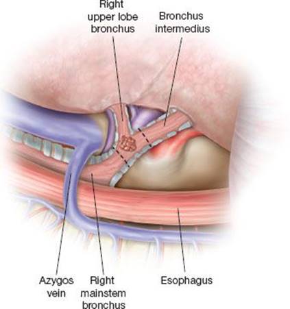
Figure 34.1 The exposure of the right mainstem bronchus, right upper lobe origin, and bronchus intermedius are shown from the posterior aspect. Tumor within the orifice of the right upper lobe is depicted, with the planned lines of airway division (dashed lines) to allow for complete resection.
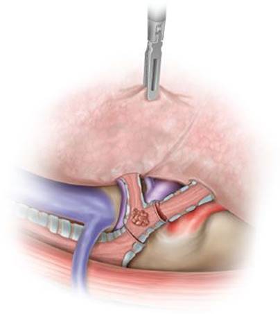
Figure 34.2 Division of the right mainstem bronchus and bronchus intermedius has been completed, allowing en bloc removal of the right upper lobe and sleeve of mainstem bronchus and the tumor within. The airway has been divided perpendicular to the long axis of the airway to facilitate reconstruction.
Anastomotic Technique
There are several points of emphasis when describing techniques used in airway reconstruction following sleeve lobectomy. These include:
Special care must be taken to maintain orientation of the proximal and distal airway segments, to avoid a “twist” or torsion at the anastomotic site. Identification and matching of the cartilaginous–membranous wall junctions will usually avoid this problem.

Figure 34.3 Partial right hilar release. The dashed line depicts the pericardial incision inferior to the right hilum, allowing the hilar structures to displace cephalad.
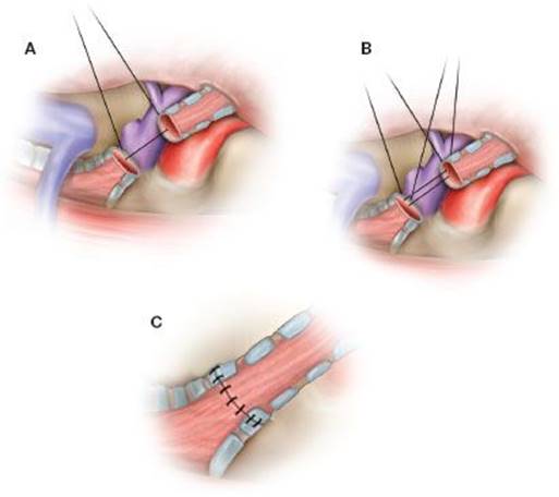
Figure 34.4 The airway reconstruction is accomplished using an open interrupted suture technique. A: The initial suture is placed at a point furthest from the operative team, with successive sutures placed progressively more anteriorly. Care is taken to avoid entanglement of subsequent sutures. B: Once all the sutures are positioned, the airway edges are brought together and the sutures are tied in reverse order from which they were placed (see text for further details). C: The completed anastomosis is carefully inspected and tested for leaks, then wrapped in autologous tissue to buttress the anastomosis and separate it from the adjacent pulmonary vessels.
Either a running or interrupted suture technique may be used, although the latter is preferred. Use of an open, interrupted anastomotic technique will allow for precise suture placement, minimize anastomotic ischemia issues, and better allow for tailoring to accommodate airway size mismatch.
Lateral traction sutures, commonly used in tracheal surgery, may be employed but are not required given the relative lack of tension at the anastomotic site.
Absorbable suture should be used to avoid postoperative granulation tissue growth at the anastomosis. 4-0 Vicryl (Ethicon, Inc., Somerville, NJ) is preferred, although 3-0 Vicryl may be used if desired for the cartilaginous portion of the airway. If a running suture technique is utilized, 4-0 PDS (Ethicon, Inc., Somerville, NJ) is preferred.
Sutures are individually placed 2 to 3 mm deep from the cut edge of the airway, and 2 to 3 mm apart, situated such the knots (once tied) lay on the outside of the airway (Fig. 34.4A,B). The individual sutures are isolated with a hemostat and affixed to the adjacent drape. It is often helpful to begin the open anastomotic technique at a point furthest away from the operating team; the subsequent individual sutures are then placed, gradually progressing anteriorly around the anastomotic “circle” 180 degrees to the point facing the operator. It is important the sutures are placed precisely, secured to the drapes after placement and not entangled. Suture placement may incorporate a cartilaginous ring, if present, but excessive incorporation of the membranous wall within the tied suture is discouraged.
Once both halves of the anastomotic “circle” are completed, the cut ends of the airway are approximated manually by bringing the cut edge of the bronchus intermedius (along with the middle and lower lobes) cephalad to the right mainstem. The individual anastomotic sutures are tied in reverse order from which they were placed, and cut as they are tied. The completed anastomosis (Fig. 34.4C) should be carefully inspected for defects or potential torsion. Inflation of the right lung at this point with submersion of the anastomotic site is recommended to detect small leaks which may require repair. Once pneumostasis of the anastomotic site is assured, a “wrap” of the anastomosis is advisable utilizing pericardial fat or a pleural or intercostal muscle flap. The wrap serves to separate the anastomosis from the divided pulmonary vessels, and buttresses the anastomotic closure with healthy, autologous tissue.
Chest drainage and thoracotomy closure are routine. It is important to replace the double-lumen endotracheal tube with a single-lumen tube to allow for immediate postoperative bronchoscopy. This allows the surgeon to carefully inspect the anastomosis and remove retained blood and secretions prior to extubation.
Isolated Bronchus Intermedius Resection
In general, the approach and technique of a bronchial sleeve resection involving the bronchus intermedius proceeds in a manner analogous to a right upper lobe sleeve resection. Because the right upper lobe remains in place, though additional care must be taken to provide adequate exposure. It is helpful to open at least the posterior aspect of the major fissure between the upper and lower lobes, and mobilize and encircle the pulmonary artery with a vessel loop. The posterior ascending and superior segmental pulmonary arterial branches “tether” the two lobes together, limiting displacement of the lobes and as such are prone to injury. Division of the bronchus intermedius above and below the area of concern must be precise to allow for safe reconstruction without impingement on the adjacent lobar orifices.
 POSTOPERATIVE MANAGEMENT
In large part, the management of patients following sleeve lobectomy mirrors that for any lobectomy patient. Appropriate pain control measures are key with the use of the epidural or intravenous narcotic supplementation to allow for early mobilization and proactive pulmonary toilet measures. Difficulty in mobilizing secretions from the distal airway segment is common, and in the case of right upper lobe sleeve resection may manifest with progressive atelectasis of the right middle and lower lobes on serial postoperative imaging. With this in mind, aggressive use of fiberoptic bronchoscopy may minimize unwanted complications related to secretion retention.
Prior to discharge, it may be advisable to assess the anastomotic site via bronchoscopy, if not already performed for pulmonary toilet. Evidence of airway necrosis or anastomotic separation is sought as a precursor to further anastomotic complications. Particularly if some anastomotic problem is identified, repeat inspection 4 to 6 weeks later may allow for gentle debridement of granulation tissue to maximize airway patency. These measures may be repeated until a mature, widely patent anastomotic site is present.
Close margins are inherent in airway surgery, and many surgeons advocate repeat inspection bronchoscopy on at least an annual basis to assess for local recurrence.
 COMPLICATIONS
A summary of morbidity and mortality rates of several recent large series describing outcomes with sleeve lobectomy is listed in Table 34.1. Mortality rates vary from 1% to 5.7%, with morbidity ranging from 14% to 49%. Prolonged air leak, retained secretions requiring bronchoscopy, pneumonia, and atrial arrhythmias predominate. Bronchial anastomotic morbidity, consisting of bronchopleural fistula, bronchovascular fistula, and anastomotic stenosis, are a group of complications largely unique to sleeve lobectomy patients. The majority of serious anastomotic complications are largely avoidable with proper patient selection and meticulous surgical anastomotic technique. However, development of a fistula, particularly involving a hilar vessel, can be catastrophic and require emergent intervention, often completion pneumonectomy. Anastomotic stenoses typically occur late, and may result from poor anastomotic technique or bronchial ischemia. They frequently can be managed with endoscopic techniques, including temporary stent placement.
TABLE 34.1
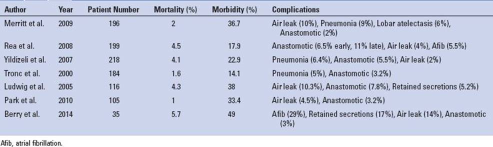
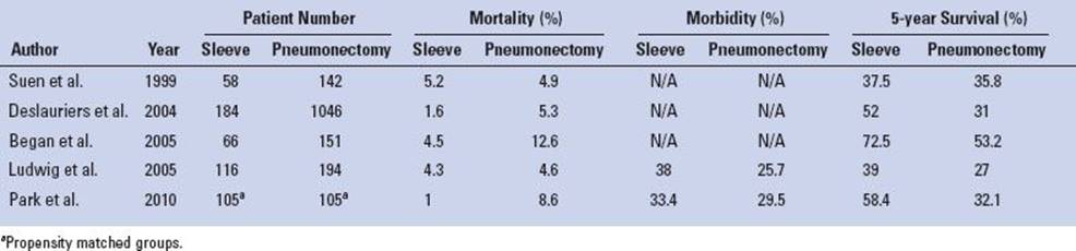
TABLE 34.2
 RESULTS
The majority of recently published series pertaining to sleeve lobectomy compare outcomes with pneumonectomy, the surgical alternative to the parenchyma-sparing sleeve resection. The studies are retrospective, and to date there has not been (nor is it likely there ever will be) publications reporting prospective, randomized data comparing the two procedures. Further, currently published studies suffer from inherent selection bias that renders direct comparisons difficult. Despite these caveats, it seems that sleeve lobectomy enjoys lower mortality and equivalent morbidity rates, and in the setting of lung cancer, comparable or improved long-term survival (Table 34.2). Data pertaining to right upper lobe sleeve resection alone are lacking. Several reports have suggested that the use of induction therapy in patients undergoing sleeve lobectomy increases the morbidity of the procedure, particularly with respect to anastomotic complications, while other studies refute this claim. The performance of sleeve lobectomy has been associated with improved quality of life and is a more cost-effective strategy when compared with pneumonectomy.
 CONCLUSIONS
Right upper lobe sleeve resection is the preferred surgical option for the majority of patients who present with malignancy or significant inflammatory stenosis of the origin or the right upper lobe or adjacent mainstem bronchus. Compared with pneumonectomy, sleeve lobectomy is associated with similar or improved postoperative outcomes. Careful patient selection and meticulous surgical technique are required for optimal outcomes.
Recommended References and Readings
Bagan P, Berna P, Pereira JC, et al. Sleeve lobectomy vs. pneumonectomy: Tumor characteristics and comparative analysis of feasibility and results. Ann Thorac Surg. 2005;80:2046–2050.
Balduyck B, Hendriks J, Lauwers P, et al. Quality of life after lung cancer surgery: A prospective pilot study comparing bronchial sleeve lobectomy with pneumonectomy. J Thorac Oncol. 2008; 3:604–608.
Berry MF, Worni M, Wang X, et al. Sleeve lobectomy for non-small cell lung cancer with N1 nodal disease does not compromise survival. Ann Thorac Surg. 2014;97(1):230–235.
Deslauriers J, Grégoire J, Jacques LF, et al. Sleeve lobectomy versus pneumonectomy for lung cancer: A comparative analysis of survival and sites or recurrences. Ann Thorac Surg. 2004;77:1152–1156.
Ferguson MK, Lehman AG. Sleeve lobectomy or pneumonectomy: Optimal management strategy using decision analysis techniques. Ann Thorac Surg. 2003;76:1782–1788.
Ludwig C, Stoelben E, Olschewski M, et al. Comparison of morbidity, 30-day mortality, and long-term survival after pneumonectomy and sleeve lobectomy for non-small cell lung carcinoma. Ann Thorac Surg. 2005;79(3):968–973.
Maurizi G, D’Andrilli A, Anile M, et al. Sleeve lobectomy compared with pneumonectomy after induction therapy for non-small cell lung cancer. J Thorac Oncol. 2013;8(5):637–643.
Merritt RE, Mathisen DJ, Wain JC, et al. Long-term results of sleeve lobectomy in the management of non-small cell lung carcinoma and low-grade neoplasms. Ann Thorac Surg. 2009;88:1574–1582.
Milman S, Kim AW, Warren WH, et al. The incidence of perioperative anastomotic complications after sleeve lobectomy is not increased after neoadjuvant chemoradiotherapy. Ann Thorac Surg. 2009;88:945–951.
Park JS, Yang HC, Kim HK, et al. Sleeve lobectomy as an alternative procedure to pneumonectomy for non-small cell lung cancer. J Thorac Oncol. 2010;5:517–520.
Rea F, Marulli G, Schiavon M, et al. A quarter of a century experience with sleeve lobectomy for non-small cell lung cancer. Eur J Cardiothorac Surg. 2008;34:488–492.
Rendina EA, Venuta F, De Giacomo T, et al. Safety and efficacy of bronchovascular reconstruction after induction chemotherapy for lung cancer. J Thorac Cardiovasc Surg. 1997;114(5):830–835.
Suen HC, Meyers BF, Guthrie T, et al. Favorable results after sleeve lobectomy or bronchoplasty for bronchial malignancies. Ann Thorac Surg. 1999;67(6):1557–1562.
Thomas CP. Conservative resection of the bronchial tree. J R Coll Surg Edinb. 1956;1:169–186.
Tronc F, Gregoire J, Rouleau J, et al. Long-term results of sleeve lobectomy for lung cancer. Eur J Cardiothorac Surg. 2000;17:550–556.
Yildizeli B, Fadel E, Mussot S, et al. Morbidity, mortality, and long-term survival after sleeve lobectomy for non-small cell lung cancer. Eur J Cardiothorac Surg. 2007;31:95–102.