
Dan J. Raz
 INDICATIONS
The rigidity of the chest wall makes it important for the thoracic surgeon to plan the surgical approach to the pleural space or mediastinum well. Thoracotomy incisions can be categorized by their location on the chest wall (anterior, lateral, and posterior), whether muscle is divided or preserved, and whether the sternum is divided, as in the hemiclamshell and clamshell approaches. Regardless of the size of the incision, it is important to plan the incision based on the location of structures that must be exposed. Even as more chest procedures are performed thoracoscopically, the minimally invasive chest surgeon must be proficient at various approaches to the lung and mediastinum. Thoracic surgeons must also be able to plan for emergency conversion from a thoracoscopic approach to a thoracotomy. Trauma surgeons must understand which thoracic incisions are useful for thoracic trauma especially when an intrathoracic subclavian artery injury is present. Finally, thoracic surgeons may be asked to provide access for spine surgery.
 PREOPERATIVE PLANNING
A number of factors are important to selecting the type of thoracotomy incision.
Location and size of the tumor: An incision should primarily be planned to facilitate exposure to the region of interest. Other structures that may need to be accessed should be considered when planning the approach, such as vascular control (e.g., in superior sulcus tumors), intrapericardial control, access to the diaphragm (if plication is anticipated), etc.
Reoperative thoracotomy: In the setting of a reoperative thoracotomy an incision in a new location may be considered to avoid lung parenchymal adhesions. Alternatively, the same incision can be used, and a rib may be excised to facilitate exposure. When adhesions are anticipated, the incision should be planned, so that extension of the incision is easy to obtain additional exposure.
Body habitus: The patient’s body habitus should be considered when planning the type of thoracotomy incision. Excellent exposure may be obtained through a small anterolateral muscle-sparing thoracotomy in thin patients; however, exposure may be more challenging in muscular men. A latissimus dorsi-sparing posterolateral thoracotomy in an obese patient may lead to seroma formation from the extensive soft tissue mobilization necessary. Finally, debilitated patients who are mostly bed bound may have fewer local wound complications with a lateral or anterior approach as opposed to a posterolateral thoracotomy.
Anticipated use of muscle flap: The thoracic surgeon should anticipate whether a muscle flap might be necessary during this operation or future operations. Intercostal muscle flaps are most easily harvested through a posterolateral thoracotomy. The latissimus muscle can be preserved and mobilized as a flap through posterolateral thoracotomy when needed to fill a pleural space. The serratus anterior muscle is sometimes used to buttress a bronchial stump alone or in addition to intercostal muscle.
Size of incision: A thoracoscopic approach results in improved pain control postoperatively, and as a result decreased respiratory complications. It is tempting to extrapolate this observation to the comparison between large and small thoracotomies. However, there is insufficient data to conclude whether the length of a thoracotomy incision results in any difference in pain control, respiratory complications, or pulmonary function. Postthoracotomy pain arises in large part as a result of intercostal neuralgia, and this may be exacerbated by greater rib spreading needed for exposure when incisions are small.
 SURGERY
Posterolateral Thoracotomy
Posterolateral thoracotomy (Fig. 8.1) is the most commonly used and versatile incision for access to the hemithorax. This incision is commonly used for pulmonary surgery, extrapleural pneumonectomy, and access to the intrathoracic esophagus, trachea, and mainstem bronchi. Most surgeons do not divide the serratus anterior muscle to avoid postoperative shoulder dysfunction. While many surgeons divide the latissimus dorsi muscle, especially for large thoracotomy incisions, the latissimus may be mobilized and retracted. This leaves open the option of future latissimus muscle flap transfer to manage an infected pleural space or bronchopleural fistula.
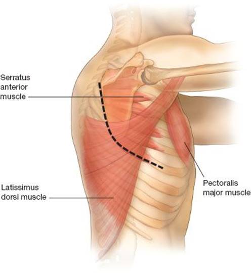
Figure 8.1 Posterolateral thoracotomy: An incision is made 1 to 2 cm below the scapular tip and continued anteriorly along the direction of the rib and posteriorly between the scapula and the spine. The latissimus dorsi is either divided or preserved and the serratus muscle is retracted anteriorly.
The patient is positioned in a lateral decubitus position and the bed is flexed, so that the hip is out of the field and the rib interspaces widened. An axillary roll is used by most surgeons to protect the brachial plexus although the need for this is questionable as some surgeons do not use an axillary roll and report no untoward consequences. The incision is centered 1 or 2 cm under the scapular tip and extended anteriorly along the rib and posteriorly at the midpoint between the medial border of the scapula and the spine. The latissimus dorsi muscle is either mobilized and preserved or divided for additional access. If the latissimus is divided, it is often helpful to mobilize it off the serratus and overlying soft tissue for ease of later closure. The serratus can usually be spared and retracted medially. The paraspinous ligament is preserved and mobilized off of the rib interspace to be entered. The trapezius and rhomboid muscles can be divided posteriorly when the incision must curve high along the scapula, such as in superior sulcus resections. The scapula is then retracted and the ribs counted. The chest is entered in the fifth interspace for pulmonary resections. Depending on the operation, one may then enter the pleural space by incising the intercostal muscle along the top of the rib, harvesting an intercostal muscle flap, or resecting a rib.
When harvesting an intercostal muscle flap, a periosteal elevator is the most effective way to harvest the muscle in the subperiosteal plane. The ribs above and below the intercostal muscle are scored and the periosteum is scraped toward the muscle. The elevator is then used to free the muscle off of the rib below and above the muscle, and then the pleura is incised to separate the muscle. This technique may result in ossification of the muscle flap with time because it leaves periosteum on the muscle, but this almost never causes a problem. Some surgeons use electrocautery to harvest the muscle flap to avoid taking the periosteum with the flap.
The rib may be shingled posteriorly at the level of the paraspinous ligament using a sliding rib cutter to prevent uncontrolled fracture of the ribs with retraction. A rib may be resected in certain circumstances such as when there is extensive pleural disease or extensive adhesions. A periosteal elevator is used to free the rib from the intercostal muscles after scoring and scraping the pleura off the bone after which the bone is resected with a rib cutter.
Once the chest retractor is inserted, the intercostal muscle beyond the incision is freed along the rib anteriorly with a freer or using electrocautery. For closure, pericostal sutures can be placed around the ribs or holes can be drilled in the inferior rib, so that the suture does not put pressure on the intercostal nerve. When a rib is resected, the intercostal muscles are reapproximated with interrupted 0-silk sutures and a rib approximator is used while tying down the sutures. The latissimus dorsi and serratus anterior muscles are then reapproximated.
Anterolateral Thoracotomy
Anterolateral thoracotomy (or “French” incision) (Fig. 8.2) is a versatile incision for access to the lung and mediastinum. The advantage of this incision is that a relatively small muscle-sparing incision can be made and access is excellent to the lung and anterior mediastinum. Pain control is better compared with posterolateral thoracotomy, and there is some evidence that postoperative pulmonary function and shoulder function are also superior. The patient is positioned similarly to posterolateral thoracotomy but may be rotated slightly posteriorly especially when the mediastinum needs to be exposed. It is helpful to insert a thoracoscope at the start of the procedure to identify the interspace to enter the pleural cavity since the ribs are not counted. This port site can be used later on as an entry site for a stapler intraoperatively and the chest tube. A skin incision is then made between the edge of the pectoralis major muscle and the latissimus dorsi. Alternatively, a skin incision may be made parallel to the lateral border of the pectoralis muscle and extended toward the axilla. One disadvantage of this approach may be greater skin anesthesia, which includes the possibility of making the nipple and breast insensate in women. In my opinion, this incision is also more challenging to extend if additional exposure is needed. The pectoralis muscle and the latissimus are spared and retracted. The serratus muscle is split along its fibers to expose the rib and intercostal space. The incision in the serratus muscle must stop before the neurovascular bundle of the long thoracic nerve to avoid damage and resultant winged scapula. The pleural space is then entered by separating the intercostal muscle off the rib as far anteriorly and posteriorly as is possible. The rib may be shingled posteriorly and anteriorly if needed for additional exposure. The rib may also be resected (“French-window” thoracotomy) if needed for exposure. For closure two pericostal sutures are placed and the serratus fibers are reapproximated.
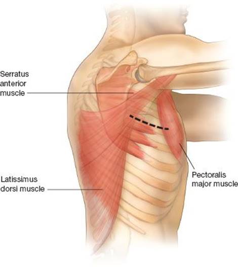
Figure 8.2 Anterolateral thoracotomy: An incision is made between the latissimus dorsi and the pectoralis muscles, which are retracted. Slips of serratus muscle are divided along their fibers, and the pleural space is entered.
Clamshell (Sternothoracotomy)
Clamshell incision (Fig. 8.3) allows for exposure of the mediastinum and bilateral pleural spaces. This incision was frequently used in the early history of cardiac surgery but was replaced by sternotomy, which is associated with less postoperative pain. The clamshell incision has had a resurgence as a result of its use in lung transplantation. The clamshell incision provides excellent access to both pleural cavities for taking down adhesions, the hilar structures, and the mediastinum for initiation of cardiopulmonary bypass when needed. This exposure also provides excellent exposure for resection of bilateral pulmonary metastases and for selected mediastinal tumors with pulmonary involvement. In addition to providing excellent exposure, the clamshell incision is more cosmetically appealing compared with median sternotomy. The incision is hidden in the inframammary crease. Compared with sternotomy, the clamshell incision has the disadvantage of more postoperative pain and limited access to the superior-anterior mediastinum.
In women, the skin incision is made along the inframammary crease and continued toward the anterior axillary line. In men, the skin incision is made just below the nipples and more closely follows the direction of the ribs. The pectoralis major muscle is exposed, and in women flaps are raised to elevate the breast tissue off the pectoralis major muscle. The Angle of Louis is used as a reliable landmark for the second rib. The pleural cavities are entered in the fourth or fifth interspace bilaterally. The internal mammary vascular pedicle is identified, suture ligated, and divided on each side of the sternum. A substernal space is developed bluntly and then the sternum is divided using an oscillating sternal saw, Lebsche knife, or Gigli saw. A Finochietto retractor is placed on either side of the sternum. The serratus anterior muscle can be divided laterally if additional exposure is needed. The intercostal muscle is divided posteriorly to allow wider retraction. The pleural reflections on the sternum are divided.

Figure 8.3 Clamshell thoracotomy: An incision is made in the inframammary fold. The internal mammary vessels are divided and the pleural spaces are entered bilaterally. The Angle of Louis is a reliable landmark for the second rib. The sternum is transected to complete the exposure.
Prior to closure, the ligated mammary pedicles are inspected for hemostasis. The ligated pedicles may be traumatized by retractors and may need to be ligated once more. Chest tubes are inserted, and the sternum is closed using two sternal wires. Three or four no. 2 Vicryl figure of eight pericostal sutures are placed on either side to approximate the intercostal space. The serratus anterior and pectoralis major muscles are approximated, followed by the subcutaneous tissue layer.
Hemiclamshell (Anterior Thoracotomy and Sternotomy)
This incision (Fig. 8.4) is useful when simultaneous access to the pleural space and mediastinum is needed, but the exposure to the pleural cavity that a sternotomy would provide is inadequate. This incision is useful for very large central lung tumors where intrapericardial control is necessary, large lung tumors with subclavian artery involvement where proximal vascular control is desirable, and large mediastinal tumors that extended into the pleural cavity such as locally advanced thymomas with pleural disease.
The patient is positioned supine with a bump under the chest and the arm out to the side. A median sternotomy incision is drawn out, and it is then angled inferiorly in the inframammary fold in women or along the fifth rib in men. I often start with thoracoscopy through the anterior thoracotomy incision in cases of locally advanced lung cancers to ensure the absence of pleural disease. The anterior thoracotomy incision is made first and the breast tissue is mobilized in women until the appropriate intercostal space is reached. Slips of pectoralis muscle and serratus muscle laterally are divided along their fibers when possible. The pleural cavity is entered typically either in the fourth or fifth interspace, depending on the location of the tumor. The internal mammary pedicle is then ligated between 2-0 silk ties. The substernal attachments are then bluntly taken down. The sternal incision is then made. A sternal saw is used to divide the sternum. It is best to saw starting from the neck, as it is easier to stay in the middle of the sternum and then angle toward the thoracotomy incision to transect the sternum. For retraction, I mostly use a Rultract Skyhook retractor as this provides excellent exposure. Alternatively, a combination of sternal and thoracotomy retractors may be used. Sternal wires and figure of eight pericostal sutures are placed for closure. The sternal wires are tied while an assistant crosses the pericostal sutures. An additional figure of eight sternal wire at the sternocostal junction may be helpful. As the intercostal spaces are widest anteriorly, it is not possible or desirable to obtain rib to rib apposition with pericostal closure. This is especially true when intercostal muscle is harvested.

Figure 8.4 Hemiclamshell thoracotomy: An incision is made in the inframammary fold and in the midline of the sternum. The pleural space is entered in the fourth or fifth interspace and the internal mammary pedicle is divided. The sternum is then divided from top to bottom.
Anterior Thoracotomy
Anterior thoracotomy has limited utility in general thoracic surgery, other than an alternative exposure for pericardial window or biopsy of mediastinal mass via anterior mediastinotomy. This incision is used commonly in minimally invasive cardiac surgery.
The patient is positioned supine with a bump under the chest with the arms padded and tucked. It is helpful to position the patient close to the edge of the bed with the padded arm tucked and secured but suspended by the draw sheet off of the bed. The angle of Louie is used as a landmark for the second rib, and the incision is made corresponding to the location of the intrathoracic target. Use of preoperative CT scan is helpful in determining the interspace to enter. Pectoralis muscle is divided along its fibers, as is serratus muscle more laterally. The interspace is then entered along with electrocautery. The rib may be shingled anteriorly, posteriorly, or both anteriorly and posteriorly depending on the circumstance and need for exposure. A small rib spreading retractor is usually sufficient to provide exposure although a number of new low profile disposable retractors have been developed for minimal access cardiac surgery. Pericostal sutures are placed for closure. As mentioned previously, rib to rib apposition should not be attempted with pericostal closure.
 POSTOPERATIVE MANAGEMENT
Postoperative Analgesia
Pain control is essential to prevent postoperative respiratory complications. This facilitates the patient’s cough and walk ability, which is vital to mobilizing pulmonary secretions and preventing postoperative pneumonia.
 Thoracic epidural: A thoracic epidural, with a mixture of local anesthetic and narcotic, is the gold standard for postoperative analgesia. Thoracic epidural anesthesia improves postthoracotomy pulmonary function and decreases sedation and hypercarbia compared to intravenous narcotics. Epidurals cannot be successfully placed in some patients with thoracic spine disease, and patients may have contraindications to an epidural due to thrombocytopenia. There are some downsides to epidural analgesia. Local analgesia may lead to sympathectomy and hypotension, requiring either vasopressor support or reduction or elimination of the local anesthetic component. Finally, in centers where a pain service is not available 24 hours a day, effective troubleshooting of a malfunctioning epidural may be challenging and lead to delay in pain control.
Paravertebral catheter: A number of thoracic surgeons place paravertebral catheters for delivery of local anesthetic, delivered through a pump. Most studies have shown this method of analgesia to be inferior to epidural analgesia although some institutional series have shown them to be equivalent. In centers where these are used, discharge from the hospital may be faster than with epidural, as oral pain medicines can be started faster.
Paravertebral blocks: Paravertebral blocks have been employed in patients who have a contraindication to epidural catheter placement. These blocks are limited by the length of action the anesthetic injected. Exparel (bupivacaine liposome injectable suspension) is a liposomal formulation of bupivacaine that lasts for up to 72 hours. While it is not FDA approved for use in blocks, it has been used off-label in some centers for paravertebral injection. Additional studies are needed to determine the efficacy of this promising anesthetic approach.
Activity Restrictions
There is little evidence to guide recommendations regarding postoperative activity restrictions such as avoidance of heavy lifting. Theoretical complications of heavy lifting or extreme arm movements involving the shoulder girdle include tearing of the pericostal sutures or muscular closure. Vigorous coughing has been shown to increase intrathoracic pressure to over 300 mm Hg. Sternal wound precautions have been better studied and scrutinized due to the problems that develop with sternal wound complications such as infection, wire breakage, and sternal nonunion. Sternal wound complications are seldom reported in series of hemiclamshell and clamshell thoracotomies. It is probably reasonable to gradually introduce shoulder girdle activity and lifting, with cessation of the activity if it produces pain. It is easier though for surgeons to provide more specific guidelines and err on the side of more conservative restrictive lifting recommendations, especially for patients who may have poor insight into their health.
 COMPLICATIONS
Pulmonary. Adequate pain control and pulmonary toilet go hand in hand to prevent atelectasis and pneumonia in the patient who has undergone thoracotomy. Educating patients on how to expectorate secretions after thoracotomy, early ambulation, and use of the incentive spirometer are the basics that usually maintain pulmonary toilet when pain is controlled. Therapeutic bronchoscopy, nasotracheal suctioning, and the use of positive expiratory pressure devices such as Metaneb or Acapella are other important adjuncts to help clear secretions and prevent pneumonia.
Chronic pain. Many surgeons underestimate the long-term discomfort associated with thoracotomy. More than two-thirds of patients still have discomfort in their thoracotomy incisions when questioned 3 months after surgery though a minority find the discomfort bothersome enough to take pain medicine. Some patients develop an ongoing chronic pain syndrome, which affects quality of life and requires pain medicines or other interventions.
Several studies have examined differences in techniques of rib closure and effects on both immediate postoperative and longer term pain control. Most studies suggest decreased pain scores in the immediate postoperative period and up to 3 months time with techniques that avoid intercostal nerve bundle compression compared with traditional pericostal closure. These techniques include intracostal closure (drilling holes in the rib for sutures), mobilization of the intercostal muscle off the rib before retraction with subsequent passage of the suture around the rib alone, and passage of the suture immediately underneath the bottom rib (avoiding the neurovascular bundle). There does not appear to be a clear advantage to one of these techniques in particular.
Adjuncts to anti-inflammatory medications and narcotics are the neuroleptics gabapentin (Neurontin) and pregabalin (Lyrica), which can be helpful in ameliorating neuropathic pain. Locally placed transdermal lidocaine patches are also effective in some patients with focal points of pain. Finally, intercostal nerve ablation with cryoablation or radiofrequency ablation may be employed for chest wall pain refractory to more conservative measures.
Positioning-related Injuries
Positioning-related injuries occur and are underreported in the surgical literature. Brachial plexus stretch from malposition of the arms has been described. While most surgeons use axillary rolls to prevent brachial plexopathy on the dependent arm, there is little evidence that this is of any benefit. Lower extremity neuralgias have been reported from placement of tape on the thighs during posterolateral thoracotomy. In patients undergoing hemiclamshell or clamshell thoracotomy, it is important not to hyperabduct the arms to avoid brachial plexopathy. It is important to pay close attention to padding pressure points and to avoid positioning limbs in a stretched position.
Lung Hernia
Herniation of lung tissue through the intercostal space is an uncommon complication that is rarely mentioned in surgical series of pulmonary surgery but is occasionally encountered. This must be considered a technical error in placement or tying of pericostal sutures. Lung hernia can present as a painful bulge or as an asymptomatic mass that expands with a breath. It is usually easily identified on physical examination and confirmed on CT scan. Lung herniation is repaired by reopening the incision and reclosing the ribs after reducing the herniated lung.
Surgical Site Infection
Wound infections after thoracotomy are exceedingly uncommon but occasionally occur. Most large surgical series of pulmonary surgery done through thoracotomy or VATS do not report any surgical site infections. Scenarios where surgical site infection becomes a concern include patients who are operated on for infectious disease that has invaded the chest wall, such as mucormycosis and Actinomycosis. These patients are often immunosuppressed and are predisposed to poor wound healing. Patients on therapeutic anticoagulation who develop postoperative hematoma may also develop local wound complications if the hematoma is large and disrupt normal wound healing or becomes infected.
Sternal Override
One unique, albeit rare, complication of this incision is sternal override where the inferior portion of the sternum is displaced anteriorly. This can be reduced or eliminated by the use of Steinmann pins or K-wires for closure. Usually sternal override does not produce cosmetic deformity in patients with the usual amount of chest wall soft tissue, and it does not cause pain.
Recommended References and Readings
Bains M, Ginsberg R, Jones WG 2nd, et al. The clamshell incision: An improved approach to bilateral pulmonary and mediastinal tumor. Ann Thorac Surg. 1994;58:30–32.
Ceppa DP, Kosinski AS, Berry MF, et al. Thoracoscopic lobectomy has increasing benefit in patients with poor pulmonary function: A Society of Thoracic Surgeons database analysis. Ann Surg.2012;256:487–493.
DiMarco AF, Oca O, Renston JP. Lung herniation: A cause of chronic chest pain following thoracotomy. Chest. 1995;107:877–879.
Garcia-Tirado J, Rieger-Reyes C. Suture techniques of the intercostal space in thoracotomy and their relationship with post-thoracotomy pain: A systematic review. Arch Bronconeumol. 2012;48:22–28.
Helms O, Mariano J, Hentz JG, et al. Intra-operative paravertebral block for postoperative analgesia in thoracotomy patients: A randomized, double-blind, placebo-controlled study. Eur J Cardiothorac Surg.2011;40:902–906.
Kinney M, Hooten W, Cassivi S, et al. Chronic post-thoracotomy pain and health-related quality of life. Ann Thorac Surg. 2012;93:1242–1247.
Lebreton G, Baste J, Thumerel M, et al. The hemiclamshell approach in thoracic surgery: Indicatitons and associated morbidity in 50 patients. Interact Cardiovasc Thorac Surg. 2009;6:965–969.
Marshall M, Carter Y. Modified French window as an alternative to thoracotomy for complex intrathoracic pathology. Ann Thorac Surg. 2009;88:685–687.
Namboothiri S, Kumar R, Menon K. Early changes in pulmonary function following thoracotomy for scoliosis correction: The effect of size of incision. Eur Spine J. 2005;14:144–148.
Wildgaard K, Ravn J, Kehlet H. Chronic post-thoracotomy pain: A critical review of pathogenic mechanisms and strategies for prevention. Eur J Cardiothorac Surg. 2009;36:170–180.