
Edie Y. Chan, M.D., Jamie Elizabeth Jones, M.D.
1 A 44-year-old man suffers a gunshot wound to his abdomen. He is hemodynamically stable and taken to the operating room. On exploration, his injuries are found to be limited to two small bowel injuries 7 cm apart, each with destruction of 70% of the bowel wall, and a through-and-through injury to the ascending colon with destruction of 30% of the bowel wall. How should these injuries be managed?
A Resection and anastomosis of the small bowel injuries and primary repair of the colon injury
B Primary repair of both the small bowel and colon injuries
C Primary repair of the small bowel injuries, primary repair of the colon injury, and creation of a diverting ileostomy
D Resection of the small bowel injuries and exteriorization of the colon injury as a colostomy
E Resection and anastomosis of all injuries
Ref.: 1
Comments
Historically, all colon injuries were treated by diversion. However, with the progression of surgical technique, resuscitative and critical care, and antibiosis, many colon injuries can be repaired primarily. Patients who are hemodynamically stable and have injuries that involve less than 50% of the circumferential bowel and no vascular disruption can undergo primary repair. In regard to small bowel injury, resection is indicated for injuries involving greater than 50% of the wall circumference, multiple injuries in a short segment, or both.
Answer
A
2 A 27-year-old woman is brought to the emergency department awake and alert after sustaining a gunshot wound to her neck. The wound is anterior to the origin of the sternocleidomastoid muscle at the angle of the mandible. The patient is asymptomatic. All of the following are correct management choices except:
A Cervical spine radiographic studies
B Mandatory neck exploration
C Four-vessel angiographic studies
D Flexible esophagoscopic examination
E Contrast-enhanced esophagographic examination
Ref.: 2
Comments
See Question 3.
Answer
B
3 Soon after the patient in Question 2 arrives in the emergency department, left hemiparesis and aphasia develop. At this time, which of the following treatments should be provided?
A Continued observation
B Repair of the carotid artery injury
C Ligation of the carotid artery injury
D Repair of the vertebral artery injury
E Systemic anticoagulation
Ref.: 2
Comments
The patient in Question 2 has suffered an injury to zone III of the neck. Zone III is defined as the area anterior to the sternocleidomastoid between the angle of the mandible and the base of the skull. Even without hard signs of vascular injury or change in neurologic status, patients with zone III injuries should routinely undergo angiography. Physical examination alone cannot be relied on to diagnose carotid artery or aerodigestive injuries in this zone. Cervical spine radiographic studies are needed to rule out any associated fracture. In addition, the esophagus should be investigated with both esophagoscopy and contrast-enhanced imaging. In Question 3, the patient has a mild neurologic deficit consistent with a carotid artery injury. The current recommendation regarding carotid artery injury is surgical repair unless complete occlusion or hemodynamic instability is present.
Answer
B
4 The upper part of the abdomen of a 42-year-old man strikes the steering wheel during a motor vehicle accident. He is hemodynamically stable. Because of positive findings on diagnostic peritoneal lavage (DPL), he undergoes exploratory laparotomy, at which time complete transection of the pancreatic neck is found. What is the most appropriate management of this injury?
A Distal pancreatectomy with oversewing and drainage of the proximal pancreatic stump
B Roux-en-Y pancreaticojejunostomy to the distal end of the pancreas with oversewing and drainage of the proximal pancreatic stump
C Primary repair and drainage of the pancreatic duct
D Whipple operation
E Total pancreatectomy
Ref.: 3
Comments
Operative management of pancreatic injuries centers on the location of the injury and whether the duct is involved. Approximately 50% of the pancreas is located on either side of the superior mesenteric artery. For pancreatic wounds with an intact duct, drainage of the area with soft closed suction drains suffices. If the main pancreatic duct is injured to the left of the mesenteric vessels, as in this patient, distal pancreatectomy with drainage of the proximal stump is indicated. The proximal pancreatic duct should be individually ligated with nonabsorbable suture if possible and the parenchymal tissue oversewn or stapled across with a stapler. The spleen should be preserved if the patient’s hemodynamic status allows. Roux-en-Y pancreaticojejunostomy to the distal end of the pancreas with oversewing of the proximal pancreatic stump carries a high rate of leakage. The Whipple procedure and total pancreatectomy would be reserved for injuries that involve extensive devitalization of the duodenum and head of the pancreas. Primary repair is technically difficult and does not address the transected pancreatic tissue.
Answer
A
5 A 30-year-old man is brought to the emergency department after being involved in a Jet Ski crash. His vital signs are stable. A high-riding prostate is noted on rectal examination. On portable pelvic radiographs he is found to have bilateral pubic rami fractures. He has not yet voided since admission. Which of the following should be the next step?
A Wait for the patient to void freely before attempting transurethral bladder catheterization.
B Initially attempt gentle transurethral bladder catheterization, but stop if resistance is encountered.
C Obtain a urethrogram before attempting transurethral bladder catheterization.
D Insert a suprapubic cystostomy tube.
E Perform computed tomography of the pelvis with three-dimensional reconstruction.
Ref.: 4
Comments
Approximately 10% of all patients with a pelvic fracture have a concomitant urethral injury. Findings on physical examination, such as blood at the meatus, a freely movable prostate, and perineal hematoma, should raise suspicion for a urethral injury. If any of these signs are present or there is a significant anterior pelvic fracture, a urethrogram should be obtained to exclude an injury before transurethral catheterization is attempted. Bladder decompression plus drainage is the mainstay of treatment of urethral injuries, either via suprapubic cystostomy for complete disruption or a with a bridging transurethral catheter for partial tears.
Answer
C
6 After a gunshot wound to the chest and the subsequent development of hemothorax, a 24-year-old man requires multiple blood transfusions, including fresh frozen plasma. On admission to the intensive care unit (ICU), the patient becomes increasingly tachypneic and begins to become hypoxic. After the airway is secured, a chest radiograph is obtained and reveals bilateral patchy infiltrates. Which of the following statements regarding this condition is true?
A This type of reaction usually develops approximately 12 to 24 hours after a transfusion.
B The mortality rate with this condition nears 50%.
C It is associated with elevated pulmonary capillary wedge pressure.
D Clinical improvement is typically seen within 2 to 8 days.
E This condition results from entrapment of activated platelets in the lung.
Ref.: 5
Comments
This clinical manifestation is most consistent with transfusion-related acute lung injury (TRALI), which is similar to acute respiratory distress syndrome (ARDS) in its findings of hypoxemia, bilateral pulmonary edema, tachycardia, and hypotension. This reaction usually develops within 2 to 6 hours after the transfusion of blood products such as platelets or fresh frozen plasma and occurs once in every 2500 to 5000 units transfused. It does differ from ARDS, however, in that elevated pulmonary capillary wedge pressure is not seen and mortality rates are between 5% and 8% versus nearly 50% with ARDS. TRALI results from the transfusion of blood containing human leukocyte antigens or by activation of the patient’s granulocytes by metabolites released during storage. Management is supportive and entails mechanical ventilation with small tidal volumes.
Answer
D
7 A 44-year-old man suffers a gunshot wound to his left thigh that results in an injury to the superficial femoral artery. The injury is repaired with a saphenous vein interposition graft within 4 hours of the injury. Although the patient had equal pulses bilaterally and was neurologically intact, 5 hours postoperatively the left distal pulses diminish and he begins to experience pain with passive dorsiflexion and extension. The left anterior compartment of the lower part of the leg has a pressure of 30 mm Hg. Which of the following statements is true regarding compartment syndrome in an extremity?
A Fractures are the cause of approximately 30% of all compartment syndromes.
B The lateral compartment of the lower part of the leg is the most commonly affected.
C A compartment pressure of 25 mm Hg negates a need for fasciotomy.
D Paresthesias are an early clinical development.
E A four-compartment fasciotomy should be performed.
Ref.: 6-8
Comments
Compartment syndrome of the extremity is an extremely important diagnosis to make as early as possible because of the significant risk for permanent limb dysfunction and potential loss. Causes of compartment syndrome include crush injury, reperfusion after a time of ischemia, and fractures, which account for 50% of cases. The anterior compartment of the lower part of the leg contains mostly type I (slow twitch) muscle fibers and is encased by dense fascia, thus making it most vulnerable to the development of ischemia. The diagnosis is largely clinical, with pain out of proportion to the findings on examination, pallor, paresthesias, diminished pulses, and tense compartments being the initial symptoms. However, because of the fact that for even after an hour of ischemia impulses can still be conducted through peripheral nerves, paresthesias are a late sign of compartment syndrome. In addition, even though compartment pressures of 30 mm Hg or higher are classically quoted, lesser pressures do not prove that there is adequate tissue perfusion, and if other signs or symptoms of compartment syndrome are present, fasciotomies should still be performed.
Answer
E
8 Regarding rhabdomyolysis:
A Acute renal failure occurs secondary to the release of myoglobin.
B An alkalotic environment promotes the formation of myoglobin casts in the renal tubules, thereby worsening the kidney damage.
C The renal failure from rhabdomyolysis typically resolves within 3 to 5 days.
D Severe hyponatremia is a frequent complication.
E Alkalinization to a pH between 8 and 9 is an important treatment goal.
Ref.: 9
Comments
Acute renal failure is the most significant complication of rhabdomyolysis and occurs when myoglobin casts are formed in the proximal renal tubules from the damaged muscle and then precipitate and form casts in the proximal renal tubules. This cast formation is thought to be inhibited by an alkalotic environment (goal pH of 6 to 7), which can be achieved by intravenous sodium bicarbonate treatment. A urine dipstick positive for blood, in conjunction with a lack of red blood cells on microscopic examination, points to a diagnosis of rhabdomyolysis. In addition, the presence of urine myoglobin and elevated serum creatinine phosphokinase is indicative of this diagnosis. Serial metabolic profiles should be obtained to monitor for hyperkalemia, the most significant and dangerous electrolyte imbalance. For patients who do suffer from acute renal insufficiency, the overall prognosis with early and aggressive volume resuscitation is good, with most patients recovering to baseline function within 2 weeks to 1 month.
Answer
A
9 An 18-year-old man is admitted to the ICU after undergoing emergency laparotomy and splenectomy. He received 12 units of red blood cells and 8 units of fresh frozen plasma. Over the course of the next 12 hours, his abdomen becomes increasingly distended and firm, and urine output decreases significantly. Which of the following statements is true regarding abdominal compartment syndrome?
A Pulmonary capillary wedge pressure is typically low.
B Functional residual capacity is increased.
C There is increased central venous return.
D Central venous pressure is increased.
E Cardiac output increases.
Ref.: 10, 11
Comments
Abdominal compartment syndrome is typically associated with elevated peak respiratory pressure, decreased urine output, hypoxia, and other deleterious physiologic effects. Overall, there is decreased venous return to the heart leading to decreased cardiac output and decreased visceral perfusion. In addition, because of the increased pressure in the abdomen, the diaphragm’s ability to contract is lessened, and pulmonary compliance and functional residual capacity are reduced. This then leads to increased pulmonary vascular resistance, which is measured as increased pulmonary capillary wedge pressure. The diagnosis of abdominal compartment syndrome is achieved primarily by clinical evaluation of the patient (vital signs and physical examination) but can be corroborated by measurement of bladder pressure. To measure bladder pressure, 50 mL of sterile saline is inserted through the Foley catheter after the bladder has been completely drained, and the distal tubing port is clamped. A transducer needle is then introduced into the specimen collecting port. A pressure of 25 mm Hg or greater is consistent with intraabdominal hypertension. Operative treatment of abdominal compartment syndrome is prompt decompressive laparotomy with temporary abdominal closure.
Answer
D
10 A 25-year-old woman is the driver of an automobile involved in a high-speed motor vehicle accident. She is 30 weeks pregnant. She complains of abdominal pain but does not have peritoneal signs. Her vital signs are stable. Which of the following statements are true regarding trauma in a pregnant patient?
A Less than 5% of all pregnancies are affected by trauma.
B The uterus is protected by the bony pelvis until the beginning of the second trimester.
C A woman of 25 weeks’ gestation will have a palpable fundal height at approximately the level of the umbilicus.
D Blood volume during pregnancy increases by approximately 30%.
E Hypotensive patients should be placed in the right lateral position.
Ref.: 12, 13
Comments
Trauma is the leading cause of death in women of childbearing age, and thus understanding the physiologic changes throughout the progression of pregnancy is imperative. However, since approximately 10% of pregnant patients are unaware of their pregnancy, a pregnancy test is recommended for all women of childbearing age early in their resuscitation. The most common cause of fetal death is, in fact, maternal death. Therefore, the focus of all initial resuscitative effort is directed toward the mother. Blood volume may increase by as much as 50% during pregnancy, which means that a patient may not have the tachycardia and hypotension usually associated with acute blood loss until almost 30% of total blood volume is lost. Although the primary and secondary surveys for a pregnant patient are virtually identical to those of a nonpregnant patient, it is important to perform a focused abdominal examination. The pelvis typically protects the uterus until about 12 weeks. At about 20 weeks’ gestation, the fundal height of the uterus approximates the umbilicus and, for every week of gestation past this stage, raises the height by roughly 1 inch. During the advanced stages of pregnancy, the uterus causes compression on the inferior vena cava, thereby leading to decreased central venous return. Hypotensive patients should be placed in the left lateral position, which even in patients with suspected spinal injury can be accomplished by securing the patient firmly to the backboard, which can then be tilted to the left. Evaluation of the fetus is accomplished by fetal heart tone monitoring and pelvic ultrasound. Tachycardia, bradycardia, and decelerations with contractions are all signs of potential fetal distress.
Answer
B
11 A 28-year-old woman is an unrestrained driver in a motor vehicle crash. She has stable vital signs and left upper quadrant tenderness without signs of peritonitis. Select the most appropriate next step in management of the abdominal pain?
A Computed tomographic (CT) scan of the abdomen and pelvis
B Diagnostic peritoneal lavage (DPL)
C Admission for observation and serial abdominal examinations
D Abdominal ultrasound
E Exploratory laparotomy
Ref.: 4
Comments
Evaluation of any trauma patient should follow the advanced trauma life support (ATLS) principles, with a complete primary and secondary survey. It is during the secondary survey that a thorough abdominal examination, including inspection and palpation, is performed. CT is the best radiographic tool for diagnosing blunt abdominal injury; it has higher sensitivity than focused assessment with sonography for trauma patients (FAST) and is noninvasive, in contradistinction to diagnostic peritoneal lavage. In addition, CT is useful for evaluating the retroperitoneum, which DPL cannot. Absolute indications for immediate laparotomy in patients with blunt abdominal trauma are abdominal distention with cardiovascular instability despite resuscitation and the presence of peritonitis.
Answer
A
12 A 58-year-old man is a restrained passenger in a high-speed motor vehicle collision. On arrival at the emergency department his pulse is 118 beats/min with a blood pressure of 90/58 mm Hg. After 2 L of lactated Ringer solution is administered, his pulse decreases to 95 and blood pressure increases to 120/62. Abdominal CT is performed and shows an isolated splenic injury with a laceration 2 cm in parenchymal depth. Which of the following statements is true regarding this type of injury?
A Approximately 60% of all splenic injuries in adults are successfully managed nonoperatively.
B The type of injury in this patient has a 5% failure rate with nonoperative management.
C This patient’s age is associated with a higher failure rate with nonoperative management.
D This patient’s initial tachycardia and hypotension preclude him from nonoperative management.
E Nonoperative management of a grade V splenic injury is associated with an approximate 25% success rate.
Ref.: 4, 14
Comments
See Question 13.
Answer
E
13 The same patient as in Question 12 is admitted for observation and serial hemoglobin tests. On hospital day 2, the patient’s heart rate increases to 120 beats/min and systolic blood pressure decreases to 100 mm Hg. His hemoglobin is now noted to be 7 g/dL. Select the next step in management.
A Transfusion of 2 units of packed red blood cells and serial hemoglobin determinations
B Angiography
C Repeated CT
D DPL
E Immediate laparotomy
Ref.: 4, 14
Comments
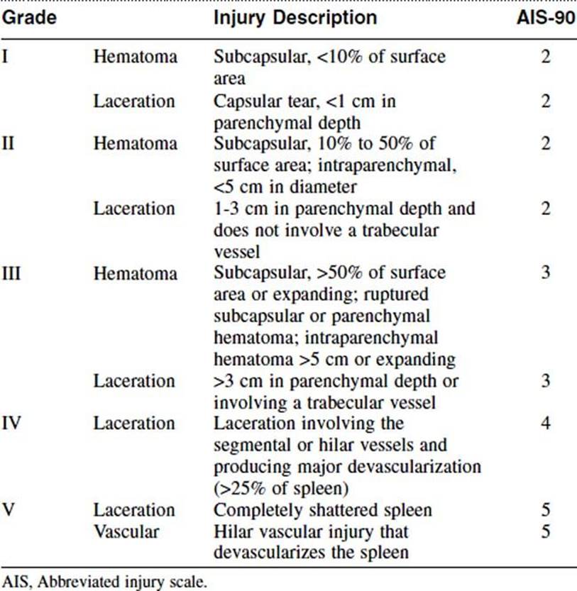
The spleen is the most commonly injured organ after blunt abdominal trauma. The patient described in this question has a grade II splenic laceration as diagnosed on CT (Table 10-1). Approximately 30% of all splenic injuries are treated operatively on arrival at the hospital. Of the remaining 60% to 70%, 80% to 90% of these are treated successfully with nonoperative management. Failure rates increase with the grade of injury, with a failure rate of 10% for grade I and II injuries, which increases to 75% for grade V injuries. Contraindications to conservative management include hemodynamic instability after adequate resuscitation, requirement for transfusion, and peritonitis. Independent risk factors for failure of nonoperative management include age older than 55 years, the presence of a pseudoaneurysm, and the amount of hemoperitoneum present on the initial CT. This patient fails conservative management because of continued hemodynamic instability and a requirement for transfusion. This necessitates laparotomy for splenectomy. The indication for angiography is the presence of an arteriovenous fistula or pseudoaneurysm on either initial or repeated CT, and it is usually performed 24 to 48 hours after admission with a splenic injury of grade III or higher.
TABLE 10-1 Spleen Organ Injury Scale—1994 Revision by the American Association for the Surgery of Trauma
Answer
E
14 A 38-year-old car mechanic is taken to the emergency department after having been pinned underneath a car. On chest radiography, multiple rib fractures are noted, as well as an air-fluid level consistent with the stomach being above the level of the left diaphragm (Figure 10-1). Which of the following statements regarding this injury is true?
A Right-sided diaphragmatic rupture is more common than left-sided rupture.
B There is a 60% incidence of coexisting pelvic fractures.
C The best initial radiographic assessment for this type of injury is FAST.
D There is a 40% incidence of normal chest radiographic findings in patients with a documented diaphragmatic injury.
E There is a 60% incidence of coexisting thoracic aortic injury.
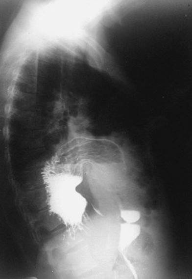
Figure 10-1 Lateral chest film showing herniation of the stomach into the left pleural cavity.
Ref.: 15
Comments
See Question 15.
Answer
D
15 For the patient in Question 14, what is the most appropriate next step in management?
A FAST
B Laparotomy
C CT scan
D Thoracotomy
E Laparoscopy
Ref.: 15
Comments
Diaphragmatic injuries/rupture occur in 3% to 5% of patients suffering major blunt abdominal trauma. Although this injury is uncommon, it is associated with a high incidence of coexistent injuries, including pelvic fractures (40%), hepatic and splenic injuries (25%), and rupture of the thoracic aorta (5%). The left side is affected three times more often than the right side. Chest radiography, despite a 40% incidence of negative findings in the face of known injury, is still the best initial diagnostic modality. The diagnostic accuracy of CT and FAST for detecting injury to the diaphragm is low. Once a diagnosis of diaphragmatic rupture has been made, treatment is operative. In this situation, in which diaphragmatic rupture is strongly suspected, laparoscopy is typically avoided because insufflation of the abdomen may cause tension pneumothorax. In the acute setting of diaphragmatic injury, laparotomy is the preferred operative approach. It allows reduction of any organs back into the abdominal cavity, as well as thorough inspection of all intraperitoneal contents. Diagnosing small diaphragmatic injuries can be difficult inasmuch as up to almost half of all patients will have normal findings on physical examination at initial evaluation. Missed diaphragmatic injuries tend to enlarge over time, which may lead to herniation and strangulation of abdominal organs. Primary repair is not usually feasible because of the rapid atrophy of diaphragmatic muscle fibers. A thoracotomy is generally performed for a chronic diaphragmatic hernia since it provides better access to the adhesions usually found in the chest.
Answer
B
16 A 30-year-old man suffers a stab wound to the right anterior aspect of his chest, approximately 3 cm inferior to the middle portion of the clavicle. Paramedics reported a weakly palpable pulse 5 minutes earlier in the ambulance, but on admission, no palpable pulses are present. His pupils are reactive. What is the initial surgical approach?
A Median sternotomy
B Right-sided clavicular incision
C Right-sided anterolateral thoracotomy
D Right-sided posterolateral thoracotomy
E Left-sided anterolateral thoracotomy
Ref.: 4, 16, 17
Comments
An emergency department thoracotomy differs from an emergency thoracotomy in that the patient is so unstable that transportation to the operating room is precluded. It is performed for a select group of patients, with the overall survival rate being dependent on the mechanism and ranging from 1.5% to 19%. It is least successful in patients with injuries caused by a blunt mechanism and is therefore usually reserved for those who initially have vital signs present but then lose these signs in the emergency department. In patients with a penetrating mechanism, emergency thoracotomy is indicated for those who lost their pulse and blood pressure either in the emergency department after initial evaluation or during transport to the emergency department. It is of highest use when cardiac tamponade or severe thoracic hemorrhage is suspected. The thoracotomy is performed via an anterolateral approach, regardless of the supposed side of injury, and allows release of pericardial tamponade, open cardiopulmonary resuscitation, and aortic cross-clamping. Injuries to the ascending aorta, innominate artery, proximal right subclavian artery, and either carotid are best managed with a median sternotomy. A right-sided clavicular incision is appropriate for a midsubclavian arterial injury. A right anterolateral approach is appropriate for injuries to the lung and chest wall. The right-sided posterolateral approach is the appropriate exposure for injuries to the trachea and midesophagus. A left posterolateral thoracotomy is used for exposure to the descending aorta and hilum.
Answer
C
17 A 17-year-old man arrives at the emergency department after having suffered a stab wound to the anterior aspect of his abdomen in the right upper quadrant. Entrance into the peritoneum is unclear on physical examination, and diagnostic peritoneal lavage (DPL) is performed. One liter of 0.9% normal saline is introduced into the abdomen, with 800 mL being retrieved. It is sent to the laboratory and returns with a value of 20,000 red blood cells/mm3. What is the most appropriate next step in management?
A CT scan of the abdomen and pelvis
B Observation with serial abdominal examinations
C Suture repair of the stab wound
D Exploratory laparotomy
E Discharge home with wet-to-dry dressing changes for the stab wound
Ref.: 4
Comments
Diagnostic peritoneal lavage is a very sensitive but nonspecific test that can be used for either blunt or penetrating trauma. In this situation, DPL is used to determine intraperitoneal injury. It is performed by introducing a catheter into the abdomen via the Seldinger technique and then infusing 1 L of 0.9% normal saline. The fluid is then returned by gravity and sent to the laboratory for analysis. For penetrating injuries, a positive DPL is defined as one with greater than 10,000 red blood cells/mm3, 500 white blood cells/mm3, or the presence of bilious/particulate material. In this patient with a positive diagnostic peritoneal lavage, the next most appropriate step in management is exploratory laparotomy.
Answer
D
18 A 21-year-old man is taken to the emergency department with a gunshot wound to the right side of his chest. The first wound is located 2 cm lateral to the right nipple, and a second wound is present just medial to the tip of the right scapula. Vital signs on initial evaluation are a heart rate of 126 beats/min and a systolic blood pressure of 88 mm Hg. A right-sided chest tube is placed, with return of 1200 mL of blood. He is resuscitated with 2 L of lactated Ringer solution, and his vital signs return to within normal limits. His chest tube output is rechecked 4 hours later, and the total amount in the collection container is 2300 mL. What is the next most appropriate step in management?
A Chest CT
B Immediate complete blood count
C Thoracotomy
D Immediate arterial blood gas analysis
E Admission to the ICU for continuous cardiac monitoring and pulse oximetry
Ref.: 4, 16
Comments
This patient has a massive hemothorax, which is defined as greater than 1500 mL of blood loss on initial placement of tube thoracostomy or loss greater than 200 mL/h for 4 hours after the initial return. Emergency thoracotomy is indicated not only for massive hemothorax but also for cardiac tamponade, major injuries to the tracheobronchial system, and injuries to the esophagus.
Answer
C
19 The head and face of 45-year-old man were assaulted with fists and feet. He arrives at the emergency department with incomprehensible responses to questions, eyes opening to pain only, and a flexor response to pain. What is this patient’s Glasgow Coma Scale (GCS) score?
A 5
B 6
C 7
D 8
E Not enough information given to calculate
Ref.: 18
Comments
See Question 20
Answer
C
20 For the patient in the previous clinical scenario, what is the next best step in management?
A CT scan of the head
B Continue to perform primary and secondary surveys
C Chest radiography
D Elevation of the head of the bed 30 degrees
E Endotracheal intubation
Ref.: 19
Comments
The first step in management for any trauma patient is assessment of the patient’s airway. It is not appropriate to continue the primary survey until the airway has not only been assessed but also either deemed secure or made secure. This patient has a Glascow Coma Scale score of 7 and is unable to protect his airway. In addition, hypoxia and hypercapnia develop in patients with severe head injury and have deleterious effects on intracranial pressure (ICP) and cerebral perfusion pressure (CPP). Obtaining a secure airway thus allows control of oxygenation and ventilation.
Answer
E
21 For the patient in Question 19, head CT shows a 2-cm subdural hematoma without a midline shift. The patient is taken to the operating room and the hematoma is evacuated. Postoperatively, he is admitted to the ICU intubated and sedated with an ICP monitor in place. Which of the following treatment measures can be used to maintain adequate CPP?
A Hyperventilation to a PCO2 of 25 mm Hg
B Mannitol, 1 g/kg intravenously for 3 days
C Hydrocortisone, 100 mg intravenously every 8 hours for 3 days
D Strict blood pressure control to a systolic range of 90 to 100 mm Hg
E Reverse Trendelenburg positioning of the bed at all times
Ref.: 4, 18, 20
Comments
The overall goal in treating patients with traumatic head injury is maintaining adequate cerebral blood flow. An estimate of this flow is obtained by calculating cerebral perfusion pressure (CPP = mean blood pressure − ICP). The goal CPP in an adult is 60 to 70 mm Hg. Therefore, patients should be aggressively volume-resuscitated to maintain adequate mean blood pressure. ICP monitors are indicated in patients with a GCS score of 3 to 8 and either (1) an abnormal finding on CT of the head or (2) any two of the following: (a) age older than 40 years, (b) posturing response to pain, or (c) systolic blood pressure less than 90 mm Hg. Overall, there is a 5% infection and 1% hematoma formation rate associated with ICP-monitoring catheters. Treatment measures used to decrease ICP include elevation of the head of the bed to 30 degrees or more, hyperventilation of patients to a PCO2 of 30 to 35 mm Hg, barbiturates, and mannitol at a dose of 1 g/kg intravenously. PCO2 should not be kept below 30 mm Hg to avoid worsening the cerebral ischemia. Steroids do not have a role in the treatment of acute traumatic head injury.
Answer
B
22 A 32-year-old construction worker is taken to the emergency department after having fallen off a roof 4 hours earlier. He has no neurologic function below cervical spine level 5. CT shows C5 and C6 anterior wedge fractures, with compromise of the spinal canal. Which of the following statements is true regarding this type of injury?
A The current treatment standard is to administer methylprednisolone intravenously on arrival at the emergency department.
B Approximately 50% of all spinal fractures occur in the cervical vertebrae.
C Neurogenic shock is characterized by hypertension with bradycardia.
D Only 15% of patients with neck pain have a true cervical spine injury.
E Methylprednisolone therapy should be continued for 72 hours after the time that the injury occurred.
Ref.: 21
Comments
Approximately 10,000 patients a year suffer a spinal cord injury. Of extreme importance in the early management of these patients is strict spinal immobilization. Seventy-five percent of all spinal fractures are located in the cervical spine. However, just 5% of patients with neck pain after trauma have a true cervical spine injury. Initial radiologic assessment is accomplished with anteroposterior, lateral, and open-mouth odontoid projections. If these films are equivocal or the C7/T1 interface is not well visualized, CT is a useful addition. In patients who do suffer a spinal cord injury, neurogenic shock can develop, which is caused by loss of sympathetic vascular tone and leads to hypotension and bradycardia. Corticosteroid use has been shown to be useful when initiated in less than 3 hours after the incident occurred. In this instance, it should be given initially as a 30-mg/kg bolus of methylprednisolone over a 1-hour period, followed by 5.4 mg/kg/h for 23 more hours. If the patient is seen, as in this case, between 3 and 8 hours after the incident, steroids should be continued for 48 hours. Steroids have not been shown to have a role in the treatment of penetrating injuries to the spinal cord.
Answer
A
23 Regarding pelvic fractures, which of the following statements is true?
A Patients with a pelvic fracture have 30% mortality if hypotension is initially present.
B A pubic symphysis diastasis of 3 cm doubles the volume of potential pelvic space for a hematoma.
C When a laparotomy is performed in a patient with an unstable pelvic fracture, it is imperative to make a large surgical incision (xiphoid to symphysis) to allow better visualization in the pelvis.
D The most common cause of death following an open pelvic fracture is overwhelming infection and sepsis.
E The external iliac artery is commonly involved in severe pelvic fractures.
Ref.: 22, 23
Comments
A pelvic fracture is diagnosed in approximately 10% to 20% of trauma patients. The overall mortality rate associated with a pelvic fracture is 10% to 15%, but this rate increases to almost 50% when hypotension, an open component, or both exist. The most common cause of death from open pelvic fractures is uncontrollable hemorrhage. The open component prevents tamponade within the pelvic space. After a pelvic fracture is diagnosed or suspected, a thorough examination of the perineum must be conducted to rule out an open component. Because a pubic symphysis diastasis of 3 cm doubles the potential space for a hematoma to form, early attempts to stabilize the pelvis are extremely important. Such stabilization can be performed through noninvasive means (wrapping the pelvis in a sheet, applying a pelvic girdle) or invasive means (external fixation, pelvic C-clamp). If a laparotomy is required because of an intraabdominal injury, a large incision below the semilunar line should be avoided. Once an incision is made below this level, the pelvic hematoma secondary to the fracture will decompress and lead to massive bleeding. If this does occur, after the other injuries have been addressed as quickly as possible, the pelvis should be packed tightly and the patient returned to the ICU for stabilization. Once stabilized, the patient can either undergo angiography or return to the operating room. The external iliac artery courses deep to the inguinal ligament and is rarely involved in pelvic fractures. The superior and inferior gluteal arteries, obturator artery, and internal pudendal artery, all branches of the internal iliac, are most frequently injured.
Answer
B
24 An 18-year-old man undergoes emergency laparotomy for multiple gunshot wounds to the abdomen. On entering the abdomen there is a large amount of hemoperitoneum. All four quadrants are packed and the packing is then removed. The injuries found are five small intestine enterotomies, a through-and-through injury to the transverse colon, and a 3-cm laceration of the infrarenal aorta. The best management of the aortic injury is:
A Primary repair
B Saphenous vein patch angioplasty
C In situ placement of a polytetrafluoroethylene (PTFE) graft
D Aortic ligation above the level of injury with a bilateral axillofemoral bypass
E Aortobifemoral bypass with a PTFE graft
Ref.: 6
Comments
Although it has been traditional teaching that artificial grafts not be used in an infected or contaminated field, this is one circumstance in which it is common practice. After débridement of the aortic injury, the large size of the defect prohibits primary repair or vein patch angioplasty. Axillofemoral and aortobifemoral bypasses are time-consuming operations that this patient will probably not tolerate. In situ placement of a PTFE graft is the preferred operation, and all care should be taken to thoroughly irrigate the abdomen before placement of the graft. In addition, the repair should be covered by omentum after completion.
Answer
C
25 A 32-year-old woman is a restrained passenger in a high-speed motor vehicle collision. In the emergency room she is found to be nontachycardic with a systolic blood pressure of 110 mm Hg. Blood is drawn for determination of the hemoglobin concentration, which is noted to be 12.2 g/dL. On FAST, a moderate amount of fluid is seen in the right upper quadrant, between the liver, kidney, and diaphragm. What is the next most appropriate step in management?
A Laparotomy
B CT scan of the abdomen and pelvis with intravenous contrast enhancement
C Angiography for embolization of the liver laceration
D DPL
E Observation with serial abdominal examinations
Ref.: 4, 24
Comments
See Question 26.
Answer
B
26 A grade IV liver laceration is diagnosed in the patient from Question 25 (Table 10-2). Her vital signs 6 hours after admission are a pulse of 100 beats/min and systolic blood pressure of 105 mm Hg. What is the next most appropriate step?
A Exploratory laparotomy
B Angiography
C Repeated FAST
D Diagnostic laparoscopy
E Repeated hemoglobin determination
TABLE 10-2 Liver Injury Scale
|
Grade |
Injury |
Description of Injury |
|
I |
Hematoma |
Subcapsular, <10% of surface area |
|
Laceration |
Capsular tear, <1 cm in parenchymal depth |
|
|
II |
Hematoma |
Subcapsular, 10% to 50% of surface area |
|
Laceration |
1-3 cm in parenchymal depth, <10 cm in length |
|
|
III |
Hematoma |
Subcapsular, >50% of surface area or expanding, ruptured subcapsular or parenchymal hematoma |
|
Laceration |
>3 cm in parenchymal depth |
|
|
IV |
Laceration |
Parenchymal disruption involving >75% of the hepatic lobe or 1-3 Couinaud segments within a single lobe |
|
V |
Laceration |
Parenchymal disruption involving >75% of the hepatic lobe or >3 Couinaud segments within a single lobe |
|
Vascular |
Juxtahepatic venous injuries (retrohepatic vena cava, central major hepatic veins) |
|
|
VI |
Vascular |
Hepatic avulsion |
From the American Association for the Surgery of Trauma, www.aast.org. Derived originally from Moore EE, Cogbill TH, Jurkovich GJ, et al: Organ injury scaling: spleen and liver, J Trauma 38:323–324, 1995.
Ref.: 4, 24
Comments
The overall success rate for nonoperative treatment of blunt hepatic injuries is about 90% for all levels of injury. Patients with grade IV and V injuries are able to be treated without surgery between 75% and 80% of the time. Requirements for nonoperative therapy include hemodynamic stability, no signs or symptoms of peritonitis, and a transfusion requirement of no more than 2 to 4 units of packed red blood cells. This patient is hemodynamically stable, and therefore further localization of the intraabdominal injury with CT initially is warranted. The initial CT not only can localize the injury but, in the case of solid organ injuries, can also provide information regarding active hemorrhage. She should be closely monitored in an intensive care setting with serial abdominal examinations and hemoglobin determinations. Angiography is a helpful adjunct to nonoperative treatment, but it is usually reserved for situations in which active extravasation or a “blush” is seen on CT. Repeated CT is advised for patients who do experience a decrease in their hemoglobin to reevaluate the liver damage and look for any active extravasation that would be amenable to angiographic embolization.
Answer
E
27 A patient with a grade V blunt liver injury is discharged home on hospital day 7 without needing operative intervention. She returns to the clinic 2 months after discharge with persistent dull continuous right upper quadrant pain. She denies any fevers or chills, and all laboratory studies, including a hepatic function panel, are within normal limits. CT of the abdomen and pelvis is performed and reveals a localized homogeneous fluid collection directly adjacent to the liver. What is the correct diagnosis and treatment?
A Hemobilia; angiography with embolization
B Biloma; CT- or ultrasound-guided percutaneous drainage
C Biloma; exploratory laparotomy with external drainage
D Hepatic necrosis; CT- or ultrasound-guided percutaneous drainage
E Hepatic necrosis; exploratory laparotomy with wide débridement and drainage
Ref.: 4, 24
Comments
Because of the increasing number of patients with significant liver lacerations being treated (successfully) nonoperatively, posttreatment complications are being encountered more often. Such complications include hemobilia, biloma, hepatic necrosis, and abscess. Hemobilia occurs when a connection exists between the biliary and arterial systems, and it is typically manifested as right upper quadrant pain, melena, and jaundice. Hemobilia can be diagnosed by CT with intravenous contrast enhancement or upper endoscopy and is usually treated by angiography with embolization. Patients with hepatic necrosis or abscess (or both) typically have right upper quadrant pain, fever, leukocytosis, and at times, localized peritonitis. It can be appreciated on CT with intravenous contrast enhancement as nonperfused liver parenchyma sometimes associated with a heterogeneous adjacent fluid collection. This condition warrants laparotomy with débridement. Bilomas occur as a result of leakage of bile and typically close spontaneously over time. The fluid collections themselves are best treated with radiologically guided percutaneous drainage when localized as it is in this patient. If the fluid collection is not amenable to percutaneous drainage, endoscopic retrograde cholangiopancreatography (ERCP) is recommended because biliary stents and sphincterotomy can reduce intrahepatic biliary pressure and increase healing.
Answer
B
28 A 22-year-old man undergoes exploratory laparotomy for a transpelvic gunshot wound. A 2-cm partial-thickness laceration is found in the proximal portion of the intraperitoneal rectum. What is the appropriate surgical management of this injury?
A Hartmann procedure—end colostomy with oversewing of the distal rectal stump
B Primary repair with a diverting loop colostomy
C Primary repair with presacral drainage
D Presacral drainage only
E Primary repair only
Ref.: 4, 25
Comments
Repair of rectal injury largely depends on location (i.e. intraperitoneal versus extraperitoneal). The posterior rectum and distal third of the anterior rectum are not serosalized, and injury in these regions is considered extraperitoneal. Nondestructive lacerations that are less than 50% of the circumference of the rectal wall should be repaired primarily after débridement of any devitalized tissue in the absence of peritonitis. This therapy differs sharply from that of extraperitoneal rectal injuries, which should be treated chiefly by fecal diversion. Presacral drainage, which historically had been used rather routinely, has been decreasing in use and has not been shown to decrease the complication rate.
Answer
E
29 All of the following are damage control treatment options for unstable patients with a ureteral injury except:
A Ligation of the injured ureter
B Percutaneous nephrostomy
C Ureteral drainage via luminal cannulation
D Transureteroureterostomy
E Placement of a bridging stent
Ref.: 4, 26
Comments
When a trauma patient is unstable on the operating room table and damage control has been initiated, time should not be spent on primary repair of a ureteral injury, thus making transureteroureterostomy inappropriate at this time. Surgical options for this type of situation consist of simple ligation of the ureter, placing a percutaneous nephrostomy through the renal parenchyma into the renal pelvis, inserting a catheter into the proximal end of the damaged ureter and bringing it out through the wound, and placing a catheter or stent in the proximal and distal ends of a small-segment ureteral injury.
Answer
D
30 Regarding orotracheal intubation in an injured patient, which of the following statements is true?
A Succinylcholine is a neuromuscular blocking agent that can cause hyperkalemia, but it can be used safely in spinal cord injury and burn patients within the first 72 hours after injury.
B During rapid-sequence intubation, preoxygenation should occur for 3 minutes via bag-valve-mask ventilation.
C Ketamine has a quick onset of action but can cause tachycardia and hypertension.
D Etomidate is contraindicated in patients with a suspected brain injury or open globe because of its side effect of increasing ICP.
E Cervical spine immobilization during intubation should be maintained with a rigid cervical collar.
Ref.: 4, 19
Comments
The most commonly used method for securing a trauma patient’s airway is orotracheal intubation. Rapid-sequence intubation consists of preoxygenating the patient for 3 minutes with bag-valve-mask ventilation, maintaining in-line cervical stabilization, applying cricoid pressure, administering paralytic/induction agents, performing laryngoscopy, and then placing an endotracheal tube. In-line cervical stabilization should be maintained with the help of an assistant, not a rigid cervical spine collar. Etomidate is a common induction agent used in trauma victims. It has a quick onset of action and is indicated for patients with a suspected brain injury or open globe because it typically does not cause an increase in ICP. Two of ketamine’s common side effects are tachycardia and increased blood pressure. However, it has a longer onset of action. Succinylcholine does cause a rise in the serum potassium concentration and can lead to severe hyperkalemia in patients with burns or spinal cord injury. However, this does not occur within the first 24 hours after the injury. Even though some studies have advocated safe use of succinylcholine following a major burn, most authors recommend avoiding its use beyond 24 hours.
Answer
B
31 An 18-year-old man was involved in a high-speed motor vehicle collision and was ejected from the vehicle. He was found approximately 100 feet from the vehicle by emergency personnel. He arrives at the emergency department hemodynamically stable. CT of the chest, abdomen, and pelvis with intravenous contrast enhancement is performed approximately 5 hours after the time of the accident. Of note, the left kidney does not enhance. What is the most appropriate treatment at this time?
A Observation only
B Laparotomy with renal artery repair
C Laparotomy with renal vein repair
D Laparotomy with left nephrectomy
E Angiography
Ref.: 26
Comments
This patient has an ischemic kidney secondary to injury to the renal vasculature. Revascularization, if attempted, should be done within 4 hours of the time of injury. Since this patient is not a candidate for revascularization, angiography is not indicated. However, as in this case, when the kidney has a prolonged warm ischemia time, laparotomy is also not necessarily indicated. Avascular kidneys should not be resected if possible. Typically, they involute over time without complication and can sometimes regain some level of function. This patient should continue to be monitored, however, with serial abdominal examinations and for signs and symptoms of bleeding.
Answer
A
32 With regard to retroperitoneal hematomas, which of the following statements is true?
A Zone 3 (pelvic hematoma) should be explored whether secondary to a blunt or penetrating traumatic injury.
B A stable hematoma in zone 1 (midline) should not be explored because treatment of vascular injuries in this zone are best managed by embolization.
C Zone 2 (perinephric hematomas) should be explored when the injury is due to a penetrating mechanism.
D The Mattox maneuver allows the right-sided abdominal organs (right colon, duodenum, right kidney) to be reflected medially for exposure of the abdominal aorta.
E All zone 1 injuries should be observed in patients with hemodynamic stability.
Ref.: 7
Comments
The most important concept to learn regarding treatment of retroperitoneal injuries is the anatomy. Zone 1 is composed of the midline retroperitoneum and is divided into supramesocolic and inframesocolic segments, with the transverse mesocolon used as the dividing line. The perinephric spaces constitute zone 2, whereas the pelvic retroperitoneum is referred to as zone 3. All zone 1 injuries should be explored, regardless of mechanism, because of the major vascular structures located there. However, zone 2 and 3 injuries are treated similarly in that they should be explored only for a penetrating mechanism. Zone 3 hematomas are most commonly associated with pelvic fractures, and treatment is directed at fixation and angiographic embolization.
Answer
C
33 An 18-year-old college football player is struck in the chest during a game and sustained multiple anterior rib fractures. His vital signs are a heart rate of 98 beats/min and an irregular systolic blood pressure of 110 mm Hg. The initial work-up for a cardiac injury includes all of the following except:
A Electrocardiogram (ECG)
B Chest radiograph
C FAST
D Troponin levels
E Echocardiogram
Ref.: 4, 27
Comments
See Question 34.
Answer
B
34 Which of the following statements is true regarding blunt cardiac trauma?
A The most commonly involved chamber of the heart is the left ventricle.
B All patients suspected of having an injury should undergo transthoracic echocardiography.
C Arrhythmias are the most common clinically significant symptoms.
D The most common arrhythmia is ventricular fibrillation.
E Approximately 5% of all patients with chest trauma have cardiac involvement.
Ref.: 4, 27
Comments
Approximately 10% to 20% of all patients with blunt chest trauma have cardiac involvement. Arrhythmias are the most common clinically significant symptom, with ST-segment and T-wave changes being found most often. The right ventricle, which is the anterior-most chamber, is the most commonly involved. Evaluation of these patients includes FAST to rule out tamponade, an ECG, and troponin levels. The ECG and troponin levels should be obtained both on admission and 8 hours afterward. Transthoracic echocardiography is indicated only in patients with hemodynamic instability or the presence of arrhythmias (as in the patient described).
Answer
C
35 A 20-year-old woman is involved in a high-speed motor vehicle accident with significant damage to the front of the car. She arrives in the emergency department with a GCS score of 15, heart rate of 102 beats/min, respiratory rate of 18 breaths/min, and a systolic blood pressure of 108 mm Hg. A chest radiograph is obtained and demonstrates a 10-cm mediastinum and deviation of the left main stem bronchus. What is the most appropriate next step in management?
A Observation
B Left-sided chest tube
C Repeated chest radiograph in 6 hours
D CT angiogram of the chest
E Transesophageal echocardiogram
Ref.: 28
Comments
A widened mediastinum (>8 cm), deviation of the left main stem bronchus, tracheal deviation, an indistinct aortic knob, and apical caps are all signs of aortic injury on chest radiography. Although a transesophageal echocardiogram can visualize the proximal descending aorta, it is limited in evaluating the ascending aorta and its arch. In addition, this is an invasive procedure that requires some level of sedation. CT angiography is an excellent screening test for aortic injury with a high negative predictive value.
Answer
D
36 A full-thickness injury to the aorta directly distal to the origin of the left subclavian artery is diagnosed in the patient in Question 35. What is the correct surgical approach for repair?
A Left supraclavicular approach
B Left infraclavicular approach
C Median sternotomy
D Left anterolateral thoracotomy
E Left posterolateral thoracotomy
Ref.: 28
Comments
A left posterolateral thoracotomy in the fourth intercostal space is the best operative approach for a descending aortic injury. Injuries to the ascending aorta, innominate artery, proximal right subclavian artery, and either carotid are best managed with a median sternotomy. A left-sided clavicular incision is appropriate for a midsubclavian arterial injury. A left anterolateral thoracotomy is used for emergency department thoracotomies because it provides access to the pericardium and thoracic aorta, thereby allowing open cardiac massage and aortic cross-clamping.
Answer
E
37 Which of the following statements regarding blunt aortic injury is true?
A It is estimated that patients with a contained rupture have a 1% per hour rate of rupture within the first 48 hours after injury.
B The proximal descending aorta is the most commonly injured and involves approximately 80% of all cases.
C Overall operative mortality after repair is between 30% and 40%.
D Paraplegia occurs after operative repair in less than 5% of patients.
E Approximately 20% of chest radiographs are falsely negative.
Ref.: 28
Comments
It is estimated that 85% of all patients with a full-thickness aortic injury die at the scene of the injury. About 60% of aortic injuries occur in the proximal descending segment, at the origin of the left subclavian artery. Other less common locations are the ascending aorta, the arch, or the level of the diaphragm. Approximately 5% to 10% of patients with an aortic injury will have normal findings on chest radiography. Operative repair should be timely because there is a 1% per hour rupture rate within the first 48 hours after injury. Operative repair is associated with 5% to 25% mortality, and paraplegia occurs in 10% of patients postoperatively.
Answer
A
38 A 14-year-old girl is an unrestrained passenger in a motor vehicle collision. She complains of right lower extremity pain. On examination, her knee is markedly swollen and has a notably limited range of motion. The foot is cool to the touch, and the dorsalis pedis and posterior tibialis pulses are absent. Radiographs of the right lower extremity show no fracture. What is the most appropriate next step in management?
A CT of the right lower extremity
B Ankle-brachial indices of both lower extremities
C Angiography
D Operative revascularization with four-compartment fasciotomy
E Operative revascularization without fasciotomy
Ref.: 7
Comments
Posterior knee dislocations are associated with a popliteal artery injury approximately 33% of the time. This type of dislocation occurs when a direct force is applied to a flexed knee. Because of the absence of extensive collateral flow around the knee joint, an injury to the popliteal artery is associated with a fairly high amputation rate, up to 20%. It is imperative to recognize and repair this type of injury early. In this patient, the lack of distal pulses warrants a direct trip to the operating room, without need for any further diagnostic testing. Because of the high rate of amputation for this type of injury, it is recommended that a fasciotomy be performed at the time of revascularization when a patient has evidence of ischemia, as in this clinical scenario.
Answer
D
39 Select the correct statement regarding flail chest:
A It occurs when three or more adjacent ribs are fractured in one place.
B Work of breathing is increased secondary to paradoxical chest wall motion.
C Patients with flail chest should be aggressively resuscitated because of the probable development of a pulmonary contusion.
D Patients with this condition should be prophylactically intubated secondary to a high likelihood of respiratory failure.
E If a patient does require mechanical ventilation, it is important to avoid the use of positive end-expiratory pressure.
Ref.: 4
Comments
Flail chest occurs when three or more adjacent ribs are fractured in at least two places. This leads to a segment of chest wall that has the opposite movement with respirations and thereby increases the patient’s work of breathing. Most often, this condition can be treated with vigilant pain control and aggressive pulmonary toilet. Pulmonary contusions often occur with flail chest and are not fully appreciated on chest radiographs until 24 to 48 hours after injury. Fluid resuscitation should be conservative so that any developing pulmonary contusions are not worsened further. However, if respiratory failure does develop and mechanical ventilation is needed, positive end-expiratory pressure is important to maintain functional residual capacity.
Answer
B
40 An 8-year-old child hits a curb with his bicycle, which causes him to flip over the handlebars. He had no initial sequelae and was monitored at home by his parents. However, 2 days after the incident, he begins having nonbilious emesis. He is brought to the emergency department and undergoes CT of the abdomen and pelvis, which demonstrates a duodenal hematoma. What is the next step in management?
A Initiation of nil per os (NPO) status and gastric decompression with a nasogastric tube
B Esophagogastroduodenoscopy to assess for luminal compromise
C Drainage of the hematoma via laparoscopy
D Drainage of the hematoma via laparotomy
E Resection of the injured portion of the duodenum with primary anastomoses
Ref.: 4, 29
Comments
Blunt injuries to the duodenum can be difficult to diagnose. Duodenal hematomas typically occur up to 3 days after injury with a gastric outlet obstruction type of clinical picture. The duodenal lumen is narrowed because of the hematoma itself and the associated edema. CT with oral contrast enhancement and upper gastrointestinal studies are useful in diagnosing this condition. If no other indication exists for exploration, treatment is conservative and consists of placement of a nasogastric tube for decompression. Typically, these hematomas and their symptoms resolve within 7 to 15 days after injury. Operative exploration is reserved for patients in whom the symptoms do not resolve within this period.
Answer
A
41 Which of the following statements regarding FAST is true?
A A 2.5-MHz convex-array transducer should be used.
B The hepatorenal space, known as the Morison pouch, is viewed between the eleventh and twelfth ribs in the right midaxillary line.
C The splenorenal space is evaluated between the ninth and eleventh ribs in the left midaxillary line.
D The bladder should preferentially by emptied before examination to allow better visualization of fluid in the pelvis.
E FAST is an important part of the primary survey.
Ref.: 30
Comments
Focused assessment for the sonographic examination of trauma patients is performed as part of the ATLS secondary survey. A 3.5-MHz convex-array transducer is used to evaluate for the presence of fluid in the abdomen. Four areas are to be examined. The first is the pericardial window, which is viewed with the transducer placed subxiphoid. The hepatorenal space is evaluated in the right midaxillary line, between the eleventh and twelfth ribs. The splenorenal space is evaluated in the right posterior axillary line, between the ninth and eleventh ribs. The last area examined is the pouch of Douglas in the pelvis. This rectouterine/rectovesical space is evaluated with the transducer placed approximately 3 cm above the pubic symphysis. A full bladder actually helps elucidate the presence of blood in this space, and Foley catheters should be placed after FAST has been performed.
Answer
B
42 An 18-year-old man arrives at the emergency department with a stab wound in the right upper quadrant of his abdomen. His initial vital signs are a heart rate of 122 beats/min and mean arterial pressure of 50 mm Hg. Two liters of lactated Ringer solution is infused. On physical examination, his abdomen is distended and tender to palpation. After the 2 L of fluid, the patient’s repeated vital signs are as follows: heart rate, 130; mean arterial pressure, 48. What is the next most appropriate step in management?
A Transfusion of 2 units of packed red blood cells
B DPL
C CT scan of the abdomen and pelvis with intravenous contrast enhancement
D Diagnostic laparoscopy
E Exploratory laparotomy
Ref.: 1, 4
Comments
This patient is hemodynamically unstable and has a penetrating wound to the abdomen. Hypotension despite initial resuscitation, evisceration, and peritoneal signs are all clear indications for emergency laparotomy in the setting of a penetrating injury. Further diagnostic testing at this time is unnecessary.
Answer
E
43 During exploratory laparotomy in a patient with multiple gunshot wounds to the abdomen, a through-and-through gunshot wound is noted in the left lobe of the liver. Brisk bleeding is seen from the bullet track. All of the following operative maneuvers for this injury are appropriate except:
A Pringle maneuver
B Tractotomy
C Omental packing
D Ligation of the proper hepatic artery
E Large mattress sutures traversing the bullet track
Ref.: 4, 24
Comments
In regard to hepatic injuries, there are three overall goals of treatment: (1) control of hemorrhage, (2) débridement of nonviable tissue, and (3) adequate drainage. Multiple operative techniques can be used to establish control of bleeding, and often a combination of these techniques are used. The Pringle maneuver is direct compression of the portal triad, either manually or with a vascular clamp. This takes a small amount of time and is helpful in identifying whether the bleeding source is from the triad, hepatic veins, or retrohepatic vena cava. It is important to keep note of how long compression is applied because these patients tend to be hypovolemic and hypothermic and as a rule do not tolerate hepatic ischemia well. Omental packing is performed by first creating a pedicle of omentum and then placing it across or in the defect. This creates a well-vascularized “packing” of the liver that also has its own natural hemostatic properties. Large mattress sutures have been used for quite some time and work by compressing the bullet track with the surrounding liver parenchyma. A tractotomy is the act of opening the already present wound to fully examine the track and identify the bleeding vessels. This then allows directed individual vessel ligation. Historically, selective hepatic artery ligation has been used and involves ligation of the hepatic artery branch to the involved lobe. Although this is still a viable option, it is associated with a fairly high rate of abscess formation and hepatic necrosis. The proper hepatic artery, however, should not be ligated.
Answer
D
44 A 58-year-old man is an unrestrained front seat passenger in a high-speed motor vehicle collision. In the emergency department he has stable vital signs, and the only finding on physical examination is a left leg that is flexed at the hip, adducted, and internally rotated. A radiograph is obtained and confirms posterior dislocation of the left hip. The next step in management is:
A Immediate closed reduction, either in the emergency department or in the operating room
B CT scan to diagnose intraarticular bone fragments
C Admission to the hospital and splinting of the left hip and thigh
D Placement of skeletal traction
E Open reduction
Ref.: 23
Comments
Posterior hip dislocations occur most commonly when direct force is applied to the knees of a person whose hips and knees are in a flexed position (i.e., sitting). Immediate closed reduction is imperative and can be performed with intravenous sedation in the emergency department or in the operating room under general anesthesia. The timing of the reduction is important to reduce risk for the development of further damage to the sciatic nerve and avascular necrosis of the femur. Avascular necrosis of the femoral head is a significant complication that often leads to total hip arthroplasty. After the hip is reduced, CT of the hip is recommended to evaluate the acetabulum and the intraarticular area for any fragments.
Answer
A
45 An 18-year-old man is involved in a boating accident and suffers a right open tibial fracture and an obvious arterial injury, with no detectable tibial nerve function. Which of the following statements about this situation is true?
A The patient’s age is a negative prognostic indicator in regard to the limb salvage rate.
B If this patient were to undergo primary amputation, at least 10 cm of the proximal end of the tibia should be preserved to facilitate the use of a prosthesis.
C A Mangled Extremity Severity Score (MESS) higher than 5 is a strong indicator that this patient would benefit from primary amputation.
D A warm ischemia time of longer than 4 hours is an absolute indication for primary amputation.
E Although the MESS is not specific in determining future functional limb status, it is highly sensitive in determining the need for primary amputation.
Ref.: 23
Comments
Mangled Extremity Severity Score is composed of four categories: skeletal/soft tissue injury, ischemia, shock, and age (Table 10-3). A score higher than 7 generally warrants primary amputation. However, no single scoring system has been shown to be highly specific or sensitive in determining the success of limb salvage or future limb function. Older age, ischemia time longer than 6 hours, hypotension, poor nutritional status, and severe coexisting injuries all have a negative impact on the chance of limb salvage. For best prosthetic fitting, 10 cm of the proximal end of the tibia is recommended.
TABLE 10-3 Mangled Extremity Severity Score
|
Component |
Points |
|
Skeletal and Soft Tissue Injury |
|
|
Low energy (stab, simple fracture, “civilian” gunshot wound) |
1 |
|
Medium energy (open or multiplex fractures, dislocation) |
2 |
|
High energy (close-range shotgun or “military” gunshot wound, crush injury) |
3 |
|
Very high energy (same as above plus gross contamination, soft tissue avulsion) |
4 |
|
Limb Ischemia (Doubled When >6 h) |
|
|
Pulse reduced or absent but perfusion normal |
1 |
|
Pulseless; paresthesias, diminished capillary refill |
2 |
|
Cool, paralyzed, insensate, numb |
3 |
|
Shock |
|
|
Systolic blood pressure always >90 mm Hg |
0 |
|
Hypotensive transiently |
1 |
|
Persistent hypotension |
2 |
|
Age (yr) |
|
|
<30 |
0 |
|
30-50 |
1 |
|
>50 |
2 |
From Johansen K, Daines M, Howey T, et al: Objective criteria accurately predict amputation following lower extremity trauma, J Trauma 30:568–573, 1990.
Answer
B
46 Select the correct statement regarding radiation exposure:
A Hematopoietic syndrome occurs after exposure to 5 to 10 Gy and is characterized by pancytopenia.
B Exposure to 8 Gy is associated with a 50% mortality rate.
C After exposure, the majority of radioactivity is contained in the patient’s clothing.
D Gastrointestinal syndrome occurs after exposure to 1 to 4 Gy and is characterized by nausea, vomiting, abdominal pain, and bloody diarrhea.
E Leukemia and solid tumors are known long-term effects of total body irradiation and appear 7 to 10 years after the exposure.
Ref.: 31
Comments
Radiation exposure is a rare but serious event. The effects of exposure are directly related to the dose. Exposure to less than 1 Gy typically produces no symptoms and is associated with 0% mortality. Exposure to greater than 8 Gy results in 100% mortality. Hematopoietic syndrome occurs after exposure to 1 to 4 Gy and is characterized by pancytopenia that develops within 48 hours of the event. Exposure to 8 to 12 Gy results in gastrointestinal syndrome, which is manifested as nausea and vomiting acutely and then, over a period of 1 to 2 weeks, as bloody diarrhea and death. Neurovascular syndrome is described after exposure to greater than 15 Gy and results in massive vasodilation, shock, and death.
Answer
C
47 An explosion occurs at a nearby construction site, and one worker near the blast is brought to the emergency department. He is covered in soot and has superficial partial-thickness burns on his left arm and hand (approximately 4% of total body surface area) and perforated tympanic membranes bilaterally. The remainder of his physical examination is negative. Select the correct statement regarding his injuries:
A After the patient’s burn wounds are cleaned and dressed and a hearing examination is performed, the patient is ready for discharge.
B An initial chest radiograph should be performed.
C An intestinal blast injury typically causes abdominal pain and occurs within the first 8 to 12 hours after the blast.
D Tympanic membrane perforation is the most significant indicator of mortality in patients with a blast injury.
E Blast lung injury occurs primarily because of air embolism induced by shock waves.
Ref.: 32
Comments
Victims involved in a blast or explosion can suffer from a myriad of injuries caused by burn, blunt, and penetrating mechanisms. Therefore, these injuries are categorized as primary, secondary, tertiary, or quaternary according to mechanism. Primary blast injury refers to damage to hollow visceral organs secondary to the blast wave itself. An example is tympanic membrane perforation, which is also a good screening tool for significant blast injuries. The majority of patients with tympanic membrane rupture heal without intervention. Other examples of primary blast injuries involve the lung and intestine. Intestinal blast injuries are rare, can occur anywhere in the small or large bowel, and typically have a delayed clinical manifestation (>24 hours after the blast). Blast lung injury is the most significant indicator of mortality and is primarily due to disruption of the alveolar septa with subsequent edema and hemorrhage induced by the shock waves. Overall, this leads to a clinical picture similar to ARDS. This injury can be complicated by pneumothorax, air emboli, and bronchopleural fistulas. Because of the significant morbidity and mortality associated with blast lung injury, patients involved in a blast or explosion should be screened with chest radiographs and otoscopic examination. This patient, who does have perforated tympanic membranes, should not only undergo chest radiography but also be observed for at least 12 hours before discharge. Secondary blast injuries occur from objects propelled by the blast wave. When the victim is thrown and strikes an inanimate object, the injuries are categorized as tertiary. Quaternary injuries are a miscellaneous category and consist of burns and crush injuries.
Answer
B
48 A 79-year-old woman is taken to the emergency department after a fall from standing. She has an obvious deformity of her right humerus, palpable radial and ulnar pulses, and numbness of the dorsal aspect of her forearm and hand. Select the correct statement:
A The nerve most likely injured is the median nerve.
B The presence of palpable distal pulses precludes the need for angiography.
C Patients with this nerve injury should not have their hand or wrist splinted because splinting can lead to markedly decreased range of motion.
D Operative intervention is indicated only for patients with an open component.
E The majority of the nerve injuries just described resolve without surgical intervention.
Ref.: 33
Comments
This patient has a fracture of the humerus, which commonly occurs after falls or motor vehicle collisions. The nerve injury described in this woman involves the radial nerve, the most commonly injured nerve when the fracture occurs in the distal third of the humerus. Approximately 70% of patients with radial nerve injury associated with fracture of the humerus experience resolution without surgical intervention. However, these patients should have their hand and wrist splinted during recovery. Operative intervention in patients with humeral fractures is indicated for an open component, as well as an inability to achieve adequate alignment with closed reduction. Arterial injuries can occur, especially with proximal humeral fractures, and should always be suspected, even in patients with palpable distal pulses.
Answer
E
References
1 Britt LD, Rushing GD. Penetrating abdominal trauma. In Cameron JL, editor: Current surgical therapy, ed 9, Philadelphia: CV Mosby, 2008.
2 Sims CA, Reilly PM. Penetrating neck trauma. In Cameron JL, editor: Current surgical therapy, ed 9, Philadelphia: CV Mosby, 2008.
3 Ledgerwood AM, Lucas CE. Blunt abdominal trauma. In Cameron JL, editor: Current surgical therapy, ed 9, Philadelphia: CV Mosby, 2008.
4 Hoyt DB, Coimbra R, Acosta J. Management of acute trauma. In Townsend CM, Beauchamp RD, Evers BM, et al, editors: Sabiston textbook of surgery: the biological basis of modern surgical practice, ed 18, Philadelphia: WB Saunders, 2008.
5 Cushing NM, Ness PM. Blood transfusion therapy. In Cameron JL, editor: Current surgical therapy, ed 9, Philadelphia: CV Mosby, 2008.
6 Zarzaur BL, Croce MA. The management of vascular trauma. In Cameron JL, editor: Current surgical therapy, ed 9, Philadelphia: CV Mosby, 2008.
7 Hirschberg A, Mattox KL. Vascular trauma. In Townsend CM, Beauchamp RD, Evers BM, et al, editors: Sabiston textbook of surgery: the biological basis of modern surgical practice, ed 18, Philadelphia: WB Saunders, 2008.
8 Feliciano DV. The management of extremity compartment syndrome. In Cameron JL, editor: Current surgical therapy, ed 9, Philadelphia: CV Mosby, 2008.
9 Mullins RJ. Acute renal failure. In Cameron JL, editor: Current surgical therapy, ed 9, Philadelphia: CV Mosby, 2008.
10 Hojman H, Rabinovici R. Abdominal compartment syndrome. In Cameron JL, editor: Current surgical therapy, ed 9, Philadelphia: CV Mosby, 2008.
11 Adams CA, Biffle WL, Cioffi WG. Surgical critical care. In Townsend CM, Beauchamp RD, Evers BM, et al, editors: Sabiston textbook of surgery: the biological basis of modern surgical practice, ed 18, Philadelphia: WB Saunders, 2008.
12 Knudson MM, Wan JJ. Reproductive system trauma. In Feliciano DV, Mattox KL, Moor EE, editors: Trauma, ed 6, New York: McGraw-Hill, 2008.
13 Mikami DJ, Beery PR, Ellison EC. Surgery in the pregnant patient. In Townsend CM, Beauchamp RD, Evers BM, et al, editors: Sabiston textbook of surgery: the biological basis of modern surgical practice, ed 18, Philadelphia: WB Saunders, 2008.
14 Edmonds RD, Peitzman AB. Injury to the spleen. In Cameron JL, editor: Current surgical therapy, ed 9, Philadelphia: CV Mosby, 2008.
15 Rudloff U, Pachter HL. Diaphragmatic injuries. In Cameron JL, editor: Current surgical therapy, ed 9, Philadelphia: CV Mosby, 2008.
16 Manaker J, Scalea TM. Emergency department thoracotomy. In Cameron JL, editor: Current surgical therapy, ed 9, Philadelphia: CV Mosby, 2008.
17 Wall MJ, Huh J, Mattox KL. Indications for and techniques of thoracotomy. In Feliciano DV, Mattox KL, Moor EE, editors: Trauma, ed 6, New York: McGraw-Hill, 2008.
18 Weingart JD. Head injuries. In Cameron JL, editor: Current surgical therapy, ed 9, Philadelphia: CV Mosby, 2008.
19 Mackersie RC, Tang JF. Airway management in the trauma patient. In Cameron JL, editor: Current surgical therapy, ed 9, Philadelphia: CV Mosby, 2008.
20 Patterson JT, Hanbali F, Franklin RL, Nauta HJW. Neurosurgery. In Townsend CM, Beauchamp RD, Evers BM, et al, editors: Sabiston textbook of surgery: the biological basis of modern surgical practice, ed 18, Philadelphia: WB Saunders, 2008.
21 Pasquale M, Li M. Spine and spinal cord injuries. In Cameron JL, editor: Current surgical therapy, ed 9, Philadelphia: CV Mosby, 2008.
22 Cryer HG. Pelvic fractures. In Cameron JL, editor: Current surgical therapy, ed 9, Philadelphia: CV Mosby, 2008.
23 Browner BD, DeAngelis JP. Emergency care of musculoskeletal injuries. In Townsend CM, Beauchamp RD, Evers BM, et al, editors: Sabiston textbook of surgery: the biological basis of modern surgical practice, ed 18, Philadelphia: WB Saunders, 2008.
24 Sonnenday CJ. Liver injury. In Cameron JL, editor: Current surgical therapy, ed 9, Philadelphia: CV Mosby, 2008.
25 Weinberg JA, Fabian TZ. Rectal injuries. In Cameron JL, editor: Current surgical therapy, ed 9, Philadelphia: CV Mosby, 2008.
26 Brandes SB, Buckman RF. Retroperitoneal injuries: Kidney and Ureter. In Cameron JL, editor: Current surgical therapy, ed 9, Philadelphia: CV Mosby, 2008.
27 Haut ER. Blunt cardiac injury. In Cameron JL, editor: Current surgical therapy, ed 9, Philadelphia: CV Mosby, 2008.
28 Safi HJ, Estrera AL, Miller CC, et al. Thoracic vasculature with emphasis on the thoracic aorta. In Townsend CM, Beauchamp RD, Evers BM, et al, editors: Sabiston textbook of surgery: the biological basis of modern surgical practice, ed 18, Philadelphia: WB Saunders, 2008.
29 Rotondo MF, Newell MA. Pancreatic and duodenal injuries. In Cameron JL, editor: Current surgical therapy, ed 9, Philadelphia: CV Mosby, 2008.
30 Dente CJ, Rozycki GS. The surgeon’s use of ultrasound in thoracoabdominal trauma. In Cameron JL, editor: Current surgical therapy, ed 9, Philadelphia: CV Mosby, 2008.
31 Lee JO, Herndon DN. Burns and radiation injury. In Feliciano DV, Mattox KL, Moor EE, editors: Trauma, ed 6, New York: McGraw-Hill, 2008.
32 Hirschberg A, Stein M. Trauma care in mass casualty incidents. In Feliciano DV, Mattox KL, Moor EE, editors: Trauma, ed 6, New York: McGraw-Hill, 2008.
33 Peterson SL, Lehman TP. Upper extremity injury. In Feliciano DV, Mattox KL, Moor EE, editors: Trauma, ed 6, New York: McGraw-Hill, 2008.