
Tina J. Hieken, M.D.
1 With regard to ultraviolet (UV) radiation, which of the following statements is true?
A Most of the UV radiation that reaches the earth is type B (UVB, wavelength of 290 to 320 nm).
B Type A UV (UVA) radiation is responsible for most of the sun damage to human skin.
C UVA is within the photoabsorption spectrum of DNA, whereas UVB is not.
D The melanin content of skin is the single best intrinsic factor for protecting skin from the harmful effects of UV radiation.
E UV radiation acts as a tumor promoter but not a tumor initiator.
Ref.: 1-3
Comments
Ultraviolet radiation comprises the middle of the electromagnetic spectrum and is divided into UVA (320 to 380 nm), UVB (290 to 320 nm), and UVC (240 to 290 nm), whereas visible light has a wavelength of 400 to 700 nm. UVC is virtually eliminated by stratospheric ozone and oxygen. Only 5% of solar UV emission is UVB, but it is the most carcinogenic part of the spectrum and is responsible for sunburn. Since UVB is partially eliminated by stratospheric ozone, a 1% decrease in stratospheric ozone increases UVB flux at the earth’s surface by about 3%. More than 95% of the sun’s UV radiation that reaches the earth’s surface is UVA. Sunbeds for indoor recreational tanning emit predominantly UVA. UV light acts both by inducing direct DNA damage and by other mechanisms, such as alteration of cellular immunity and DNA repair mechanisms. Although UVB and UVC radiation is within the photoabsorption spectrum of DNA, UVA radiation contributes to the development of skin cancers mainly via non-DNA targets. It penetrates more deeply and affects dermal fibroblasts, which results in photoaging. Recent data suggest that UVA may also directly affect DNA; experimental studies have shown UVA-induced development of the characteristic carcinogenic photoproduct (cyclobutane pyrimidine dimer) seen classically with DNA damage secondary to UVB. Melanin is the most important factor in protecting the skin from the harmful effects of UV light. Tightly woven clothing, sunscreen use, and avoidance of the sun also offer protection against the harmful effects of UV radiation. Ultraviolet radiation can act as both a tumor initiator and a tumor promoter.
Answer
D
2 Which of the following statements regarding genetic predisposition to skin cancer is not true?
A About 10% of cases of malignant melanoma are familial.
B Familial melanoma is characterized by an earlier age at onset, multiple primary tumors, and a frequent association with multiple dysplastic nevi.
C The p16/CDKN2A tumor suppressor gene, located on chromosome 9, is implicated in 90% of cases of familial melanoma.
D Mutations of PTC, a tumor suppressor gene, are responsible for most cases of basal cell nevus syndrome.
E The penetrance of p16/CDKN2A gene mutations exhibits geographic variation.
Ref.: 2, 4, 5
Comments
Approximately 10% of melanoma patients have a family history of melanoma. Factors that increase the risk for melanoma include the presence of dysplastic nevus syndrome (familial atypical mole and melanoma syndrome or atypical nevus syndrome), a clinical syndrome distinguished by the presence of numerous large dysplastic nevi usually over the trunk, xeroderma pigmentosum (characterized by mutations in genes responsible for the fidelity of DNA repair), familial retinoblastoma, and a family history of melanoma. Individuals at high risk include those with two or more first-degree relatives with melanoma, two relatives of any degree if one exhibits signs of dysplastic nevus syndrome, and those with three relatives of any degree with melanoma. As with other familial cancers, familial melanoma is characterized by earlier age at onset and multiple tumors. Germline mutations in the CDKN2A gene, which encodes the proteins p16/INK4A and p14ARF, are the most common cause of inherited risk for melanoma with high penetrance and may be identified in up to 40% of all melanoma families with three or more affected individuals. The penetrance of this gene is variable and varies significantly with geography, with melanoma penetrance of 0.13 (0.58) in Europe, 0.5 (0.76) in the United States, and 0.32 (0.91) in Australia by the age of 50 (80). Although commercial testing is available, testing may be premature at this time because the risk for other cancers in CDKN2A families and factors that modify penetrance are not yet well described. In addition, the lack of correlation between CDKN2A gene carriers and the phenotypic dysplastic nevus syndrome may falsely reassure family members. Mutations of the CDK4 gene have been identified in a few melanoma kindreds. The most frequently identified gene that predisposes to melanoma is the MC1R gene associated with red hair and freckles. Variations in this gene are associated with an elevated risk for melanoma, even in patients without red hair, but such variations impart a weak susceptibility to melanoma (low penetrance) in white populations. The deleterious effect of MC1R variations is amplified by high sun exposure. Mutations in PTC, the human homologue of the Drosophila patched gene, have been identified in most patients with basal cell nevus (Gorlin) syndrome. Mutations have also been identified in a few sporadically occurring basal cell carcinomas.
Answer
C
3 A 25-year-old woman is evaluated for a left axillary abscess. She states that she has had problems with recurrent infections in this location over the past few years. On examination of the axilla she has a 3-cm fluctuant area with overlying erythema and a few adjacent pustules. Which of the following is not true regarding her condition?
A It is an infection of the apocrine glands.
B Staphylococci and streptococci are the predominant organisms isolated.
C The axilla, areola, groin, perineum, perianal, and periumbilical areas are usually involved.
D The lesions begin with slight subcutaneous induration and progress to suppuration and cellulitis.
E Definitive treatment necessitates radical excision with split-thickness skin grafting or open wound packing.
Ref.: 6
Comments
Hidradenitis suppurativa is an acneiform infection that involves the apocrine glands and occurs most frequently in the axillae and groin. The initial symptoms may be suppuration and cellulitis or a chronic condition characterized by coalescing cutaneous nodules with a surrounding fibrous reaction. Successful treatment varies with the individual. In some patients, cure is achieved with improved hygiene. Others respond to high doses of oral or topical antibiotics. Incision and drainage of an acute abscess may be required. Occasionally, in extensive and chronic cases, radical excision and reconstruction with split-thickness skin grafts, flaps, or open-wound packing are required. In older patients this disease is uncommon, and neoplasm should be excluded.
Answer
E
4 Which of the following is true regarding benign cystic lesions of the skin?
A An epidermal inclusion cyst lacks a fully mature epidermis with a granular cell layer.
B The wall of a trichilemmal cyst, usually located on the scalp, is characterized by an epidermal lining that includes a granular cell layer.
C The most common location of a ganglion cyst is on the dorsal aspect of the wrist.
D Malignant degeneration may occur in a dermoid cyst.
E A pilonidal cyst results from infection in a congenital coccygeal sinus.
Ref.: 7-9
Comments
A number of cystic lesions occur in the skin. Complete excision of each of the lesions listed is curative, whereas incomplete excision may lead to recurrence. When infection is present, primary incision plus drainage with secondary excision is preferred. The diagnosis can often be determined from the history and location of the cyst. Epidermal inclusion cysts, the most common type of cutaneous cyst, have a completely mature epidermis with a granular layer. The creamy material in the center of these cysts is keratin from desquamated cells. The wall of a trichilemmal cyst, the second most common type and often found on the scalp, does not have a granular layer. Ganglions are composed of connective tissue from the synovial membrane of a joint or tendon sheath and contain thick jellylike mucinous material similar in composition to synovial fluid. Ganglions commonly occur over the tendons of the wrist, hands, and feet and may be congenital, related to trauma, or a result of arthritic conditions. They are more common in females. Sixty percent of ganglions occur on the dorsal aspect of the wrist and arise in the region of the scapholunate ligament. Asymptomatic ganglions may be treated expectantly. If treatment is needed, the initial approach can be aspiration with a large-bore needle, with or without steroid injection. Failure of this approach necessitates surgical excision, which should include removal of the pedicle of the ganglion from its origin at the involved joint or tendon sheath. Dermoid cysts are found along the body fusion planes and usually occur over the midline abdominal and sacral regions, over the occiput, and on the nose. Malignant degeneration has not been reported. Although in the past it was thought that pilonidal cysts result from penetration of a congenital coccygeal sinus by an ingrown hair, which sets the stage for infection and cyst formation, most now believe that pilonidal cysts are acquired. They result from embedded hairs in the intergluteal cleft but may occur at other locations and are more common in hirsute persons. They are two to four times more common in males than females.
Answer
C
5 A 32-year-old man has multiple soft tissue masses over his trunk and extremities. He is noted to have axillary freckling and café au lait spots. Which of the following is not true regarding his condition?
A It is associated with an increased risk for the development of central nervous system (CNS) tumors and lymphoma.
B A malignant peripheral nerve sheath tumor (PNST) will develop in 50% of affected individuals.
C Malignant PNSTs in these patients are more often multiple and occur at a younger age than do their sporadically occurring counterparts.
D The gene responsible for this disorder is inherited in an autosomal dominant fashion.
E It is associated with an increased risk for the development of nonneurogenic soft tissue sarcomas.
Ref.: 10-12
Comments
Neurofibromatosis (NF) is a multisystem genetic disorder with characteristic cutaneous, neurologic, and bony manifestations. Neurofibromatosis type 1 (NF1, von Recklinghausen disease) is an autosomal dominant disorder estimated to affect 1 in 3000 individuals. The NF1 gene, located on chromosome 17q11.2, encodes a protein, neurofibromin, that is important in neuroectodermal differentiation and cardiac development. NF1 patients may have café au lait spots (six or more spots >5 mm in children younger than 10 years or >15 mm in adults); neurofibromas (two or more); axillary or inguinal freckling; Lisch nodules (iris hamartomas, two or more); optic nerve gliomas; sphenoid dysplasia or long-bone abnormalities; cutaneous, subcutaneous, and visceral plexiform neurofibromas; and a first-degree relative with NF1. The presence of two or more of these eight characteristics confirms the clinical diagnosis of NF1. The most common tumor is a neurofibroma (a benign PNST), and benign schwannomas and neurilemomas may also be present. Although about half of malignant PNSTs develop in patients with NF1, affected individuals have a 3% to 15% lifetime risk for the development of malignant tumors, including CNS tumors, Wilms tumor, soft tissue sarcomas, and lymphomas, as well as malignant PNSTs. These tumors often occur in association with major peripheral nerve trunks. Malignant tumors appear as enlarging soft tissue masses, variably associated with pain and other neurologic symptoms. NF1-associated malignant PNSTs may be multiple and tend to occur at a younger age than do their sporadic counterparts. Positron emission tomography (PET) may help differentiate benign neurofibromas and schwannomas from malignant tumors.
Answer
B
6 Risk factors for the development of soft tissue sarcoma include all of the following except:
A Retinoblastoma
B Li-Fraumeni syndrome
C von Hippel-Lindau syndrome
D Lymphedema
E External beam radiation
Ref.: 12-15
Comments
Inherited syndromes, including retinoblastoma (also associated with osteosarcoma), Li-Fraumeni syndrome (also leukemia and brain, breast, and adrenocortical cancers), and neurofibromatosis, confer an increased risk for soft tissue sarcoma. Ionizing radiation is a risk factor for soft tissue sarcomas, and such tumors tend to behave in an aggressive fashion. Chronic lymphedema also predisposes to soft tissue sarcoma in the affected extremity, predominantly angiosarcoma. von Hippel-Lindau syndrome is associated with renal cell carcinoma, as well as pheochromocytomas and hemangioblastomas (benign CNS tumors).
Answer
C
7 A 42-year-old woman has a mass in the posterior aspect of the upper part of her arm that was first noted 3 months earlier. It is not painful and she has no associated symptoms. Magnetic resonance imaging (MRI) demonstrates a 5-cm neoplasm arising from the triceps. The best next step in the management of this patient is:
A PET–computed tomography (CT)
B Fine-needle aspiration (FNA) biopsy
C Percutaneous core needle biopsy
D Incisional biopsy
E Excisional biopsy
Ref.: 13, 14, 16, 17
Comments
See Question 8.
Answer
C
8 After biopsy, a high-grade malignant fibrous histiocytoma is diagnosed in the patient in Question 7. Which of the following is true regarding this condition?
A Postoperative adjuvant radiotherapy improves outcome.
B Preoperative chemotherapy improves outcome.
C Lymph node dissection should be performed at the time of definitive surgical treatment.
D Grade is a more important predictor of outcome than tumor size and location.
E Muscle compartment resection is necessary to maximize the chance for cure.
Ref.: 13, 14, 16, 17
Comments
Soft tissue sarcomas account for less than 1% of adult and 15% of pediatric malignancies. Estimates of the incidence of soft tissue sarcoma in the United States in 2009 are 10,660 new cases and 3820 deaths. The extremities are the most common site (>40%). There are more than 50 histologic types of soft tissue sarcoma, with liposarcoma, malignant fibrous histiocytoma, and leiomyosarcoma being the most common. With expert pathologic review, 80% to 90% of extremity soft tissue sarcomas can be diagnosed by percutaneous core needle biopsy. For lesions smaller than 3 cm, complete excision is an appropriate diagnostic procedure. When an incisional biopsy is necessary to achieve a diagnosis or when excising smaller tumors, it is important to plan the incision properly and avoid unduly contaminating tissue planes to not interfere with definitive surgical treatment. An incision oriented on the long axis of the limb is preferred and should be carried out so that the incision and remainder of the surgical field may be completely resected at the time of definitive surgical treatment. For the majority of soft tissue sarcomas in adults, complete surgical resection is the mainstay of treatment. Principles of surgical treatment include resection with approximately 2-cm margins of normal tissue (except vital structures) and avoidance of enucleation. Excision and amputation of muscle groups are no longer primary treatment modalities for most patients. Sarcomas rarely metastasize to lymph nodes. Postoperative adjuvant radiotherapy is beneficial in improving local control in patients with high-grade, large and deep tumors, whereas it is probably unnecessary for patients with small (<5 cm), superficial, low-grade tumors treated by complete resection (microscopically negative margins). Preoperative radiotherapy permits a lower administered dose with a smaller treated field but is associated with a higher incidence of postoperative wound complications, and treatment proceeds without knowledge of the final surgical histopathology. Preoperative radiotherapy is preferred for patients with marginally resectable, very large, high-grade tumors to maximize the chance of a microscopically margin-negative resection and functional preservation of the limb. With the exception of rhabdomyosarcoma and Ewing sarcoma, neoadjuvant chemotherapy is not generally beneficial. Limited data suggest that neoadjuvant chemotherapy may be justified in carefully selected high-risk patients with large, high-grade tumors. In terms of distant recurrence and disease-specific survival, tumor size and tumor grade are equally important independent predictors of outcome.
Answer
A
9 With regard to basal cell carcinoma, which of the following statements is true?
A It originates from the deep dermal appendages.
B Intermittent intense exposure to UV light is a greater risk factor than exposure at a low dose per episode of a similar total dose.
C Fifty percent occur on the head and neck.
D The risk for a second basal cell carcinoma is lower for men with index tumors on the trunk.
E Superficial basal cell carcinoma is the most common type.
Ref.: 18, 19
Comments
Basal cell carcinoma is the most common malignancy in the United States and accounts for about 80% of all skin cancers. Basal cell carcinoma originates from the pluripotential basal keratinocytes of the epidermis and from hair follicles, not from the dermis. Exposure to UV radiation is a major risk factor for basal cell carcinoma, especially recreational exposure to the sun during childhood and adolescence. Although cutaneous squamous cell carcinomaappears to be strongly related to cumulative sun exposure, the relationship between exposure to UV radiation and risk for basal cell carcinoma, like melanoma, is more complex. The timing, pattern, and amount of exposure are significant. Other risk factors are fair skin, light-colored hair and eyes, topical arsenic exposure, and immunosuppression. Eighty percent occur on the head or neck. The most common type of basal cell carcinoma is the nodular form, which accounts for 60% of cases, and it appears as a classic domed, pearly papule with surface telangiectasia (E-Figure 12-1; E-Figures throughout this chapter are available online at www.expertconsult.com). Other types of basal cell carcinoma include superficial (15%), which usually appears as a minimally raised pink-red patch or papule, and morpheaform (sclerosing, infiltrative), which appears as a white scarlike plaque with indistinct margins. Some basal cell carcinomas are pigmented. Basal cell carcinomas rarely metastasize, but if they are neglected or recurrent, they can be locally destructive and require extensive local treatment and reconstruction. After an initial diagnosis of basal cell carcinoma, the risk for a second tumor is elevated tenfold. Male gender, truncal carcinomas, and older age increase risk for the development of subsequent basal cell carcinomas.

E-Figure 12-1 Nodular basal cell carcinoma.
Answer
B
10 A 75-year-old man has a newly noted, raised 1.5-cm pearly nodule with surface telangiectasia on the cheek. What is the next most appropriate step in his care?
A Punch biopsy
B Topical imiquimod
C Curettage
D Surgical excision
E Radiation therapy
Ref.: 18, 19
Comments
Basal cell carcinoma may be treated surgically or nonsurgically. Surgical approaches include curettage, electrodesiccation, cryosurgery, excision, and Mohs micrographic surgery. The latter two have the benefit of histologic evaluation of the excised tumor. The cure rate after surgical excision is greater than 99% for primary lesions of any size on the neck, trunk, and extremities. Surgical excision is less efficacious for larger lesions of the head unless frozen section control of margins or Mohs surgery (fixation in vivo with repeated horizontal frozen section and excision to microscopic negative margins) is performed. Although recent randomized trial data show no significant difference in recurrence with primary or recurrent facial tumors, Mohs surgery is often used for these tumors and is probably beneficial for larger, poorly defined lesions in anatomically critical areas of the face. Treatment without histologic evaluation is acceptable for small low-risk lesions. Nonsurgical approaches include radiotherapy, topical and injectable therapy, and photodynamic therapy. Radiotherapy is useful for tumors in difficult-to-treat locations and unresectable tumors, but it is potentially carcinogenic, has inferior cosmesis and efficacy, and is best avoided in patients younger than 60 years. Topical therapy includes 5-fluorouracil and imiquimod. Imiquimod, a nonspecific immune response modifier, was approved by the Food and Drug Administration (FDA) in 2004 for the treatment of superficial basal cell carcinomas smaller than 2.0 cm in nonimmunocompromised adults. Therapy 5 days per week for 6 weeks results in histologic clearance rates of greater than 80%.
Answer
D
11 A fair-skinned 68-year-old woman has a sharply demarcated 2-cm ulcerated skin lesion in an old burn scar on her forearm. What is the most appropriate treatment for this patient?
A Topical chemotherapy
B Topical biologic therapy
C Surgical excision with frozen section
D Mohs micrographic surgery
E Radiotherapy
Ref.: 2, 18
Comments
Cutaneous squamous cell carcinoma appears most frequently on sun-exposed areas, with two thirds occurring on the head or neck; typical locations include exposed portion of the ears, the lower lip at the vermillion border, the paranasal areas, the maxillary skin, and the dorsum of the hands (E-Figures 12-2 and 12-3). Risk factors include fair skin and light eyes, prior actinic keratosis, xeroderma pigmentosum, and exposure to nitrates, arsenicals, and hydrocarbons, as well as chronic excessive sun exposure, immunosuppression, previous trauma, and burns. The aggressiveness of these cancers is related to the underlying cause, location, and size of the lesion and is increased in lesions arising in areas of previous burns (Marjolin ulcer) or trauma and in lesions of the lips and perineum. Excision of cutaneous squamous cell carcinoma, generally with margins of 4 to 5 mm, should be accompanied by frozen section evaluation of the surgical margins. These tumors are radiosensitive. Surgery is preferred for tumors arising in scarred, traumatized, or previously irradiated skin. Large lesions may require adjuvant radiotherapy after surgical excision. Mohs surgery may be used for lesions with clinically indistinct margins. Regional lymph node dissection for squamous cell carcinoma is performed for clinically evident (palpable) disease.
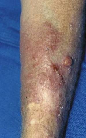
E-Figure 12-2 Multiple squamous cell carcinomas.
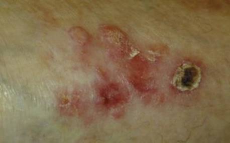
E-Figure 12-3 Squamous cell carcinoma.
Answer
C
12 Which of the following is a potential premalignant precursor of melanoma?
A Keratoacanthoma
B Actinic keratosis
C Seborrheic keratosis
D Dysplastic nevus
E Bowen disease
Ref.: 2, 18
Comments
Keratoacanthomas, characterized by rapid growth, rolled edges, and a crater filled with keratin, can mimic either squamous or basal cell carcinoma in appearance (E-Figure 12-4). Although they often grow rapidly and then involute over a period of several months, biopsy is usually performed. Actinic (solar) keratosis and cutaneous horns are premalignant lesions found on the sun-exposed areas of skin in fair-skinned individuals, more commonly in those with prolonged exposure to the sun or to carcinogens (E-Figure 12-5). These lesions may be treated with cryotherapy or topical agents. Raised lesions or lesions resistant to this treatment should be excised, although some may involute spontaneously. The likelihood of progression to squamous cell carcinoma is low (estimated to be 1 in 1000), with an associated lifetime risk of 5% to 10%. Bowen disease is cutaneous squamous cell carcinoma in situ. About 10% of these lesions progress to invasive squamous cell carcinoma. They should be excised completely to negative margins. The presence of dysplastic nevi confers an increased risk for the development of melanoma. In addition, some dysplastic nevi represent true precursors of melanoma and may progress to invasive melanoma if untreated. They are usually reddish to brown, have scalloped edges and variegated pigmentation, are generally larger than 6 mm in diameter, and often appear on the trunk and other non–sun-exposed regions of the body.
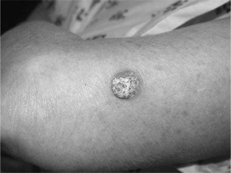
E-Figure 12-4 Keratoacanthoma.

E-Figure 12-5 Actinic keratosis.
Answer
D
13 A 65-year-old man has a rapidly growing red-blue nodule on his left forearm. The remainder of his physical examination is normal. Biopsy demonstrates Merkel cell (neuroendocrine) carcinoma of the skin. Appropriate care for this patient includes all of the following except:
A Chest radiograph
B Wide local excision with 2- to 3-cm margins
C Sentinel lymph node biopsy with selective lymph node dissection
D Axillary lymph node dissection
E Postoperative adjuvant radiotherapy
Ref.: 2, 18, 20
Comments
Merkel cell (neuroendocrine) carcinoma of the skin is derived from neuroectoderm and is manifested as a rapidly growing pink to red to blue to violaceous firm nodule, often in elderly patients. This tumor is histologically indistinguishable from small cell carcinoma of pulmonary origin, and a chest radiograph should be obtained to exclude metastases from a lung primary. After biopsy confirmation of the diagnosis, primary treatment consists of wide excision with 2- to 3-cm margins and histologic confirmation of negative margins. Although approximately 30% of patients initially have palpable regional lymph nodes, up to 70% of the remainder relapse in the regional lymph nodes within 2 years of diagnosis without nodal treatment. Current National Comprehensive Cancer Network (NCCN) guidelines recommend sentinel lymph node biopsy with selective lymph node dissection for patients with clinically node-negative Merkel cell carcinoma; approximately 30% of patients will be found to have occult metastatic disease. Immunohistochemistry with pancytokeratin AE1/AE3, cytokeratin 20, and chromogranin A helps detect micrometastatic disease. This approach improves locoregional disease control, and sentinel node status is a significant indicator of prognosis. Adjuvant radiotherapy is usually given to the primary site and may include the draining lymphatics and regional nodal basin for patients with node-positive disease. This improves locoregional control over surgical treatment alone, and some studies have suggested a survival benefit as well. Overall survival is poor, with mortality rates of 50% to 80% overall and 30% at 5 years after diagnosis.
Answer
D
14 Regarding the epidemiology of melanoma, which of the following is not true?
A The incidence of melanoma is increasing more rapidly than that of any other solid tumor.
B The increased incidence of melanoma is mainly due to an increased diagnosis of early lesions, whereas the incidence of thicker tumors is declining.
C The death rate from cutaneous melanoma is increasing.
D Before the age of 45 years, the incidence of melanoma is greater in females than in males.
E More than 20% of melanomas are diagnosed in individuals younger than 40 years.
Ref.: 2, 16, 20-22
Comments
For past several decades, the incidence of melanoma has been increasing more rapidly than that of any other solid tumor. Analysis of recent Surveillance, Epidemiology, and End Results (SEER) data shows a continued increasing incidence of 3% to 6% per year in the United States, with the death rate from melanoma increasing as well, although at a lower rate of less than 1% per year. The greatest increase continues to be in older men. This increase applies to all thickness groups, including tumors greater than 4 mm, and to all histologic types, thus implying that the increase in incidence is not just due to increased screening and that surveillance and early detection efforts have not led to a measurable reduction in the incidence of unfavorable melanomas. While the age of the U.S. population continues to increase, with the median age at diagnosis increasing from 40 to older than 50 years over the past generation, melanoma is still diagnosed in a substantial proportion of patients at a young age. Melanoma is second only to adult leukemia among cancers in terms of years of productive life lost. Melanoma is more common in women younger than 40 to 45 years and more common in men thereafter.
Answer
B
15 Which of the following is not a risk factor for melanoma?
A Total nevus count
B Fair skin
C Natural red or blonde hair
D Prior blistering sunburn
E Cigarette smoking
Ref.: 2, 3, 21
Comments
In addition to the well-established risk associated with solar UV radiation in susceptible individuals, especially intense intermittent exposure, blistering childhood sunburns also elevate the risk for development of melanoma. Constitutional risk factors include fair skin, red or blonde hair, blue or green eye color, the presence of many nevi, raised nevi, and dysplastic nevi. Patients with xeroderma pigmentosum have an elevated risk for the development of both melanoma and nonmelanoma skin cancer, as do immunocompromised patients and those with a previous history of nonmelanoma skin cancer and individuals with a family history of melanoma.
Answer
E
16 The preferred diagnostic biopsy method for pigmented skin lesions is:
A Punch biopsy
B Incisional biopsy
C Shave biopsy
D Excisional biopsy
E Excision with 0.5-cm margins
Ref.: 2, 21, 23
Comments
Information from diagnostic biopsy guides treatment and provides important prognostic and staging information for patients with newly diagnosed melanoma. A properly performed diagnostic biopsy is of crucial importance. Excisional biopsy (with 1 mm of normal surrounding skin) to remove the entire visible lesion is recommended. The excision should not be extended to permit more cosmetic closure because this may lead to unnecessarily extensive subsequent surgery. Ideally, the biopsy scar should be oriented to be most compatible with definitive treatment should the lesion prove to be melanoma (usually in the longitudinal axis for extremity lesions and perpendicular to the underlying muscle fibers for truncal and head and neck lesions). Excisional biopsy is not always performed or possible. Shave biopsy is discouraged for the diagnosis of pigmented lesions because the deep margin is often positive. Punch or incisional biopsy may be performed for large lesions or those in anatomically constrained areas and should include the full thickness of the skin and the most elevated and the darkest portions of the lesion. Patients in whom the diagnosis is made by less than excisional biopsy may require additional treatment and should be counseled that the final diagnosis may differ from that arrived at by biopsy. Regardless of the biopsy type, pathologic review by a dermatopathologist with an interest in pigmented lesions improves diagnostic accuracy, minimizes diagnostic error, and facilitates optimal patient care. The biopsy report should include tumor thickness (millimeters), histologic type, presence or absence of ulceration, Clark level, mitoses per square millimeter, peripheral and deep margin status, tumor location, and comments on the presence or absence of regression, tumor-infiltrating lymphocytes, vertical growth phase, angiolymphatic invasion, neurotropism, and microsatellitosis.
Answer
D
17 Which of the following is a component of current American Joint Commission for Cancer (AJCC) tumor staging for melanoma:
A Presence or absence of regression
B Presence or absence of lymphovascular invasion
C Level of invasion
D Tumor diameter
E Tumor mitotic rate
Ref.: 24
Comments
Adoption of mitotic rate in the classification of thin melanomas is one of the key changes in the new (2009, effective January 2010) AJCC staging system for cutaneous melanoma. The level of invasion is no longer used in AJCC tumor staging, except when the mitotic rate (described per square millimeter of tissue) cannot be obtained. Melanomas 1 mm or less in thickness are now classified as “a” for lesions with fewer than 1 mitosis/mm2 that are nonulcerated and as “b” for lesions with 1 or more mitoses/mm2 or ulceration. Previously, melanomas 1 mm or less in thickness but level IV or V, as well as ulcerated melanomas, were categorized as T1b. As in the last staging iteration, ulceration differentiates “a” from “b” lesions in all other thickness categories. The AJCC recommends that the mitotic count per square millimeter of tissue be enumerated on the standardized pathology report for review by clinicians and reported by cancer registrars to national databases because the mitotic rate has prognostic significance as a continuous variable.
Answer
E
18 The most common histologic type of melanoma is:
A Superficial spreading
B Nodular
C Lentigo maligna
D Acral lentiginous
E Desmoplastic
Ref.: 2
Comments
Superficial spreading melanoma is the most common type; it accounts for 70% of melanomas and is characterized by some degree of radial growth (E-Figure 12-6). Nodular melanoma, the next most common type, accounts for about 15% of melanomas and is characterized by vertical growth with a minimal to absent radial growth phase (E-Figure 12-7). Lentigo maligna melanoma, about 10% of melanomas, is characterized by an extensive radial growth phase, most commonly occurs on sun-exposed body areas in older patients, and is generally diagnosed at a thinner stage (E-Figure 12-8). Acral lentiginous melanoma is the most common type of melanoma in nonwhite individuals and is usually darkly pigmented (E-Figure 12-9). The prognosis depends on the thickness of the lesion, not the histologic subtype per se.
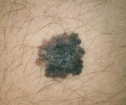
E-Figure 12-6 Superficial spreading melanoma.
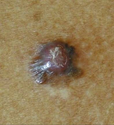
E-Figure 12-7 Nodular melanoma.

E-Figure 12-8 Lentigo maligna melanoma.
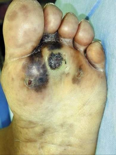
E-Figure 12-9 Acral lentiginous melanoma.
Answer
A
19 Adverse prognostic factors for clinically localized (stage I and II) melanoma include all of the following except:
A Older age
B Male gender
C Increasing tumor thickness
D Tumor regression
E Tumor ulceration
Ref.: 2, 25
Comments
Older age is a predictor of poorer survival as established in numerous studies and verified in retrospective reviews of recent clinical trials. Male patients fare worse than females. Tumor site (extremity better than the trunk or head or neck) is also prognostic. Tumor thickness is the most important predictor of outcome in patients with clinically localized melanoma, followed by ulceration (Figure 12-1). Although approximately half of melanomas show some degree of regression, the influence of regression on outcome is unclear; reports are variable regarding its significance, with some suggesting a favorable and others an adverse influence on outcome.

Figure 12-1 Observed (diamonds) and predicted (solid line) 10-year mortality rate in patients with clinically localized melanoma. This is based on a mathematical model derived from the American Joint Committee on Cancer melanoma database of 15,230 patients.
(From Balch CM, Soong SJ, Gerschenwald JE, et al: Prognostic factors analysis of 17,600 melanoma patients: validation of the American Joint Committee on Cancer melanoma staging system, J Clin Oncol19:3622–3634, 2001.)
Answer
D
20 A 33-year-old woman has a 2.1-mm-thick, nonulcerated nodular melanoma on her right thigh. The results of physical examination are otherwise normal. What is the most appropriate treatment?
A Wide local excision with 1.0-cm margins
B Wide local excision with 2.0-cm margins
C Wide local excision with 2.0-cm margins and sentinel lymph node biopsy
D Wide local excision with 2.0-cm margins, sentinel lymph node biopsy with frozen section, and possible inguinofemoral lymph node dissection
E Wide local excision with 3.0-cm margins, sentinel lymph node biopsy with frozen section, and possible inguinofemoral lymph node dissection
Ref.: 2, 20, 21, 23, 24
Comments
Treatment of melanoma involves complete excision of all skin and subcutaneous tissue down to the underlying fascia for a defined distance from the biopsy site, scar, or margins of the pigmented lesion, depending on tumor thickness. Recommended margins, based on data from five randomized clinical trials, are 0.5 cm for melanoma in situ, 1.0 cm for thin melanomas, and 2.0 cm for melanomas thicker than 1.0 mm. Wider margins are considered for lesions thicker than 4 mm and those with satellite lesions. Margins may be adjusted for concerns regarding cosmesis and function (Table 12-1). Lymphatic mapping(lymphoscintigraphy), sentinel lymph node biopsy, and selective lymph node dissection are recommended for patients with melanomas greater than 1.0 mm in thickness. Sentinel lymph node biopsy may be recommended for thicker melanomas, mainly those 0.76 mm or thinner, Clark level IV or V, ulcerated, diagnosed by incomplete biopsy, exhibiting regression, with a high mitotic rate, and occurring in younger patients. The likelihood of finding a positive sentinel lymph node is most strongly predicted by tumor thickness, with minimal risk for melanomas thinner than 0.76 mm, 5% for melanomas 0.76 to 1.0 mm, 8% to 10% for melanomas 1.01 to 1.5 mm, 18% to 30% for melanomas 1.51 to 4.0 mm, and 30% to 40% for melanomas thicker than 4.0 mm. Although use of the mitotic rate to classify thin melanomas as T1b was based on survival data, some data suggest that the likelihood of occult nodal disease increases with increasing mitotic rate. The use of immunohistochemistry (with at least one melanoma-specific marker such as HMB-45, Melan-A, or MART-1) will increase the sentinel lymph node positivity rate by approximately 10% over hematoxylin-eosin staining alone. Nomograms that incorporate patient age, site, thickness, level, and ulceration have been developed to help predict the likelihood of a positive sentinel lymph node. The current AJCC melanoma staging committee states that “sentinel node staging [is] encouraged for standard patient care” and that “staging with sentinel node technology should be required as entry criterion for all melanoma patients presenting with clinical stage IB and II disease before entry into clinical trials involving new surgical techniques or adjuvant therapy.” Frozen section examination of melanoma sentinel lymph nodes has been abandoned because of its low sensitivity (50%) and concern that tissue destruction may preclude an accurate diagnosis from permanent sections.
TABLE 12-1 Recommended Margins for Wide Local Excision of the Primary Site
|
Thickness |
Margin |
Note |
|
Melanoma in situ |
5 mm |
Head and neck: consider preoperative margin assessment |
|
Melanoma <1 mm |
1 cm |
|
|
Melanoma 1-4 mm |
2 cm |
1 cm acceptable in limited anatomic locations |
|
Melanoma >4 mm |
2 cm |
Consider 3 cm if easily obtained |
Answer
C
21 The patient in Question 20 is found to have metastatic melanoma in an inguinal sentinel lymph node. Which of the following statements regarding her condition is incorrect?
A On completion lymphadenectomy, the likelihood of nonsentinel lymph node metastases is 10% to 30%.
B Completion lymph node dissection should be performed.
C Adequate inguinofemoral lymph node dissection implies removal of a minimum of seven to eight lymph nodes.
D Adjuvant radiation therapy is beneficial for patients with more than three involved lymph nodes and when there is breach of the nodal capsule with bulky nodal disease.
E Completion lymph node dissection is unnecessary for micrometastatic disease (<0.2-mm tumor within the sentinel lymph node).
Ref.: 2, 20, 21, 23, 26-28
Comments
As yet there have been no prognostic factors or nomograms that accurately and reproducibly identify a subset of sentinel lymph node–positive melanoma patients without risk of harboring additional metastatic lymph nodes. Completion lymph node dissection should be performed for all sentinel node–positive patients. The goals of surgery include control of regional disease and improved survival. The majority of patients who undergo completion lymph node dissection will not be found to have additional positive lymph nodes. However, examination of these nonsentinel lymph nodes is by necessity less rigorous than the stepwise examination of the sentinel lymph node, so small foci of disease may be missed. Unlike breast cancer, there is no lower limit of the size of metastatic melanoma within a lymph node that defines a positive lymph node. Retrospective studies suggest that patients with nodal disease less than 0.1 mm are at substantially increased risk for relapse versus sentinel node–negative patients. Standard surgical oncology teaching defines complete lymph node dissections on the basis of anatomic boundaries. Recommendations based on expert opinion have suggested a minimum number of lymph nodes per region to define a complete lymph node dissection. A recent review by the Sidney Melanoma Unit based on more than 2000 regional lymph node dissections for melanoma suggested following standard anatomic guidelines consisting of minimum (and target mean) lymph node counts of 10 (21) for the axilla, 7 (14) for inguinal and ilioinguinal dissections, and 20 (39) for cervical dissections involving four or more levels . The 2010 NCCN guidelines recommend complete lymph node dissection based on proper anatomic boundaries, which they recommend be dictated into the operative note, but they do not set a target number of lymph nodes. A recent meta-analysis of sentinel lymph node biopsy–guided selective lymph node dissection versus observation with therapeutic lymph node dissection for nodal relapse demonstrated a significantly higher risk for death (hazard ratio of 1.6) for patients in the therapeutic lymph node dissection group. Adjuvant radiotherapy is considered for most patients with multiple cervical lymph node metastases and for those with bulky, large (metastatic nodes >3 cm in size) or extracapsular disease at any site. Even though no published randomized trials have compared surgery alone with surgery and postoperative radiotherapy, retrospective single-institution studies suggest improved regional control and disease-free survival.
Answer
E
22 Whether a deep (pelvic) lymph node dissection is performed with inguinofemoral lymph node dissection may be based on all of the following except:
A Findings on pelvic CT or PET-CT
B Palpable versus micrometastatic nodal disease
C The number of positive nodes in the superficial compartment
D Primary tumor ulceration
E The status of Cloquet’s node
Ref.: 20, 21, 23, 27, 29
Comments
The appropriate extent of groin dissection in patients with metastatic melanoma to the lymph nodes remains controversial. Isolated pelvic nodal disease is seen in the absence of superficial inguinal lymph node involvement and should be sought on lymphoscintigraphy performed for sentinel node identification, but this is uncommon. Some surgical oncologists perform radical ilioinguinal lymph node dissection for all patients deemed candidates for inguinal node dissection, whereas others take a selective approach. Proponents of routine radical ilioinguinal lymph node dissection note that a third of patients have additional disease only in the iliac and obturator nodes with no further inguinal nodal disease and that there are no clear indicators to help the surgeon select patients who will not benefit from pelvic lymph node dissection. Selective addition of deep pelvic lymph node dissection is based on the likelihood of finding involved nodes, which is increased when dissection is performed for macrometastatic inguinal disease and when a greater number of inguinal nodes are involved. Preoperative imaging results (CT or PET-CT) may also be used in conjunction with the clinical findings to help guide the extent of dissection. Cloquet’s node is the most inferior node in the iliac chain; evaluation of this node may be used to determine the potential value of deep pelvic node dissection that encompasses the obturator and hypogastric nodes in addition to the iliac nodes up to the level of bifurcation of the common iliac vessels. Complications of either procedure include seroma, lymphocele, lymphatic fistula, and lymphedema. There is no evidence that the more extensive operation carries greater morbidity.
Answer
D
23 Adjuvant systemic therapy for node-positive melanoma patients may include any of the following except:
A Adriamycin-based chemotherapy
B High-dose interferon alfa-2b
C Enrollment in a clinical antiangiogenic agent trial
D Enrollment in a clinical immunotherapy trial
E Observation
Ref.: 2, 20, 30
Comments
Numerous randomized clinical trials of chemotherapy, nonspecific immune stimulants, and vaccines for high-risk stage II and stage III melanoma have been conducted, but most have been underpowered and yielded negative results. The FDA-approved adjuvant therapy for node-positive melanoma patients is high-dose interferon alfa-2b, 20 million IU/m2 5 days per week intravenously for 4 weeks, followed by 10 million IU/m2 subcutaneously 3 days per week for 48 weeks. Although randomized controlled clinical trials have demonstrated improved disease-free survival with adjuvant interferon alfa-2b, there is a very modest effect on overall survival, and associated side effects of the treatment are significant. Newer agents under study in the adjuvant setting include antiangiogenic therapy such as bevacizumab and the anti–CTLA-4 antibody ipilimumab. Because of the lack of effective adjuvant therapy with low toxicity, observation, either as a control arm of a clinical trial or in the absence of clinical trial enrollment, is acceptable. Doxorubicin (Adriamycin) is not effective for the treatment of melanoma.
Answer
A
24 Which of the following is the most significant prognostic factor for patients with node-positive (stage III) melanoma?
A Nodal size
B Number of involved lymph nodes
C Tumor thickness
D Tumor ulceration
E Patient gender
Ref.: 23-25
Comments
The most significant prognostic factor for patients with node-positive melanoma is the number of involved lymph nodes. The next most significant prognostic factor is nodal tumor burden (microscopic or clinically occult versus macroscopic or clinically apparent), followed by primary tumor ulceration and thickness. Nodal size is not a component of staging, nor has it been shown to have significant independent prognostic value. Gender does not have a significant effect on outcome in patients with node-positive melanoma.
Answer
B
25 A 52-year-old man is evaluated for a palpable left inguinal lymph node 7 years after wide local excision of a thin melanoma on the ipsilateral right calf. No sentinel lymph node biopsy or other staging was done at the time of his initial diagnosis. What should be the next step in the care of this patient?
A Excisional biopsy
B Excisional biopsy with frozen section examination followed by immediate inguinofemoral lymph node dissection
C Total-body PET-CT per melanoma protocol
D FNA biopsy (cytology)
E Measurement of serum S-100 protein and lactate dehydrogenase (LDH)
Ref.: 2, 31
Comments
Recurrence in the regional lymph node basin is the most common site of initial recurrence after treatment by wide local excision alone and can occur even in patients with thin primary melanomas and many years after the initial diagnosis. Evaluation of clinically palpable lymph nodes is most expeditiously performed by FNA, with or without ultrasound guidance. Core needle biopsy or excisional biopsy can be done if the FNA is negative or nondiagnostic. Frozen section evaluation of lymph node metastases of melanoma has low sensitivity, and freezing the tissue may destroy areas with metastatic disease; the diagnosis is generally deferred to evaluation of the permanent section. Complete regional lymphadenectomy will lead to durable long-term survival in approximately half of such patients. Once the diagnosis of nodal melanoma recurrence is established, given that the remainder of the patient’s physical examination is unremarkable, a metastatic work-up that includes liver function tests, PET-CT, and MRI of the head is appropriate before surgery to exclude the presence of clinically unsuspected metastatic disease. In this clinical scenario, evaluation with PET-CT is estimated to change treatment in 15% to 30% of patients.
Answer
D
26 Which of the following is not indicated for the initial treatment of in transit metastasis from cutaneous melanoma?
A Excision
B Injection
C Laser treatment
D Heated limb perfusion
E Amputation
Ref.: 2, 21, 23, 24, 32
Comments
In transit metastases develop in about 2% of patients with cutaneous melanoma and appear as visible dermal or subcutaneous tumor nodules between the primary site and regional draining nodal basin. They arise from intralymphatic tumor spread. After evaluation for metastatic disease, treatment depends on the extent of disease. Limited in transit metastases amenable to excision may be treated by wide excision with 1-cm margins. Other local therapy options include laser ablation with healing by secondary intention and local immunotherapy with bacille Calmette-Guérin (BCG) or interferon alfa injection or topical application of imiquimod cream. Injection results in a response in approximately 80% of lesions, sometimes in neighboring lesions as well, and the toxicity is predominantly local (erythema, edema, ulceration). For extensive disease, isolated limb perfusion with a pump oxygenator circuit and regional chemotherapy (most commonly melphalan and sometimes the addition of tumor necrosis factor-α), with or without concomitant regional lymph node dissection, has limited indications but may be useful for locoregional control of disease. This involves surgical placement of arterial and venous cannulas in the affected extremity, which is excluded from the general circulation with a tourniquet. The reported complete response rate is 40% to 80%, but the toxicity may be considerable and includes lymphedema, compartment syndrome, and neuropathy. Responses may not be durable, with 50% recurrence rates 18 months after infusion. Recently, isolated limb infusion with percutaneously placed cannulas has been proposed as an alternative, less morbid therapy with complete responses rates of approximately 30%. With these available therapies, major amputation is rarely required as primary therapy for in transit disease. As secondary treatment, amputation may provide palliation, or regional control, for unmanageable progressive recurrences when other approaches fail. Indications are usually intractable pain with loss of limb function (often associated with tumor fungation, bleeding, infection, gangrene, and severe lymphedema) in patients whose life expectancy is at least 3 months and who have reasonable performance status otherwise. In the absence of nodal metastases, patients with in transit disease have 5- and 10-year overall survival rates of 69% and 52%, whereas those with concomitant regional nodal metastases have diminished 5- and 10-year overall survival rates of 46% and 33%.
Answer
E
27 A 37-year-old man is evaluated for a 4-month history of anemia and intermittent abdominal discomfort and distention 7 years after treatment of a stage I melanoma on his right forearm. The results of physical examination are normal, colonoscopy is negative, and CT demonstrates an area of invaginated jejunal mesentery with an adjacent dilated loop of small bowel. What is the next most appropriate step in the management of this patient?
A Exploration and small bowel resection
B Video capsule endoscopy
C Systemic biochemotherapy
D Whole-body PET-CT
E Air contrast small bowel barium study
Ref.: 2, 21, 23, 24, 32
Comments
The small intestine is the most common site of gastrointestinal tract metastases from cutaneous melanoma, which are present in a high proportion of patients with melanoma at autopsy. Symptomatic patients most commonly have abdominal pain, chronic gastrointestinal bleeding, obstruction, and weight loss. Polypoid tumors arising from the submucosa may act as the lead point for an intussusception, as in this case. Surgical resection of solitary and even multiple small intestinal metastases is associated with improved survival over nonoperative therapy and is effective treatment of associated obstruction. With complete resection of small intestinal disease, median survival times of 4 years are reported. Since this patient is symptomatic without evidence of other disease by physical examination or CT of the abdomen and pelvis, further preoperative radiologic work-up is unlikely to alter the need for or benefit of surgery. Although the disease recurs in most patients at multiple sites and they are best treated with systemic therapy, those who are suitable candidates for resection of limited metastatic disease may experience long-term disease-free survival. In general, for stage IV melanoma the site of the metastatic disease and the serum LDH level correlate with prognosis. One-year survival rates for those with cutaneous, subcutaneous, or distant nodal metastases (M1a) versus those with lung metastases (M1b) versus those with any other visceral metastases or any metastasis with an elevated serum LDH level (M1c) are estimated to be 62%, 53%, and 33%, respectively. Factors reported to be associated with improved outcome after resection of melanoma metastases include initial disease stage, disease-free interval after treatment of the primary melanoma, initial site of metastasis, solitary site of disease, and complete resection. Even when not performed with curative intent, palliative surgical treatment of metastatic melanoma may be beneficial for the treatment of symptomatic patients.
Answer
A
References
1 Tran TT, Schulman J, Fisher DE. UV and pigmentation: molecular mechanisms and social controversies. Pigment Cell Melanoma Res. 2008;21:509-516.
2 Urist MM, Soong S-J. Melanoma and Cutaneous Malignancies. In Townsend CM, Beauchamp RD, Evers BM, et al, editors: Sabiston textbook of surgery: the biological basis of modern surgical practice, ed 18, Philadelphia: WB Saunders, 2008.
3 Tyrrell RM. Ultraviolet protection. In: Lejeuene FJ, Chaudhuri PK, Das Gupta TK, editors. Malignant melanoma: medical and surgical treatment. New York: McGraw-Hill, 1994.
4 Bishop DT, Demenais F, Iles MM, et al. Genome-wide association study identifies three loci associated with melanoma risk. Nat Genet. 2009;41:920-925.
5 Bishop DT, Demenais F, Goldstein AM, et al. Geographical variation in the penetrance of CDKN2A mutations for melanoma. J Natl Cancer Inst. 2002;94:894-903.
6 Velasco AL, Dunlap WW. Pilonidal disease and hidradenitis. Surg Clin North Am. 2009;89:689-701.
7 Netscher D, Fiore N. Hand surgery. In Townsend CM, Beauchamp RD, Evers BM, et al, editors: Sabiston textbook of surgery: the biological basis of modern surgical practice, ed 18, Philadelphia: WB Saunders, 2008.
8 Cole P, Heller L, Bullocks J, et al. The skin and subcutaneous tissue. In Brunicardi FC, Andersen DK, Billiar TR, et al, editors: Schwartz’s principles of surgery, ed 9, New York: McGraw-Hill, 2010.
9 Rakinic J. Modern Management of Pilonidal Disease. In Cameron JL, editor: Current surgical therapy, ed 9, Philadelphia: CV Mosby, 2008.
10 Benz MR, Czernin J, Dry SM, et al. Quantitative F18-fluorodeoxyglucose positron emission tomography accurately characterizes peripheral nerve sheath tumors as malignant or benign. Cancer. 2010;116:451-458.
11 Pletcher BA. Neurofibromatosis Type 1. In eMedicine Clinical Knowledge Base, Neurology. Updated December 2009 www.emedicine.com Accessed January 31, 2010
12 Das Gupta TK, Chaudhuri PK, editors. Tumors of the soft tissues, ed 2, Norwalk, Conn: Appleton & Lange, 1998.
13 Singer S, Canter RJ. Soft tissue sarcoma. In Cameron JL, editor: Current surgical therapy, ed 9, Philadelphia: CV Mosby, 2008.
14 Singer S. Soft tissue sarcomas. In Townsend CM, Beauchamp RD, Evers BM, et al, editors: Sabiston textbook of surgery: the biological basis of modern surgical practice, ed 18, Philadelphia: WB Saunders, 2008.
15 Hieken TJ. Genetic testing for cancer susceptibility. In: Saclarides TJ, Millikan KW, Godellas CV, editors. Surgical oncology: an algorithmic approach. New York: Springer-Verlag, 2003.
16 Jemal A, Siegel R, Ward E, et al. Cancer statistics, 2009. CA Cancer J Clin. 2009;59:225-249.
17 Hueman MT, Thornton K, Herman JM, et al. Management of extremity soft tissue sarcomas. Surg Clin North Am. 2008;88:539-557.
18 Kazin R, Shermak MA. Skin and soft tissue. In Cameron JL, editor: Current surgical therapy, ed 9, Philadelphia: CV Mosby, 2008.
19 Rubin AI, Chen EH, Ratner D. Basal-cell carcinoma. N Engl J Med. 2005;353:2262-2269.
20 Hieken TJ. The role of sentinel node biopsy in skin cancer, ed 3. In eMedicine Clinical Knowledge Base, Dermatology. Updated March 2009 www.emedicine.com Accessed January 31, 2010
21 Faries MB, Morton DL. Cutaneous Melanoma. In Cameron JL, editor: Current surgical therapy, ed 9, Philadelphia: CV Mosby, 2008.
22 Criscione VD, Weinstock MA. Melanoma thickness trends in the United States, 1988-2006. J Invest Derm. 2010;130:793-797.
23 Lange JR, Balch CM. Cutaneous melanoma. In Cameron JL, editor: Current surgical therapy, ed 8, Philadelphia: CV Mosby, 2004.
24 Balch CM, Gershenwald JE, Soong SJ, et al. Final version of 2009 AJCC melanoma staging and classification. J Clin Oncol. 2009;27:6199-6206.
25 Balch CM, Soong SJ, Gershenwald JE, et al. Prognostic factors analysis of 17,600 melanoma patients: validation of the American Joint Committee on Cancer melanoma staging system. J Clin Oncol. 2001;19:3622-3644.
26 Ollila DW, Ashburn JH, Amos KD, et al. Metastatic melanoma cells in the sentinel node cannot be ignored. J Am Coll Surg. 2009;208:924-930.
27 Spillane AJ, Cheung BL, Stretch JR, et al. Proposed quality standards for regional lymph node dissections in patients with melanoma. Ann Surg. 2009;249:473-480.
28 Pasquali S, Mocellin S, Campaña LG, et al. Early (sentinel lymph node biopsy-guided) versus delayed lymphadenectomy in melanoma patients with lymph node metastases: personal experience and literature meta-analysis. Cancer. 2010;116:1201-1209.
29 Santinami M, Carbone A, Crippa F, et al. Radical dissection after positive groin sentinel biopsy in melanoma patients: rate of further positive nodes. Melanoma Res. 2009;19:112-118.
30 Eggermont AMM, Testori A, Marsden J, et al. Utility of adjuvant systemic therapy in melanoma. Ann Oncol. 2009;20(Suppl 6):vi30-vi34.
31 Brady MS, Akhurst T, Spanknebel K, et al. Utility of preoperative (18)F fluorodeoxyglucose–positron emission tomography scanning in high-risk melanoma patients. Ann Surg Oncol. 2006;13:525-532.
32 Wargo JA, Tanabe K. Surgical management of melanoma. Hematol Oncol Clin North Am. 2009;23:565-581.