
Tricia Moo-Young, M.D., Richard A. Prinz, M.D.
1 With regard to calcium homeostasis, which of the following statements is false?
A Calcium is the most abundant cation in human beings.
B Approximately 50% of calcium is free or ionized and is metabolically active.
C Hypoalbuminemia can make the measured total calcium concentration appear artificially low.
D Hypoventilation can decrease ionized calcium levels and thus exacerbate symptoms of hypocalcemia.
E Calcium is bound to citrate and is biologically inactive.
Ref.: 1, 2
Comments
Calcium is the most abundant cation in the human body. As much as 99% is stored in the musculoskeletal system. The remainder is present in serum and exists in three forms: (1) 45% is bound to albumin and is biologically inert, (2) 50% is ionized and metabolically active, and (3) a small percentage is complexed with citrate and also is biologically inactive. Hypoalbuminemia means that more of the total serum calcium will be free and metabolically active. Although total serum calcium may be low, the patient may not be metabolically hypocalcemic. Ionized calcium levels are inversely affected by the pH of blood. A 1-unit rise in pH will decrease the ionized calcium level by 0.36 mmol/L. Hypoventilation would cause a drop in pH and thus a subsequent rise in the ionized calcium level.
Answer
D
2 Vitamin D synthesis begins in the skin keratinocytes. What is the next step of activation in vitamin D synthesis?
A Hydroxylation in the kidney to yield 1,25-dihydroxyvitamin D
B Hydroxylation in the liver to yield 25-hydroxyvitamin D
C Decarboxylation in the liver to yield 25-hydroxyvitamin D
D Decarboxylation in the kidney to yield 25-hydroxyvitamin D
E Decarboxylation in the periphery to yield 25-hydroxyvitamin D
Ref.: 1, 2
Comments
Vitamin D synthesis begins in the keratinocytes of the skin. Subsequently, hydroxylation occurs in the liver to yield 25-hydroxyvitamin D. The final step in conversion of vitamin D to its active form occurs in the kidney, where a second hydroxylation reaction takes place to yield 1,25-dihydroxyvitamin D. Sunlight plays a key role in the initial synthesis step in the skin. Persons who are not exposed to sunlight require supplemental vitamin D through dietary intake.
Answer
B
3 Calcitonin helps mediate calcium homeostasis by which of the following actions?
A Stimulates osteoblast-mediated bone formation and inhibits renal resorption of calcium and phosphate
B Directly inhibits secretion of parathyroid hormone (PTH)
C Inhibits intestinal absorption of calcium
D Stimulates hydroxylation of vitamin D
E Stimulates osteoclast-mediated bone resorption
Ref.: 1, 2
Comments
In humans, the parathyroid glands are derived from the branchial pouches. The superior parathyroid arises from the fourth branchial pouch, and the inferior parathyroid originates from the third. The percentage of patients with ectopic glands ranges between 2.5% and 22%. The position of the superior parathyroid is most consistent. By contrast, the inferior gland can be located in the thymus up to 15% of the time. The inconsistent position of the inferior gland is believed to be the result of its longer migratory path from the third branchial pouch. An undescended inferior parathyroid can be located above the superior gland at the base of the skull or angle of the mandible. The frequency of intrathyroidal glands is relatively rare (0.5% to 3%). Calcitonin is produced by the parafollicular cells (C cells) of the thyroid gland. It helps lower ionized calcium levels in primarily two ways. First, it inhibits osteoclast-mediated bone resorption. Second, it inhibits resorption of calcium and phosphate by the kidney. Calcitonin has no direct effects on intestinal absorption or osteoblast-mediated bone formation.
Answer
A
4 With regard to PTH, which of the following statements is incorrect?
A PTH directly stimulates increased intestinal absorption of calcium.
B PTH stimulates osteoclast resorption of calcium and phosphate.
C PTH cells express G protein–coupled calcium-sensing receptors.
D PTH inhibits calcium excretion at the distal convoluted tubule of the kidney.
E PTH enhances renally mediated hydroxylation of 25-hydroxyvitamin D.
Ref.: 1, 2
Comments
Parathyroid hormone has a variety of actions and targets to help increase serum calcium levels. The parathyroid cells express a G protein–coupled membrane receptor that senses serum calcium levels. When calcium levels fall below appropriate levels, the receptor stimulates the release of PTH into the circulation. PTH has a half-life of about 2 to 4 minutes in the circulation. Before its rapid clearance, it first targets osteoclasts and stimulates them to resorb calcium and phosphate from bone. In the kidney, PTH blocks calcium excretion at the distal convoluted tubule. It indirectly promotes intestinal absorption of calcium by enhancing the renally mediated activation of vitamin D.
Answer
A
5 All of the following conditions can cause hypercalcemia in patients with normal parathyroid function except:
A Malignancy
B Sarcoidosis
C Lithium
D Cirrhosis
E Tuberculosis
Ref.: 1, 2
Comments
A number of conditions can cause hypercalcemia, including granulomatous disorders such as sarcoidosis, tuberculosis, and histoplasmosis. Medications can falsely elevate the serum calcium level. Examples include thiazide diuretics, lithium, and vitamin A or D. Other conditions include Paget disease, immobilization, and malignancy. In hospitalized patients, malignancy is the most common cause of hypercalcemia.
Answer
D
6 Routine work-up of a patient with suspected primary hyperparathyroidism (PHPT) includes all of the following except:
A Serum 1,25-dihydroxyvitamin D levels
B 24-hour urine calcium
C Intact PTH (iPTH)
D Serum calcium level
E Detailed physical examination and history
Ref.: 1, 2
Comments
In a patient with suspected PHPT, the minimum testing that should be performed includes intact parathyroid hormone, serum calcium, blood urea nitrogen, creatinine, and vitamin D levels. It is not necessary to measure PTH-related protein (PTHrp) unless metastatic cancer is suspected. It is important to document normal renal function before the interpretation of parathyroid function tests. Urinary calcium measurements need not be done routinely, except in patients who have a family history of hypercalcemia and no previous history of normal calcium levels. In these patients, 24-hour urine calcium excretion should be measured to exclude benign familial hypercalcemia hypocalciuria.
Answer
B
7 All of the following are consistent with the diagnosis of secondary hyperparathyroidism except:
A Elevated serum phosphate level
B Normal serum calcium level
C Vitamin D deficiency
D Elevated PTH level
E Calcitonin is the drug of choice for initial treatment
Ref.: 1, 2
Comments
Secondary hyperparathyroidism most commonly occurs in patients with a history of chronic renal failure. The pathophysiology of this condition is multifactorial but it is believed to be the result of chronic hyperphosphatemia and deficiency in active vitamin D because of loss of renal tissue. Patients will commonly have an elevated PTH level and normal serum calcium. In such a setting, vitamin D levels should be measured and, if low, treated for a minimum of 6 weeks with supplemental vitamin D. Some of these patients are managed medically with the use of calcimimetic agents such as cinacalcet. This medication works by binding the calcium-sensing receptors on the chief cells of the parathyroid gland and increasing its sensitivity to extracellular calcium. Calcitonin has no pharmacologic role in treating secondary hyperparathyroidism.
Answer
E
8 All of the following are indications for surgical treatment of secondary hyperparathyroidism except:
A Calcium-phosphate product of less than 70
B Uremic pruritus
C Osteitis fibrosa cystica
D Calciphylaxis
E Tumoral calcinosis
Ref.: 1, 2
Comments
Secondary hyperparathyroidism is most commonly managed medically with the use of calcimimetic agents, phosphate binders, adequate calcium intake, and vitamin D replacement. Surgical treatment is indicated in patients with (1) renal osteodystrophy, (2) calciphylaxis, (3) calcium-phosphate product of greater than 70, (4) soft tissue calcium deposition and tumoral calcinosis, and (5) calcium level greater than 11 mg/dL with an inappropriately high level of PTH. Renal osteodystrophy is a major issue in hemodialysis patients. The aluminum present in the dialysate bath accumulates in bone and contributes to the development of osteomalacia. Osteitis fibrosa cystica, a type of renal osteodystrophy, is characterized by marrow fibrosis and increased bone turnover. Bone cysts, osteopenia, and decreased bone strength develop. To halt progression of this disease process, these patients with secondary hyperparathyroidism are treated surgically. Calciphylaxis is a rare vascular disorder in which calcium is deposited in the media of small to medium-sized arteries. As a result, ischemic damage to the dermal and epidermal structures develops. The ulcerated lesions are extremely painful and can become infected with subsequent sepsis and eventually death. Patients with early signs of calciphylaxis should undergo urgent parathyroidectomy, although there is some evidence that aggressive management of serum calcium and parathyroid levels with cinacalcet may be beneficial. Care should be taken in wound care management because aggressive débridement can lead to chronic nonhealing wounds since wound healing is very poor in these patients. Uremic pruritus is characterized by severe itching that is thought to result from increased deposition of calcium salt in the dermis without the visible lesions of calciphylaxis. Parathyroidectomy seems to alleviate these symptoms and halts progression to the more serious skin and vascular complications seen with calciphylaxis.
Answer
A
9 With regard to PHPT, which of the following statements is true?
A PHPT is more common in men than in women.
B A common feature of PHPT is polyuria.
C A history of nephrolithiasis is present in 80% of patients with PHPT.
D Five percent of patients with PHPT can have multiple glands affected.
E Familial hypercalcemic hypocalciuria is associated with PHPT.
Ref.: 1, 2
Comments
Primary hyperparathyroidism is a relatively common disorder that affects 0.3% of the human population, most commonly women. The exact cause of PHPT is unknown. In 80% of patients only a single adenoma is present, but multiple adenomas or hyperplasia can be present in up to 15% to 20%. Patients with PHPT can have symptomatic or “asymptomatic” disease. Some degree of renal dysfunction is present in up to 80% of patients. Nephrolithiasis, however, is far less common, with an incidence of approximately 20% to 25%. The clinical manifestations of PHPT vary widely across patients, but if a detailed history is taken, many will complain of polydipsia and polyuria from the calciuresis associated with the disease. Although PHPT occurs sporadically in the majority of patients, in a small percentage it is part of a familial syndrome. Multiple endocrine neoplasia type I (MEN-I) results from a germline mutation in the menin gene located on chromosome 11q12-13. Patients with MEN-I are susceptible to the development of pancreatic neuroendocrine tumors, pituitary adenomas, and PHPT. MEN-IIA is an autosomal dominantly inherited condition caused by a germline mutation on chromosome 11 that is associated with PHPT, pheochromocytoma, and medullary thyroid cancer. Patients with familial jaw tumor syndrome have a higher risk for the development of parathyroid carcinoma. Familial hypercalcemic hypocalciuria is associated with elevated calcium levels and low urinary excretion of calcium. The primary defect is abnormal sensing of calcium in blood by the parathyroid gland and the renal tubules, which causes inappropriate secretion of PTH and excessive renal reabsorption of calcium.
Answer
B
10 In 2002, the National Institutes of Health (NIH) released a consensus statement outlining indications for the surgical treatment of patients with asymptomatic hyperparathyroidism. All of the following are part of the criteria for surgical treatment except:
A Creatinine clearance reduced by greater than 30% in comparison to age-matched subjects
B PHPT in a patient younger than 50 years
C Calcium elevated to greater than 1 to 1.6 mg/dL above normal
D Osteitis fibrosa cystica
E 24-hour urinary calcium excretion greater than 150 mg/day
Ref.: 1, 2
Comments
The 2002 National Institutes of Health Consensus Conference defined “asymptomatic” hyperparathyroidism as the absence of bone, neurologic, gastrointestinal, or renal complaints associated with the disease. In their statement they outlined criteria for surgical referral in patients with “asymptomatic” disease (Box 17-1).

BOX 17-1 National Institutes of Health Criteria for Parathyroidectomy
Serum calcium >1-1.6 mg/dL above normal
Nephrolithiasis
Creatinine clearance reduced by 30% with age-matched control
Age <50 yr
History of life-threatening hypercalcemia
Neuromuscular symptoms (ataxia, proximal muscle weakness, hyperreflexia)
Reduction in bone mass >2 standard deviation below matched controls
24-hour urine calcium excretion elevated to >400 mg/day
Answer
E
11 A 54-year-old woman has proximal muscle weakness, polyuria, and a depressed mood. Laboratory work-up reveals a serum calcium level of 11.2 mg/dL and a PTH level of 110 ng/L. Which of the following is the least sensitive preoperative localization study to identify an abnormal parathyroid gland?
A Magnetic resonance imaging (MRI)
B Single-photon emission computed tomography (SPECT)
C Technetium-99m–labeled sestamibi scan
D Neck ultrasound
E Standard computed tomography (CT)
Ref.: 1, 2
Comments
Although all of the these imaging studies have been used to identify the location of a parathyroid adenoma, magnetic resonance imaging is the least sensitive of those listed. Routine preoperative localization in patients with PHPT includes neck ultrasound and technetium-99m–labeled sestamibi scan. Sestamibi scan has a reported sensitivity as high as 90%. Ultrasound is slightly less sensitive (75%), but the ease of in-office use makes it a useful tool for the general surgeon. SPECT, when used with planar sestamibi, is very good at locating potential ectopic glands such as those in the mediastinum. MRI is the least sensitive of the other listed modalities but, when used along with CT, can be helpful in locating ectopic glands.
Answer
A
12 Of the following, which patient is the least likely to have multigland disease?
A A 65-year-old lady with a PTH level of 110 ng/L and calcium level of 10.5 mg/dL
B A 22-year-old woman with a PTH level of 140 ng/L, a calcium level of 10.1 mg/dL, and MEN-I
C A 75-year-old man with a 10-year history of renal failure
D A 44-year-old woman with a diagnosis of secondary hyperparathyroidism
E A 39-year-old woman 6 years after a gastric bypass for morbid obesity
Ref.: 3, 4
Comments
No study has yet identified a reliable predictor of which patients with sporadic hyperparathyroidism will have multigland disease. The exception is in familial, secondary, and tertiary hyperparathyroidism. Because of the nearly uniform incidence of four-gland hyperplasia, all these patients are managed with bilateral neck exploration and either total parathyroidectomy with autotransplantation or three-and-a-half gland parathyroidectomy. Although some surgeons believe that patients with higher preoperative PTH or calcium levels (or both) are more likely to have multigland disease, this has not proved to be true in clinical studies. Intraoperative PTH (IOPTH) monitoring is used to determine whether all hyperfunctioning tissue has been removed. Several different criteria for the interpretation of IOPTH have been published (Table 17-1). Studies comparing these various criteria and their ability to predict multigland disease have been controversial. The pitfalls of the various criteria include false-positive results that lead to unnecessary bilateral neck exploration versus false-negative results in which the presence of multigland disease is not recognized. The best clinical marker of single-gland disease is concordant preoperative imaging in combination with appropriate correction of IOPTH levels.
TABLE 17-1 Criteria for Parathyroid Hormone Monitoring to Predict Operative Success
|
Miami Criteria |
≥50% decline from the highest (either preincision or preexcision) value within 10 minutes of gland removal |
|
Vienna Criteria |
≥50% decline from the preincision value within 10 minutes of gland removal |
|
Halle Criteria |
Decay into the low normal range (PTH ≤35 ng/L) within 15 minutes |
|
Rome Criteria |
≥50% decline from the highest preexcision level, and/or PTH concentration within the reference range at 20 minutes after excision, and/or ≤7.5 ng/L lower than the value at 10 minutes after excision |
Answer
A
13 A 55-year-old woman with a diagnosis of hyperparathyroidism wishes to undergo minimally invasive parathyroidectomy (MIP). Which of the following would preclude a patient from being a candidate for this approach?
A PHPT
B Lack of preoperative localization
C Preoperative imaging of a solitary lesion on only one of two localization studies
D Previous neck surgery
E Secondary hyperparathyroidism
Ref.: 1, 2
Comments
Minimally invasive parathyroidectomy is the preferred approach in patients who have a solitary lesion that is imaged conclusively by ultrasound, sestamibi, or a combination of both scans. It is advisable to use intraoperative parathyroid hormone determination to document an appropriate drop in PTH levels after removal of the suspected gland. Previous neck surgery or lack of concordant imaging on two types of studies is not a contraindication to attempting MIP. Patients in who no localization has been successful should not generally be offered MIP. Patients suspected of having multigland disease are managed by four-gland exploration, although surgeons at some centers are advocating exploration via a minimally invasive approach.
Answer
B
14 A pregnant mother in her first trimester comes to her clinician’s office with a diagnosis of PHPT. What is the correct management?
A Parathyroidectomy during the second trimester
B Parathyroidectomy during the third trimester
C Prescribing a calcimimetic agent to help reduce hypercalcemia until after delivery, when definitive surgery can be offered safely
D Close observation and parathyroidectomy following delivery
E Weekly injections of calcitonin until delivery, when definitive surgery can be offered safely
Ref.: 1, 2
Comments
Hyperparathyroidism during pregnancy is often unrecognized and is associated with a 3.5-fold increase in miscarriage. Loss of the pregnancy most often occurs during the late second trimester. The incidence of hyperparathyroidism in pregnancy is 0.7%. Maternal complications include hyperemesis, nephrolithiasis, and pancreatitis. Fetal complications include spontaneous abortion and growth retardation. In those who reach delivery, neonatal complications include hypocalcemic crisis within the first few days of life. Calcimimetic medications have not been used in the setting of hyperparathyroidism in pregnancy. Calcitonin has no role in the management of hyperparathyroidism.
Answer
A
15 A patient is undergoing directed exploration for PHPT. A single large parathyroid gland is found adjacent to the left superior thyroid pole. The preincision PTH level was 300 ng/L, and the pre–pedicle clamp level was 400 ng/L. Five and ten minutes after removal of the gland, the PTH level is measured to be 200 ng/L. All are appropriate next steps in the management of this patient except:
A Repeated PTH measurement
B Frozen section confirmation of the removed parathyroid gland
C Four-gland exploration
D Drawing blood from the contralateral jugular vein to determine the PTH level
E Conclusion of the operation given the 50% drop from the highest pre-removal level
Ref.: 1, 2
Comments
Although this patient did experience a 50% drop from the highest pre-removal level, 200 ng/L is still quite elevated. When the surgeon is going to accept a 50% drop, the final level should either approach normal or be following a kinetic trend toward normal. A preexcision level that is within a few-fold elevation of the upper limit of normal should demonstrate normal kinetics and rapidly approach normal levels within two to three half-lives of the hormone after the adenoma is removed. The first steps in this situation should be to repeat the PTH level 15 to 20 minutes following gland removal and send off a frozen section for confirmation of the candidate gland removed. At this point it would be appropriate to either await the results of these two tests or proceed with four-gland exploration. The use of intraoperative parathyroid hormone monitoring has revolutionized how parathyroid surgery is performed. It has allowed surgeons to perform directed operations and, before leaving the operating room, document whether biochemical cure has been achieved with relative certainty. There is wide variability in the literature on what constitutes appropriate use and interpretation of IOPTH monitoring. The most commonly used criteria include determination of a preincision PTH level, a stimulated (pre–pedicle ligation) level, and subsequent postremoval levels at 5-minute intervals until a normal level is achieved. Alternatively, some surgeons require only greater than a 50% drop in the PTH level from the highest pre-removal level (preincision or pre–pedicle ligation level). They do not require that the final level determined be within the normal range. In patients with secondary or tertiary hyperparathyroidism and stimulation levels as high as 2000 ng/L, a 90% decrease has been suggested as being indicative of cure.
Answer
E
16 The patient in Question 15 has a repeated PTH value of 200 ng/mL. Frozen section of the removed left superior candidate gland shows “hyperplastic parathyroid tissue.” On further exploration, no gland can be identified in the left inferior location. The right side is explored, both glands are located, and “hyperplastic parathyroid tissue” is confirmed by frozen section. Blood is drawn from the left internal jugular vein to determine the PTH level, which is found to be 600 ng/L. All of the following include appropriate steps in the management of this patient except:
A Cervical thymectomy
B Exploration of the left carotid sheath
C Left thyroid lobectomy
D Left lateral neck dissection
E Exploration of the retroesophageal space
Ref.: 1, 2
Comments
The left inferior parathyroid gland has not been identified in this patient. Frozen section has confirmed “hyperplastic parathyroid tissue” in the three identified glands. Failure to find a normal parathyroid gland among those identified should alert the surgeon that the patient probably has four-gland hyperplasia. Failure to find the remaining gland places the patient at high risk for persistent hyperparathyroidism. Most lower parathyroid glands are found in proximity to the lower thyroid pole. If not found in this location, the thyrothymic ligament and thymus should be explored. If the IOPTH level does not normalize after this maneuver, intraoperative ultrasound of the left thyroid lobe versus left lobectomy should be performed. Finally, central neck dissection and exploration of the left carotid sheath are performed. The procedure is terminated after these steps, and further imaging and localization studies should be undertaken if the patient has persistent hyperparathyroidism.
Answer
D
17 The most common location for a missed adenoma in patients undergoing reoperative parathyroid surgery is in:
A The thymus
B A normal upper position
C A normal lower position
D The tracheoesophageal groove
E The carotid sheath
Ref.: 5
Comments
Based on review of a series of more than 200 patients undergoing reoperative surgery for a missed adenoma, the most common location in which the missing gland was found was the tracheoesophageal groove (27%). The remaining locations included the anterior mediastinum/thymus (18%), the normal upper position (13%), the normal lower position (12%), intrathyroidal (10%), undescended parathyroids(8%), and the carotid sheath (4%). Failure to identify parathyroid pathology during an operation can occur in various settings, including the presence of an intrathyroidal adenoma, failure of the surgeon to expose the abnormal gland in its standard position, failure to recognize multigland disease, and failure to recognize an intrathymic adenoma.
Answer
D
18 All of the following have been associated with an increased risk for hungry bone syndrome after parathyroidectomy except:
A Graves disease
B Tertiary hyperparathyroidism
C Preoperative PTH level
D Age
E Large single adenomas
Ref.: 1, 2
Comments
“Hungry bone syndrome” is characterized by postparathyroidectomy hypocalcemia and hypophosphatemia. Patients most at risk are those with four-gland hyperplasia from secondary or tertiary hyperparathyroidism. The postoperative calcium level in these patients can drop critically low and necessitate intravenous calcium supplementation. During this period both serum calcium and phosphate levels must be monitored closely. In some patients it can take more than 4 to 5 days for serum calcium and phosphate levels to stabilize. Other patients shown to have increased risk for this condition are those who are older or have concomitant thyrotoxicosis or a large single adenoma. The preoperative PTH level has not been found to be an independent predictor of whether “hungry bone syndrome” will develop postoperatively.
Answer
C
19 A patient with chronic renal failure comes to the emergency department complaining of increasing confusion, muscle weakness, nausea, vomiting, and fatigue. The serum calcium level is 12.4 mg/dL. The first step in management of this patient should be:
A Emergency parathyroidectomy
B Aggressive intravenous hydration
C Initiation of furosemide infusion
D Continuous calcitonin infusion
E Initiation of bisphosphonates
Ref.: 6
Comments
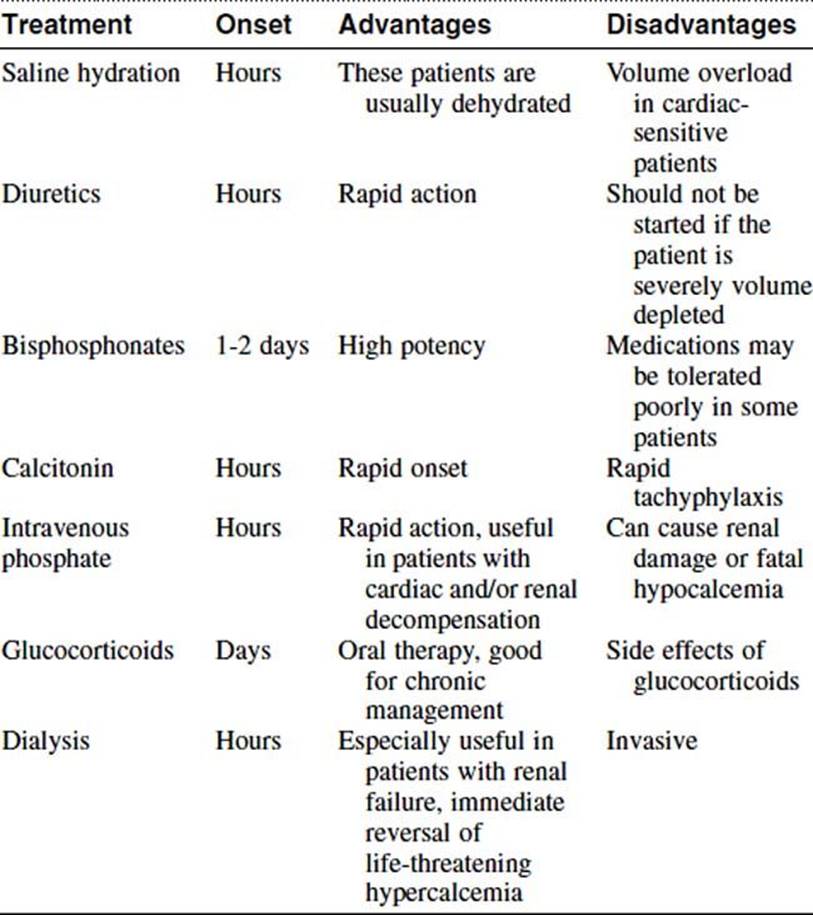
The first step in the management of this patient with hypercalcemia should be aggressive immediate hydration. This should be done before giving loop diuretics. Along with intravenous hydration, salt loading should be initiated to help induce a natural diuresis. Table 17-2 lists therapies used to treat hypercalcemic crisis, along with the onset of action, advantages, and disadvantages of each treatment.
TABLE 17-2 Management Options for Hypercalcemic Crisis
Answer
B
20 Which of the following is not a sign or symptom of hypocalcemia?
A Shortened QT interval
B Trousseau sign
C Circumoral numbness
D Anxiety
E Laryngospasm
Ref.: 1, 2
Comments
Postoperative hypocalcemia following parathyroidectomy is not unusual. The majority of patients are asymptomatic and identified only when routine postoperative laboratory tests are obtained. Symptomatic patients will most commonly complain of circumoral numbness and tingling in their extremities. These two features are early signs of hypocalcemia. On examination these patients may have carpopedal spasm elicited by occlusion of blood flow to the forearm (Trousseau sign) or contraction of the facial muscles elicited by tapping on the facial nerve (Chvostek sign). However nearly 20% of the general population has a positive Chvostek sign. If the condition is allowed to worsen, tetany, laryngeal stridor, or tonic-clonic seizures can develop, all of which can be fatal. These patients should be treated immediately with intravenous calcium. In asymptomatic patients, oral calcium and vitamin D should be initiated promptly.
Answer
A
21 A 34-year-old woman has undergone three operations for hyperparathyroidism. She states that in each previous operation the surgeon has removed pathologically confirmed hyperplastic parathyroid tissue in the central portion of her neck and nodules within various muscles of her neck. Nonetheless, she continues to have elevated parathyroid levels associated with progressive bone loss. What is the most likely diagnosis in this patient?
A Missed parathyroid adenoma
B Multigland disease
C Parathyromatosis
D Familial hyperparathyroidism
E Vitamin D deficiency
Ref.: 1, 2
Comments
This scenario is a classic description of a patient with parathyromatosis. This rare condition is manifested clinically as recurrent or persistent hyperparathyroidism following multiple attempts at resection. On exploration, patients will have several small nodules of hyperfunctioning parathyroid tissue throughout the neck and possibly the mediastinum. It can be difficult to distinguish this condition from parathyroid carcinoma or an “atypical adenoma.” Parathyromatosis is believed to result from either a low-grade parathyroid malignancy, fracture of the parathyroid adenoma capsule at the original procedure, or overgrowth of embryologic rests of parathyroid tissue. Management of these patients involves either serial debulking of the disease when it can be radiographically identified or pharmacologic treatment. Patients are rarely cured with surgery.
Answer
C
22 A 54-year-old woman arrives for surgical evaluation with a PTH level of 280 ng/L and serum calcium level of 14.5 mg/dL. She has a past medical history of mild renal failure (creatinine, 1.9 mg/dL) and jaw tumor syndrome. The most likely diagnosis in this patient is:
A Parathyromatosis
B Missed adenoma
C Parathyroid carcinoma
D Secondary hyperparathyroidism
E Tertiary hyperparathyroidism
Ref.: 1, 2
Comments
A preoperative serum calcium level greater than 14 mg/dL, a palpable nodule, and adherence to surrounding tissues have all been found to be useful predictors of parathyroid carcinoma. Additionally, patients with familial hyperparathyroidism and jaw tumor syndrome have an increased risk for parathyroid cancer. Parathyroid carcinoma is rare and represents just 1% of all cases of PHPT. The prognosis is extremely poor, and most patients have advanced disease when initially seen. Parathyroid carcinoma should be suspected in a patient with a preoperative calcium level greater than 14 mg/dL or a serum PTH level two to three times the upper limit of normal. Intraoperatively, parathyroid cancers are typically large and less brown than a benign parathyroid adenoma. Invasion or adherence to the surrounding tissues should also raise suspicion for parathyroid carcinoma. Appropriate surgical management of this condition entails en bloc resection of the tumor with ipsilateral thyroid lobectomy. It is not necessary to perform a total thyroidectomy. A compartment-oriented lymph node dissection is necessary only if there is gross evidence of lymph node involvement.
Answer
C
23 Which of the following is not part of the standard work-up of a patient with persistent or recurrent hyperparathyroidism?
A Selective venous sampling
B SPECT
C Technetium-Tc-99m-sestamibi scanning
D Ultrasound
E Positron emission tomography (PET)
Ref.: 1, 2, 7
Comments
Persistent hypercalcemia is defined as hypercalcemia that fails to correct after parathyroidectomy. Recurrent hyperparathyroidism occurs less frequently and is defined by the development of hypercalcemia following a 6- to 12-month period of normocalcemia. Preoperative localization is the mainstay of treatment algorithms for patients with persistent or recurrent hyperparathyroidism. The tests available can be divided into those that are noninvasive versus invasive. The noninvasive tests include ultrasound, sestamibi scanning, single-photon emission computed tomography, magnetic resonance imaging, and computed tomography. Of these, CT and MRI are the least useful and carry the lowest sensitivity. Invasive testing includes selective venous sampling and, more recently, intraoperative radioprobe guidance. Patients receive intravenous technetium-99m the morning of surgery, and then the radioprobe is used intraoperatively to identify the parathyroid gland. One limitation of this technology is a high background level, which can prevent identification of an intrathyroidal parathyroid gland. Positron emission tomography has yet to have any defined role in parathyroid localization.
Answer
E
References
1 Sosa JA, Udelsman R. The parathyroid glands. In Townsend CM, Beauchamp RD, Evers BM, et al, editors: Sabiston textbook of surgery: the biological basis of modern surgical practice, ed 18, Philadelphia: WB Saunders, 2008.
2 Lal G, Clark OH. Thyroid, parathyroid and adrenal. In Brunicardi FC, Andersen DK, Billiar TR, et al, editors: Schwartz’s principles of surgery, ed 9, New York: McGraw-Hill, 2010.
3 Riss P, Kaczirek K, Heinz G, et al. A “defined baseline” in PTH monitoring increases surigcal success in patients with multiple gland disease. Surgery. 2007;142:398-404.
4 Lombardi CP, Raffaelli M, Traini E, et al. Intraoperative PTH monitoring during parathyroidectomy: the need for stricter criteria to detect multiglandular disease. Lagenbecks Arch Surg. 2008;393:639-645.
5 Jaskowiak N, Norton JA, Alexander HR, et al. A prospective trail evaluating a standard approach to reoperation for missed parathyroid adenoma. Ann Surg. 1996;224:308-320.
6 Fauci AS, Kasper DL, Longo DL, et al. Harrison’s principles of internal medicine, ed 17. New York: McGraw-Hill; 2008.
7 Chen H, Sippel RS, Schaefer S. The effectiveness of radioguided parathyroidectomy in patients with negative technetium Tc 99m-sestamibi scans. Arch Surg. 2009;144:643-648.