
Jacquelyn Turner, M.D., Theodore J. Saclarides, M.D.
A
Small Bowel
1 With regard to ileostomy physiology, which of the following statements is true?
A Daily output from an established ileostomy is approximately 1500 mL.
B Ileostomy output can increase by 50% at times of dietary indiscretion.
C With dehydration, the concentration of sodium output from the ileostomy rises.
D When compared with normal ileal fluid, ileostomy effluent contains a 100-fold increase in the number of aerobes and a 2500-fold increase in the number of coliform bacteria.
E The microbiologic flora of ileostomy output is similar to that of normal ileal fluid.
Ref.: 1
Comments
The daily output from an established ileostomy is 500 to 800 mL. Although there is a great deal of variation in daily output among individuals, the output in a given patient varies only about 20% with changes in diet or with episodes of gastroenteritis. The usual ileostomy sodium concentration is 115 mEq/L, although the concentration rises and falls with changes in total body sodium. With dehydration, the sodium concentration falls and the potassium level rises as a result of the ability of the terminal ileum to conserve sodium in times of salt depletion. Normally, the sodium-to-potassium ratio is about 12 : 1. The microbiologic flora of ileostomy output is markedly different from that of normal ileal fluid. The total number of bacteria is 80 times greater, and there is a 100-fold increase in the number of aerobes, a 2500-fold increase in the number of coliform bacteria, and an increase in the number of total anaerobes.
Answer
D
2 Which of the following statements about small bowel motility is true?
A Oral feeding stimulates the production of migrating motor complexes (MMCs).
B If motility is impaired, absorption of nutrients is similarly affected.
C MMCs are peristaltic contractions occurring at 10- to 20-minute intervals.
D Vagotomy-induced diarrhea is the result of increased secretion secondary to denervation.
E Segmental bowel resection causes a temporary interruption of MMCs, but the clinical results are usually insignificant.
Ref.: 2
Comments
Migrating motor complexes are propagated aboral peristaltic contractions occurring at 90-minute intervals. The activity fronts of MMCs usually originate high in the stomach, propagate distally, and end in the ileum, usually at the midileal level. Oral feeding inhibits MMCs, which results in irregular, nonpropagating contractions throughout most of the small intestine. This postprandial inhibition may persist for 3 to 4 hours after a meal and is most pronounced with lipids. Although this motility pattern is disorganized, there is distal progression of chyme. Absorption is not affected by intestinal motility. Enteral feedings can therefore be used safely and efficiently in postoperative patients in whom motility may be altered.
Both gastric and small bowel motility can be affected by exogenous conditions. The small bowel is less sensitive than the stomach to general anesthesia and laparotomy, each of which decreases the frequency of MMCs. The frequency of MMCs returns to normal within 6 to 24 hours in the absence of peritonitis or abscess formation. The tone of the stomach is affected more than that of the small bowel by general anesthesia and laparotomy, at times taking longer than 24 hours to normalize. This may explain the occurrence of postoperative nausea and emesis. Vagotomy-induced diarrhea is a result of persistence of the sustained, organized wave of MMCs during the postprandial state.
Segmental small bowel resection or denervation temporarily reduces the frequency of MMCs, with a resultant temporary impairment of motility. Resection or denervation does not, however, produce long-term sequelae, provided that intestinal length is not sacrificed.
Answer
E
3 During an operation for presumed appendicitis, the appendix is found to be normal. The terminal ileum, however, is markedly thickened and feels rubbery to firm. Its serosa is erythematous and inflamed, and several loops of apparently normal small intestine are adherent to it. The terminal ileum mesentery is thickened, with fat growing about the bowel circumference. Which of the following is the most likely diagnosis?

(Courtesy of Mary R. Schwartz, M.D., Baylor College of Medicine.)
A Crohn’s disease of the terminal ileum
B Perforated Meckel diverticulum
C Ulcerative colitis
D Ileocecal tuberculosis
E Acute ileitis
Ref.: 3, 4
Comments
Crohn’s disease is the most common primary disease of the small intestine that requires surgery. Its incidence is highest in the United States, England, and Scandinavia. Crohn’s disease is three times more common in Jews than in non-Jews, more common in whites than in nonwhites, and slightly more common in males than in females. It occurs in all age groups but is most frequently diagnosed in young adults. The distribution of involvement is such that 30% of patients have disease limited to the small intestine and 20% to the colon. About 50% have both small and large intestinal involvement. Diseased segments may be separated by normal bowel (i.e., skip areas). Isolated involvement of the esophagus, stomach, or duodenum does occur but is rare. Crohn’s disease can have an acute manifestation, and when it involves the terminal ileum, it may clinically resemble appendicitis. Involved segments of bowel may have a characteristic gross appearance. The mesenteric fat “creeps” over the serosa; the mesentery is thickened, dull, and rubbery; and it may contain lymph nodes as large as 4 cm in diameter. Not infrequently, partial obstruction of the involved segment can produce dilation of the proximal part of the bowel. Enteric fistulas to adjacent viscera may also be seen, such as to the bladder, vagina, or bowel. Acute ileitis may clinically mimic appendicitis and grossly appear as inflammation of the terminal ileum. The operative findings, however, do not resemble those of advanced Crohn’s disease.
Meckel diverticulitis can mimic appendicitis clinically, but the inflammatory process is located approximately 50 cm proximal to the ileocecal valve, and the bowel wall and mesenteric changes seen with Crohn’s disease are not present. Tuberculosis of the terminal ileum—rare in the United States—can produce scarring and stenosis of the distal ileum and enlargement of the mesenteric lymph nodes. Demonstration of caseation and acid-fast bacilli on biopsy of a mesenteric lymph node confirms the diagnosis. There may also be miliary seeding of the peritoneal cavity, seen as tiny disseminated white spots on the serosa and peritoneum. Ulcerative colitis is confined to the large bowel, and any associated pain can usually be distinguished from that of appendicitis.
Answer
A
4 During exploratory surgery for presumed appendicitis, the cecum and appendix are found to be normal. The terminal 50 cm of ileum, however, is inflamed, beefy red, and slightly edematous. It is soft, and there is no proximal ileal distention. Which of the following is the most appropriate operative choice?
A Appendectomy
B Resection of involved ileum and the appendix
C Placement of irrigation catheters and appendectomy
D Closure without appendectomy or ileal resection
E Bypass ileo-ascending colostomy
Ref.: 3, 4
Comments
When acute regional enteritis of the terminal ileum is encountered during exploration for presumed appendicitis, the appropriateness of appendectomy is somewhat controversial. The incidence of enterocutaneous fistula after surgery in patients with Crohn’s disease is high, but the fistulas usually arise from the diseased ileum, not the appendiceal stump. In addition, 90% of patients in whom acute regional enteritis is found at surgery do not progress to chronic Crohn’s disease. Symptoms resolve without sequelae. Therefore, if the stump of the appendix is not involved, most surgeons favor performance of an appendectomy. This step ameliorates the dilemma of the differential diagnosis if right lower abdominal pain develops at a later date. When acute regional enteritis is encountered, as in this clinical setting (i.e., without evidence of obstruction or fistula formation), the ileum should not be resected.
Answer
A
5 A 27-year-old man with a long-standing history of Crohn’s disease is noted to have several of the extraintestinal manifestations of Crohn’s disease, including erythema nodosum, arthritis, ankylosing spondylitis, anemia, and past episodes of pancreatitis. During evaluation of his right lower quadrant pain, he is found to have a segment of thickened ileum causing obstruction. Which of his extraintestinal manifestations of Crohn’s disease would you not expect to subside after resecting the involved segment of bowel?
A Erythema nodosum
B Arthritis
C Ankylosing spondylitis
D Anemia
E Pancreatitis
Ref.: 3
Comments
The extraintestinal manifestations of Crohn’s disease are listed in Box 21-1 but are not a primary indication for surgery in patients with this disease. Indications for surgery include obstruction, perforation, fistulas causing malabsorption, fistula to the urinary tract, cancer, and perianal disease. However, if the involved bowel was resected, most extraintestinal manifestations subside except for ankylosing spondylitis and hepatic complications. Bowel resection in patients with Crohn’s disease should be limited to the offending segment. If adjacent areas of bowel are affected but are not the cause of a complication such as perforation, obstruction, and fistula formation, that segment of the bowel should be spared. Obstruction is the most common indication for surgical therapy in patients with Crohn’s disease. Options for obstructed segments of the bowel include segmental resection and primary anastomosis, strictureplasty, and bypass procedures. It should be kept in mind that repeated wide resections of small bowel could lead to short gut syndrome. Strictureplasty is beneficial in patients with multiple short areas of narrowing over long segments of bowel or in patients who have previously undergone small bowel resection. Perforation occurs in 15% to 20% of patients and usually results in the formation of a contained abscess, phlegmon, or an internal fistula to the bowel, bladder, or vagina. Enterocutaneous fistulas rarely occur in patients not previously operated on, but they are common after surgery. Free perforations into the peritoneal cavity are rare. When they do occur, they are generally on the antimesenteric border of the distal ileum, proximal to a stenotic lesion. Frank hemorrhage is rare, but it can occur if an ulcer erodes into a large blood vessel. Perirectal abscesses or fistulas develop in up to 30% of patients with Crohn’s disease of the small bowel, usually without evidence of communication with the diseased segment of small bowel. Patients with Crohn’s disease have an increased risk for the development of cancer in comparison with the general population, but the risk for colon cancer does not approach the level seen in patients with chronic ulcerative colitis. This difference may be related to the shorter period between diagnosis and colectomy for Crohn’s disease than for ulcerative colitis. The risk, however, is not considered high enough to warrant prophylactic resection. Most cases of small bowel cancer associated with Crohn’s disease have occurred in patients with long-standing disease and have appeared in a previously bypassed segment of bowel. They may also be formed at the site of a small bowel stricture.

BOX 21-1 Extraintestinal Manifestations of Crohn’s Disease
Skin
Erythema multiforme
Erythema nodosum
Pyoderma gangrenosum
Eyes
Iritis
Uveitis
Conjunctivitis
Joints
Peripheral arthritis
Ankylosing spondylitis
Blood
Anemia
Thrombocytosis
Phlebothrombosis
Arterial thrombosis
Liver
Nonspecific triad inflammation
Sclerosing cholangitis
Kidneys
Nephrotic syndrome
Amyloidosis
Pancreas
Pancreatitis
General
Amyloidosis
Answer
C
6 Regarding the microscopic appearance of Crohn’s disease, which of the following statements is true?
A The disease is confined to the mucosa.
B The disease is confined to the mucosa and submucosa.
C Granulomas demonstrating caseation without acid-fast bacilli confirm the diagnosis.
D Submucosal fibrosis occurs secondary to bacterial invasion.
E Marked lymphangiectasia is a prominent microscopic feature.
Ref.: 3, 4
Comments
Several microscopic features characterize but are nonspecific for Crohn’s disease. These features progress from an early to a late phase of involvement and can be described as a granulomatous fibrotic inflammation progressing through all layers of the bowel wall. In the early phase, edema of the entire bowel wall is seen, accompanied by lymphangiectasia and hyperemia associated with an increased proportion of goblet cells in an otherwise normal mucosa.
In the intermediate phase, thickening is caused by fibrosis of the submucosal and subserosal areas of the bowel. Focal mucosal ulcers become numerous, and in 60% of patients, sarcoid-like granulomas appear, particularly in the submucosa, subserosa, and regional lymph nodes. These granulomas contain epithelioid giant cells, do not caseate, and do not contain acid-fast bacilli. The absence of granulomas does not exclude the diagnosis of Crohn’s disease. Lymphangiectasia remains visible throughout the intermediate and late phases.
In the late phase, the dense fibrosis exceeds that expected from the simple healing of an inflammatory insult and produces a fixed stenosis and partial obstruction of the lumen. The mucosa is denuded over wide areas, with occasional islands of intact mucosal cells (pseudopolyps). Glands deep in the mucosa resemble those of the pyloric region and are termed aberrant pyloric glands or Brunner gland metaplasia. The ulcers can be deep, and progression through the bowel wall may occur, sometimes resulting in fistula formation.
Answer
E
7 A 17-year-old boy has persistent right lower quadrant pain as his only complaint. He has had intermittent cramping abdominal pain and normal bowel movements. He has undergone an appendectomy in the past. An upper gastrointestinal (GI) radiograph is shown in Figure 21-1. What is the next best step in his management?
A Nasogastric tube, steroids, and intravenous fluids
B Exploratory laparotomy with strictureplasty
C Exploratory laparotomy with ileocolic bypass
D Colonoscopy with intubation and biopsy of the terminal ileum
E Exploratory laparotomy with segmental resection

Figure 21-1 Upper gastrointestinal radiograph showing the classic string sign where luminal narrowing affects the passage of contrast material. Normal mucosal folds are not seen, and edema, or thickening, of the bowel wall separates the involved segment from the adjacent bowel.
Ref.: 3, 4
Comments
See Question 8.
Answer
E
8 The pathology from the patient in Question 7 reveals Crohn’s disease. Which of the following statements is true of the etiology of this disease?
A The primary pathologic mechanism is progressive, obstructive lymphangitis.
B Crohn’s disease is a form of sarcoidosis limited to the GI tract.
C A mouse footpad virus has been identified as the etiologic agent.
D The disease is the result of a local hypersensitivity reaction.
E The cause is unknown.
Ref.: 3, 4
Comments
Despite extensive investigation, the cause of Crohn’s disease is unknown. The possibility of a transmissible agent has emerged as a result of work demonstrating the development of granulomatous lesions in the mouse footpad following injection of intestinal homogenates obtained from patients with Crohn’s disease. These results, however, have been difficult to reproduce, and their precise meaning requires further investigation. Although the granulomas associated with sarcoidosis and Crohn’s disease are similar, Kveim test results, positive in 80% of patients with active sarcoidosis, are almost always negative in those with Crohn’s disease. It is generally thought that the immunologic alterations and psychosomatic manifestations seen in patients with Crohn’s disease reflect responses to the disease rather than indicate its cause.
An upper gastrointestinal series of x-ray films with small bowel follow-through studies, as well as a barium enema with reflux into the terminal ileum, should be obtained when evaluating patients suspected of having Crohn’s disease. Barium enema alone is not sufficient for determining the extent of disease. Luminal narrowing of the terminal ileum as a result of acute edema or chronic fibrosis of the bowel wall produces the string sign of Kantor seen on barium examination. Thickening of the bowel wall and mesentery increases the space between adjacent loops of bowel and may give the impression of extraluminal abscess formation. Fistulas may be seen but they are often obscured by adjacent loops of bowel. The mucosal pattern may be markedly distorted, and skip areas of diseased bowel with intervening normal bowel segments may also be detected.
Up to 90% of all patients with Crohn’s disease ultimately need an operation. Because Crohn’s disease is panintestinal and typically recurrent, surgery is not curative (all tissue at risk for Crohn’s disease cannot be removed). Therefore, surgery is reserved for treating the complications of Crohn’s disease, not to cure the disease. Whichever operation the surgeon chooses to perform, the foremost goal is preservation of intestinal length whenever possible. Most surgeons resect only grossly diseased bowel. Neither the use of frozen section microscopic examination to assess resection margins nor excision of involved mesenteric lymph nodes has been conclusively shown to improve the long-term course of the disease. Simple bypass and bypass with exclusion are no longer used routinely. The bypassed segment often continues to be a source of active disease, and it is prone to the development of bacterial overgrowth, obstruction, perforation, and possibly malignant transformation. Bypass is reserved for elderly or poor-risk patients, for patients with obstructive gastroduodenal disease (treated with gastrojejunostomy), for patients who have previously undergone extensive small bowel resection, and for instances in which resection would be too risky because of fixation to adjacent structures. Multiple fibrotic strictures in a patient who has undergone previous resections can be treated with strictureplasty in an attempt to conserve bowel length. In this patient, a diagnosis has not been established. He does not have evidence of bowel obstruction, so a nasogastric tube would be of little use. Recurrence of symptoms after surgery occurs in up to 50% of patients, and the yearly rate for reoperation remains constant at approximately 15%.
Answer
E
9 Regarding the clinical manifestations of Crohn’s disease, which of the following statements is true?
A Most patients are initially seen in an acute stage with pain, nausea, and diarrhea.
B Bloody diarrhea is an infrequent symptom.
C Bloody diarrhea almost always produces anemia.
D Steatorrhea is present as a result of pancreatic involvement.
E Fever and signs of systemic toxicity are common.
Ref.: 3, 4
Comments
Only 10% of patients with Crohn’s disease are initially seen in an acute stage and with symptoms similar to those of appendicitis. In most instances, the onset is insidious, with intermittent pain or discomfort being the most frequent and sometimes the only symptom. The pain is often precipitated by a dietary indiscretion. With advanced disease, the pain may become associated with signs and symptoms of partial obstruction. Constant, localized pain, especially if associated with a palpable mass, suggests the presence of an abscess or bowel fistula.
Diarrhea is the next most frequent symptom, and unlike the diarrhea in patients with chronic ulcerative colitis, it rarely contains mucus, pus, or blood. Diarrhea is the result of several factors. The inflamed segment of small bowel has a decreased capacity to absorb intestinal contents. In addition, the obstruction produced by this involved segment alters the absorptive capacity of the proximal part of the bowel. Decreased absorption of bile salts in the terminal ileum leads to bile salt–induced damage to the absorptive cells of the colonic mucosa and produces a choleretic diarrhea.
One third of patients initially have fever and one half experience weight loss, weakness, and easy fatigability. Although the diarrhea is usually nonbloody, persistent occult loss of blood frequently produces anemia, which may be aggravated by deficiency of vitamin B12. Hypoproteinemia occurs because of increased loss of protein from the inflamed bowel mucosa. Vitamin and mineral deficiencies are the results of decreased ingestion, altered metabolism, and decreased absorption.
Answer
B
10 A 26-year-old woman with a history of Crohn’s disease is experiencing a Crohn’s flare-up. She is 6 weeks pregnant. Which of the following is true regarding the use of corticosteroids in patients with inflammatory bowel disease?
A Corticosteroids are unsafe to use in pregnant patients with an acute flare-up of Crohn’s disease.
B Corticosteroids effectively maintain remission of Crohn’s colitis and ulcerative colitis.
C Corticosteroids used in enema (topical) form are not absorbed into the systemic circulation and therefore have no systemic side effects.
D Therapy every other day is effective in these patients.
E Intravenous corticosteroids and adrenocorticotropic hormone (ACTH) are equally effective in patients with acute severe ulcerative colitis that is refractory to oral treatment.
Ref.: 5
Comments
The use of steroids in patients with an acute flare-up of Crohn’s colitis or ulcerative colitis during pregnancy has been shown to be not only effective but also safe for the mother and fetus. The same statements apply to sulfasalazine.
Corticosteroids have never been shown to maintain remission of Crohn’s colitis or ulcerative colitis. Sulfasalazine and the newer 5-acetylsalicylic acid (5-ASA) products, olsalazine and coated 5-ASA, are effective in maintaining remission of only ulcerative colitis.
Topical steroids in foam or enema preparations may be absorbed in small amounts (10% to 20%). Alternate-day dosing has not been effective in most patients with inflammatory bowel disease.
Intravenous ACTH is preferred instead of intravenous hydrocortisone by some, but controversy still exists regarding whether ACTH is more effective, even for previously untreated ulcerative colitis. An ACTH dose of 40 to 60 units over an 8-hour period appears to be as effective as 300 to 400 mg/day of hydrocortisone.
The duration of steroid therapy varies, depending on the severity of the disease, but it should always be tapered on an individual basis, with the goal of discontinuation. Many patients (10% to 15%) are kept on a low maintenance dose when complete elimination leads to flare-up. However, steroid therapy should not be continued as maintenance in patients who have achieved complete remission. Failure to achieve remission after 2 months of administering more than 15 mg of prednisone may be considered an indication for an operation.
Answer
E
11 For patients with inflammatory bowel disease refractory to medical treatment, nutritional support may influence the course of disease. Which of the following statements is true?
A Bowel rest and parenteral nutrition are the primary therapy for Crohn’s colitis.
B Total parenteral nutrition (TPN) helps prevent the need for total colectomy in patients with ulcerative colitis.
C In patients with Crohn’s ileitis, TPN is superior to enteral nutrition for providing adequate caloric replacement.
D In those with Crohn’s disease and a high-output fistula, TPN promotes closure of the fistula.
E An elemental diet is the primary therapy for exacerbation of Crohn’s disease.
Ref.: 6
Comments
Total parenteral nutrition has no role as primary therapy for ulcerative colitis, but it may help maintain a satisfactory nutritional state during bowel rest. TPN does not prevent the need for colectomy in refractory cases. The role of TPN in patients with Crohn’s colitis is not well established, but in those with Crohn’s colitis and small bowel involvement, TPN may induce remission and promote fistula closure. Elemental diets have been shown by some to be effective in inducing remission of active Crohn’s disease. The patient’s tolerance may be poor, however, and the results are not superior to those obtained with corticosteroids and sulfasalazine. Peripheral intravenous alimentation rarely provides adequate caloric replacement and may induce venous sclerosis and phlebitis.
Answer
D
12 A 30-year-old woman has a bowel obstruction secondary to Crohn’s disease. She has undergone multiple previous small bowel resections. At laparotomy, multiple strictures are noted throughout her bowel. Which of the following statements is true?
A Strictureplasty should be considered only for patients with an isolated stricture.
B Segmental bowel resections are preferable to stricturoplasty for the current laparotomy.
C Anastomotic leakage and fistula formation following strictureplasty have been seen in 50% of cases.
D Restricture at the strictureplasty site has been seen in less than 5% of patients.
E Because residual disease is left behind, reoperation for Crohn’s disease is more likely with strictureplasty than with bowel resection.
Ref.: 1
Comments
Strictureplasty for Crohn’s disease was first performed in 1981. Experience since then has shown it to be a safe alternative to resection in properly selected patients. Strictureplasty should be considered in any patient who has had extensive previous resections of diseased bowel and in whom further resection might create short bowel syndrome. Multiple strictures can be treated safely at a single laparotomy. The entire small bowel must be inspected to avoid overlooking strictures that are not obvious. This can be accomplished by passing, via a proximal enterostomy, a long intestinal tube with the balloon inflated to a diameter of 2 cm through the entire length of small bowel. Ideally, fibrotic rather than acute edematous strictures are treated. A longitudinal incision is made over the stricture and extended for 2 cm proximally and distally beyond the stricture. The enterotomy is then closed transversely. If a stricture is encountered at a patient’s first surgery, resection rather than strictureplasty is preferable because it eliminates diseased bowel and establishes the diagnosis. Patients treated by strictureplasty have been compared with patients treated by resection. The need for reoperation at the original site is similar. Postoperative complications are infrequent. At the Cleveland Clinic, anastomotic leakage, abscesses, or fistulas have occurred in 9% of patients treated by strictureplasty. Restricture at the strictureplasty site occurred in only 2%.
Answer
D
13 A 54-year-old man is being assessed for colicky abdominal pain and occasional nonbilious emesis. He denies fevers and does not have leukocytosis. He has a history of melanoma that was resected from his arm 5 years earlier. His upper GI radiograph is shown in Figure 21-2. What is the next best step in this patient’s management?
A Barium enema with pneumatic decompression
B Exploratory laparotomy and manual reduction
C Exploratory laparotomy, manual reduction, and resection of the involved segment
D Nasogastric tube placement, intravenous fluid, and a trial of nonoperative management
E Exploratory laparotomy and intestinal bypass

Figure 21-2 Upper gastrointestinal tract.
(Courtesy of Melvyn H. Schreiber, M.D., The University of Texas Medical Branch.)
Ref.: 3, 7
Comments
Intussusception is the telescoping of one portion of an intestinal segment onto the lumen of an adjacent segment (Figure 21-3). It is commonly seen in children and is the most frequent cause of pediatric bowel obstruction. It is rare in adults, in whom it accounts for less than 5% of cases of bowel obstruction. Most cases of intussusception in adults are caused by a lead point, whereas only 8% to 20% are idiopathic. Causes of intussusception in adults include inflammatory bowel disease, adhesions, Meckel diverticulum, neoplasms, and intestinal tubes. Barium enema is useful for intussusception in children because it is diagnostic and therapeutic. However, barium enemas and pneumatic decompression are not useful in adults. Surgery is the mainstay for symptomatic adult intussusception. A formal bowel resection with oncologic principles is warranted when malignancy is suspected, such as in this patient with a history of melanoma. Melanoma can metastasize to the small intestine.

Figure 21-3 Intussusception.
(Courtesy of Steven Williams, M.D., Nampa, Idaho.)
Answer
C
14 A 26-year-old man arrives at the emergency department with a complaint of recurrent, colicky, midabdominal pain. Physical examination reveals a palpable abdominal mass and several areas of increased pigmentation on his lips, palms, and soles. He states that his father had a colon polyp removed several years ago. Computed tomography (CT) of the abdomen was performed (Figure 21-4). Which of the following is the most likely diagnosis?
A Familial polyposis with malignant degeneration
B Gardner syndrome with intussusception
C Peutz-Jeghers syndrome (PJS) with intussusception
D Symptomatic Crohn’s disease
E Idiopathic intussusception

Figure 21-4 Abdominal computed tomographic scan.
Ref.: 3, 4
Comments
Peutz-Jeghers syndrome is an autosomal dominant familial disease characterized by intestinal polyposis and mucocutaneous hyperpigmentation. The polyps are hamartomas that are most frequently located in the jejunum and ileum, but they can also be found in the stomach, duodenum, colon, and rectum. It is generally believed that their malignant potential is extremely low. PJS can cause intussusception or hemorrhage. Up to one third of patients initially have abdominal pain and a palpable mass. Surgery is indicated for obstruction or bleeding and should be limited to conservative resection of the involved portion of bowel rather than attempt to resect all polyps.
Answer
C
15 A 60-year-old alcoholic man has a 24-hour history of nausea and vomiting, abdominal pain, distention, and decreased passage of stool and flatus. He underwent abdominoperineal resection of the rectum for cancer 18 months earlier, along with postoperative irradiation and chemotherapy. Examination reveals a distended, diffusely tender, tympanitic abdomen. Which of the following is the least likely diagnosis?
A Pancreatitis with ileus
B Adhesive bowel obstruction
C Bowel obstruction caused by extrinsic compression
D Bowel obstruction secondary to radiation injury
E Alcoholic hepatitis with ascites
Ref.: 3, 4
Comments
The classic manifestation of bowel obstruction is the triad of nausea and vomiting, cramping abdominal pain, and decreased passage of stool and flatus. However, nonobstructive conditions may have similar findings.
Ileus is temporary paralysis of the bowel caused by metabolic or neurologic factors. Electrolyte disorders (particularly hypokalemia) may cause paralytic ileus by disrupting the normal electrical activity of intestinal nerves and muscles. Neural reflexes that inhibit intestinal motor activity may be caused by distention of a hollow viscus (ureter), retroperitoneal processes (e.g., hemorrhage, pancreatitis, or spinal fracture), or peritonitis.
Mechanical bowel obstruction may be the result of a process extrinsic to the bowel wall, intrinsic to the bowel wall, or within the lumen of the bowel. Extrinsic lesions cause obstruction by kinking or compression of the lumen of the bowel. Intra-abdominal adhesions form in up to 90% of patients after abdominal surgery and may also follow intra-abdominal inflammatory conditions (e.g., diverticulitis or abscess). Adhesions may cause a fixed bend in the bowel or a tight band crossing a segment of bowel, or they can act as a focus for the bowel to twist on itself (volvulus). Adhesions are the most common cause of small bowel obstruction in adults. The other two major categories of extrinsic bowel obstruction are hernias and masses. Hernias may be external (inguinal, femoral, ventral, or perineal) or internal (caused by congenital or surgical defects in the mesentery). Obstructing masses may be neoplastic (primary malignancy, carcinomatosis, or desmoid tumor) or inflammatory (phlegmon or abscess). Intrinsic lesions may be congenital (atresia or duplication), anastomotic or inflammatory (Crohn’s disease, radiation injury, or recovered ischemic bowel), or neoplastic (adenocarcinoma, melanoma, lymphoma, or sarcoma). Intraluminal lesions may cause bowel obstruction by acting as a lead point for intussusception or by acting as masslike intraluminal contents (large gallstone, bezoar, inspissated barium, stool, or foreign object).
Although abdominal distention may be because of ascites, its onset is much more gradual than 24 hours, and it is usually painless.
Answer
E
16 Which of the following statements is true regarding the radiographic appearance of small bowel obstruction?
A Gas within the small bowel is distinguished from gas within the colon by luminal lines perpendicular to the bowel wall. The small bowel lines partially cross the lumen, whereas the colonic lines completely cross the lumen.
B Ileus may be difficult to distinguish from small bowel obstruction, because both conditions can produce gaseous distention of the bowel with air-fluid levels.
C The “string of pearls” sign refers to a series of radiolucent images in the small bowel representing the gallstones of gallstone ileus.
D A gasless abdomen seen on plain films rules out small bowel obstruction.
E The distinction between complete and partial small bowel obstructions during the early stages is made by assessing the colonic gas pattern.
Ref.: 3, 4
Comments
Plain radiographs of the abdomen are useful for evaluating patients with a possible diagnosis of small bowel obstruction. Gas-filled loops of small bowel are typically seen in the central portion of the abdomen. The presence of both dilated (>4 cm) and normal-diameter (<2 cm) small bowel is typical of small bowel obstruction. Small bowel loops are recognized by the valvulae conniventes (plicae circulares), which are visible as lines that completely cross the lumen. Colonic loops are usually located peripherally and have lines within them that only partially cross the lumen (plicae semilunares and haustra).
Air-fluid levels are seen with both small bowel obstruction and ileus. They are apparent only on upright or decubitus views, which allow gravity to be directed perpendicular to the x-ray beam, with pooling of intestinal fluid in the dependent portion of the bowel. The string of pearls sign is a series of small radiolucent circles seen when a small amount of air at the top of an air-fluid level is broken by several valvulae conniventes in a row.
A gasless abdomen may be seen in patients with small bowel obstruction. It may be the result of decompression of the obstructed proximal part of the bowel by emesis or nasogastric suctioning, or there may be completely fluid-filled bowel with no visible air.
Distinction between partial and complete bowel obstructions is important. A partial obstruction is present when the patient is able to pass some gas and liquid stool beyond the obstruction. Radiographically, this condition is recognized by gas seen in decompressed bowel distal to the transition point. In contrast, complete small bowel obstruction is manifested as an absence of flatus and stool (obstipation), and no gas is seen distal to the dilated proximal part of the bowel. Early, complete bowel obstruction may be confused with partial obstruction when distal gas and stool, present before the obstruction developed, have not yet been evacuated.
Answer
B
17 Which of the following is true regarding the initial treatment of patients with acute, complete small bowel obstruction?
A Immediate surgery is warranted as soon as the diagnosis is made.
B Nasogastric decompression for 24 hours allows spontaneous resolution of complete bowel obstruction in most patients.
C The presence of fever, tachycardia, localized pain, or leukocytosis suggests strangulation and warrants prompt surgery.
D All patients with complete small bowel obstruction require blood and plasma for resuscitation.
E If a small bowel resection must be performed, a stoma and mucous fistula are necessary because an anastomosis is subject to nonhealing in the face of obstruction.
Ref.: 3, 4
Comments
Timing an operation for a small bowel obstruction requires considerable clinical judgment. The duration of initial resuscitation must be balanced against the need to prevent gangrene by prompt intervention. Severe intravascular volume depletion can occur as a result of fluid sequestration (as much as 6 L) in the lumen of the bowel and peritoneal cavity. Sodium, chloride, and potassium depletion frequently accompanies bowel obstruction. Blood loss is unusual unless strangulation is present. Before induction of general anesthesia, fluid and electrolyte replacement should be instituted with isotonic saline solution to normalize the heart rate, blood pressure, and urine output. Potassium repletion should begin once adequate urine output is established. Surgery is delayed until the patient is stabilized. Nasogastric decompression is an important component of supportive therapy, nausea and vomiting are controlled by this measure, and the risk for aspiration is reduced. Swallowed air is evacuated, thus further limiting intestinal distention.
In patients with adhesive partial bowel obstruction and no signs of strangulation (i.e., fever, tachycardia, localized abdominal pain, or leukocytosis), a 24- to 48-hour period of bowel rest and nasogastric decompression is warranted. In most patients, the obstruction resolves spontaneously. Delay in surgical intervention for complete small bowel obstruction is not recommended (beyond the period of resuscitation) because the possibility of strangulation is higher than with partial bowel obstruction.
There is no increase in the anastomotic leakage rate of small bowel anastomoses in urgent versus elective small bowel resections, provided that the segment of bowel used for the anastomosis is healthy. Therefore, a proximal stoma and mucous fistula are seldom necessary following small bowel resection for obstruction.
Answer
C
18 An 85-year-old woman has severe abdominal pain and abdominal distention. She is tachycardic, oliguric, and acidotic. She underwent abdominal radiographs, which showed pneumobilia and a mass (Figure 21-5). What is the best surgical management for this patient during exploratory laparotomy?
A Resection of the mass
B “Milking” the mass distally past the obstruction
C Enterotomy and removal of the mass
D Cholecystectomy, enterotomy, and removal of the mass
E Hepaticojejunostomy

Figure 21-5 Abdominal radiograph showing pneumobilia and a mass (arrow).
Ref.: 3
Comments
This patient has gallstone ileus. Gallstone ileus accounts for 1% of all intestinal obstructions. It is caused by the passage of a large stone through a biliary enteric fistula, thus producing a bowel obstruction. The most common manifestation of gallstone ileus includes nausea, vomiting, and abdominal pain. About 50% of patients will have gallbladder-related symptoms. Plain abdominal radiographs can reveal pneumobilia, dilated loops of small bowel, and a calcified stone outside the gallbladder. The most common site of obstruction in patients with gallstone ileus is the terminal ileum because of the narrow lumen at the ileocecal junction.
Gallstone ileus is treated surgically. Obstruction is relieved by milking the stone in a retrograde fashion and removing it through a proximal enterotomy. The segment of bowel at the site of impaction should be inspected for evidence of ischemia and necrosis. If ischemic compromise has occurred, the ischemic bowel should be resected. Takedown of the biliary-enteric fistula and cholecystectomy can be done during the initial laparotomy. However, in patients who are not able to tolerate a prolonged operation, the fistula can be addressed at a second laparotomy.
Answer
C
19 A woman is undergoing open incisional hernia repair through a previous cesarean section incision. During the operation this structure (seen below) is noted about 60 cm from the ileocecal valve. What is true regarding this incidental finding?

A They are true diverticula.
B They are found in various anatomic forms and clinical manifestations in 50% of the population.
C Pancreatic tissue is the most common ectopic tissue found in this diverticula.
D Most complications occur in the elderly.
E Diverticulitis is the most common complication.
Ref.: 3, 4
Comments
A Meckel diverticulum, the most frequently encountered diverticulum involving the small intestine, occurs in 2% to 4% of the general population. It is a true diverticulum and arises from the antimesenteric border of the ileum, 50 to 75 cm from the ileocecal valve. The diverticulum is a result of abnormal regression of the vitelline duct. Frequently, there is a persistent band of tissue extending from the tip of the diverticulum to the umbilicus.
The diverticulum may contain ectopic gastric mucosa capable of producing peptic ulceration and bleeding in adjacent ileal mucosa. This ectopic gastric mucosa can be visualized with 99mTc-labeled scans. Gastric tissue is the most common ectopic tissue and is found in 50%. Pancreatic and colonic tissue may also be found in the diverticulum less commonly. Clinical problems are most often seen in the pediatric population. The most frequent complications are bleeding, intussusception, and obstruction. The latter is generally caused by volvulus or twisting around the persistent band. The least common complication is diverticulitis, which is clinically manifested as lower abdominal pain and is usually diagnosed as appendicitis. Therapy consists of diverticulectomy for uncomplicated diverticulitis and segmental ileal resection for bleeding or for complicated diverticulitis. Prophylactic diverticulectomy for an incidentally found Meckel diverticulum is controversial. Some clinicians report that diverticulectomy is not generally performed when a diverticulum is found incidentally unless there is evidence of ectopic gastric mucosa or the neck of the diverticulum is narrow. The rate of complications from a Meckel diverticulum is about 6.4% over a lifetime. Other clinicians argue that postoperative complications after prophylactic removal are low (<2%) and that it should therefore be removed.
Answer
A
20 Concerning duodenal, jejunal, and ileal diverticula, which of the following statements is true?
A Duodenal diverticula are true diverticula.
B Duodenal diverticula are often multiple, whereas jejunal diverticula are often solitary.
C Asymptomatic duodenal diverticula should be resected to avoid potentially serious complications.
D Asymptomatic jejunal diverticula do not require therapy.
E Duodenal diverticula usually cause symptoms and are found during specific work-up.
Ref.: 4
Comments
Most duodenal, jejunal, and ileum diverticula are asymptomatic and found incidentally. Diverticula of the duodenum, jejunum, and ileum are false (pulsion) diverticula and lack muscularis propria. Duodenal diverticula are usually solitary and project medially toward the head of the pancreas. Although most are asymptomatic, 10% of patients have nonspecific epigastric symptoms, such as bleeding and perforation. In instances of perforation, the local site should be drained. Gastrojejunostomy is the operation most often applicable, although biliary decompression is occasionally necessary. In instances of bleeding without inflammation, diverticulectomy is indicated, either from a dorsal approach using the Kocher maneuver or via a duodenotomy.
Jejunal and ileal diverticula are often multiple and project from the mesenteric border of the bowel into the leaves of the mesentery. This type of diverticulum is more common in the jejunum than in the ileum. The usual treatment of symptomatic diverticula in these areas is segmental resection. Asymptomatic diverticula of the duodenum, jejunum, or ileum do not require therapy.
Answer
D
21 What is the most common finding with small bowel tumors?
A Hematemesis
B Perforation
C Abdominal pain
D Intussusception
E Anemia
Ref.: 3
Comments
See Question 22.
Answer
C
22 What is the most common benign small bowel tumor?
A Lipoma
B Gastrointestinal stromal tumor (GIST)
C Hamartoma
D Hemangioma
E Adenoma
Ref.: 3, 8
Comments
The most frequent symptom of small bowel tumors is abdominal pain. Common benign lesions of the small intestine include gastrointestinal stromal tumors, adenoma, and hemangioma. The most common benign small bowel tumor is an adenoma. The most common symptomatic small bowel tumors are GISTs. Symptoms associated with small bowel tumors are often vague and nonspecific such as anorexia, dyspepsia, and abdominal pain. Patients may also have signs of obstruction that are usually related to intussusception. Occult bleeding is likewise a common initial symptom. When small bowel neoplasms are suspected, a barium small bowel follow-through study is indicated and is generally diagnostic.
GISTs arise from the interstitial cells of Cajal. They express CD117 and CD34. These tumors grow intramurally and can cause obstruction. At times, these tumors can reach considerable size and outgrow their blood supply, which results in GI bleeding. Mitotic rates higher than five per 50 high-power fields increase the risk for recurrence. Lipomas are included in the GIST category.
There are three types of small bowel adenomas: adenomatous polyps, villous adenomas, and Brunner gland adenomas. Most of these lesions are asymptomatic and found incidentally at the time of autopsy. Adenomas are usually manifested as bleeding or obstruction. The treatment of choice is segmental resection. Polypectomy may be performed in the duodenum if the tumor is histologically benign. Hamartomas of the small bowel are a part of PJS. Patients have 1- to 2-mm pigmented lesions located in the circumoral region of the face, buccal mucosa, palms, soles, and perianal region, as well as in the jejunum, ileum, and less commonly the stomach, colon, and rectum. The most common initial symptom is colicky abdominal pain, usually as a result of intermittent intussusception.
Hemangiomas are malformations consisting of a submucosal proliferation of blood vessels. They can occur anywhere along the GI tract, with the jejunum being the most common small bowel segment involved. Bleeding is the most frequent symptom. Angiography and 99mTc-labeled red blood cell scanning are useful diagnostic studies.
When identified, small bowel tumors should be excised because of the risk for complications, to establish the diagnosis, and to exclude cancer. Very small lesions can be excised with an enterotomy and primary closure performed. Usually, segmental resection with primary anastomosis is needed.
Answer
E
23 What is the most common primary malignant small bowel tumor?
A Leiomyosarcoma
B Adenocarcinoma
C Carcinoid
D Lymphoma
E GIST
Ref.: 3, 4
Comments
See Question 24.
Answer
B
24 What is the most common finding in patients with malignant small bowel tumors?
A Hematemesis
B Perforation
C Abdominal pain
D Intussusception
E Anemia
Ref.: 3, 4
Comments
Malignant tumors of the small bowel account for 2% of all GI malignancies. The most frequent primary type is adenocarcinoma, followed in decreasing frequency by carcinoid, GIST, and lymphoma. Although adenocarcinoma occurs with equal frequency in the duodenum, jejunum, and ileum, the other types tend to occur most often in the ileum. In contrast to benign lesions, malignant lesions of the small intestine are usually accompanied by pain and weight loss. Other clinical manifestations may include diarrhea, obstruction, or chronic blood loss with anemia. Obstruction from malignant lesions is usually because of tumor infiltration and adhesion. The preferred therapy is wide resection with regional lymphadenectomy. For each entity, survival is dependent on a number of factors and is variable, but in general, GISTs have a 5-year survival rate that ranges from 7% to 56%, lymphomas have a 5-year survival rate of about 40%, and adenocarcinoma has the lowest (about 20%). Postoperative chemotherapy and radiation therapy can be useful in treating a patient with lymphoma but are not useful adjuncts for adenocarcinoma or sarcoma. Histiocytic lymphoma may develop in patients with long-standing celiac sprue and has a worse prognosis than do conventional small bowel lymphomas. The Mediterranean-type lymphoma, a variant associated with monoclonal alpha heavy chains and a dense plasma cell tumor infiltration, also carries a bad prognosis.
Answer
C
25 A 54-year-old man reports a 2-month history of abdominal pain and significant weight loss. He has undergone upper endoscopy, lower endoscopy, and CT, all of which had normal findings. On a barium upper GI study with small bowel follow-through, he was noted to have a mass in his midileum. At surgical exploration he is found to have a carcinoid tumor on frozen section in the midileum. Which statement is true regarding his condition?
A The prognosis is related to tumor size, location, and histologic pattern.
B The cell of origin is the Kupffer cell.
C The rectum is the most common site of origin.
D Carcinoid tumors are usually easily palpable on external physical examination of the bowel.
E Resection is not indicated in patients with metastatic disease.
Ref.: 2-4
Comments
The origin of carcinoid tumors is the Kulchitsky cell, which is thought to arise from the neural crest. Carcinoids can occur anywhere in the GI tract. The most frequent site is the appendix, followed by the ileum and rectum. Extraintestinal sites include the bronchus and ovarries. Small bowel carcinoid tumors tend to be multiple in 30% of cases, and a second GI tumor of another histologic type can be found in 30% of patients. The prognosis is a function of the size of the tumor and its site of origin. Ileal carcinoids tend to metastasize more commonly than do those that originate in the appendix.
The usual submucosal location of carcinoid tumors often makes them difficult to find on radiographic examination or with cursory palpation during exploratory laparotomy. The tumors may incite an intense fibrotic reaction in the surrounding soft tissue and mesentery, which can cause luminal narrowing. Mesenteric lymph node and liver metastases can be large in comparison with the primary tumor. Tumors less than 1 cm in diameter and without demonstrable metastases can be treated by excision or segmental resection. Those larger than 1 cm or with regional metastases should be excised widely. The excision should include right hemicolectomy for lesions of the distal ileum and appendix. For patients with metastases (local or distant) and in whom carcinoid syndrome is present, removal of the primary tumor and debulking of metastatic disease can provide considerable palliation.
Answer
A
26 A 54-year-old man reports a 2-month history of abdominal pain, diarrhea, flushing, palpitations, and significant weight loss. He underwent upper endoscopy and lower endoscopy, the findings of which were normal. CT demonstrated several hepatic lesions. Percutaneous biopsy of one of the liver lesions revealed carcinoid tumor. On a barium upper GI study with small bowel follow-through, he was noted to have a mass in his midileum. Which statement is true regarding his condition?
A Cardiac manifestations occur early and primarily affect the mitral and aortic valves.
B Cutaneous phenomena, such as flushing, are the most characteristic and frequently recognized manifestations.
C Diarrhea is a significant complaint in less than 30% of patients.
D The most useful diagnostic test for suspected carcinoid syndrome is determination of serum serotonin levels.
E Carcinoid syndrome does not develop in patients with normal serotonin levels.
Ref.: 2-4
Comments
Episodic manifestations of carcinoid syndrome include flushing, diarrhea, and asthma. The cutaneous manifestations are the most common and consist of episodes of flushing of the face, neck, arms, and upper part of the trunk, occasionally accompanied by vasomotor collapse. Diarrhea is significant in more than 80% of patients and is usually sudden in onset, watery, and accompanied by cramping pain and borborygmi. Asthmatic attacks occur in 25% of patients. Manifestations of long-standing involvement include the development of facial hyperemia with telangiectases on the cheeks, nose, and forehead; development of the cutaneous lesions of pellagra; and valvular heart disease. The valves most commonly involved are the tricuspid and pulmonic, although the mitral and aortic valves are sometimes affected. Peripheral edema is present in about 70% of patients and can occur in the absence of valvular disease.
Functioning carcinoid tumors divert up to 60% of dietary tryptophan into the production of serotonin, thereby contributing to the development of pellagra and protein deficiency. Serotonin is metabolized in the liver to 5-hydroxyindolacetic acid (5-HIAA), which is excreted in urine. For this reason, the most useful diagnostic test in patients suspected of having a carcinoid tumor is determination of 5-HIAA levels in a 24-hour collection of urine. 5-HIAA is inactive and does not cause carcinoid syndrome. It is produced by release of serotonin into the systemic circulation either by liver metastases or by tumors located outside the portal distribution. Although it is generally believed that patients with carcinoid syndrome have tumors that produce serotonin, the role of serotonin in mediation of the syndrome is not clear. Not all patients with elevated production of serotonin have the syndrome. Some patients with the syndrome have normal levels of 5-HIAA in urine, and injection of pure serotonin does not create all of the manifestations of the disease. It is likely that carcinoid tumors have the capacity to produce a number of biologically active peptides, which accounts for the variability of the syndrome and discrepancies between a patient’s serotonin levels and the clinical findings. Other substances produced by carcinoid tumors include histamine, dopamine, kallikrein, substance P, prostaglandins, and neuropeptide K. Treatment of carcinoid crisis include intravenous octreotide, intravenous antihistamine, and hydrocortisone.
Answer
B
27 On abdominal exploration for a suspected carcinoid tumor, a 2 cm mass is found at the terminal ileum. No liver lesions were detected on preoperative imaging or with intraoperative palpation. What is the best treatment option for this patient?
A Segmental resection
B Medical therapy with octreotide
C Resection of the terminal ileum with preservation of the ileocecal valve
D Right hemicolectomy with wide resection of the terminal ileum
E Neoadjuvant therapy with streptozotocin and 5-fluorouracil
Ref.: 3
Comments
Treatment of small bowel carcinoid is based on tumor size and the presence or absence of metastatic disease. Segmental resection is adequate for tumors smaller than 1 cm without regional lymph node metastasis. Wide excision is indicated for lesions larger than 1 cm, multiple tumors, or regional lymph node metastasis. Right hemicolectomy is indicated for lesions of the terminal ileum. Debulking is indicated for metastatic carcinoid tumors. This may involve liver resection. Hepatic artery ligation or percutaneous embolization has also produced good results in controlling the carcinoid symptoms produced by liver metastasis.
Medical therapy for patients with malignant carcinoid is directed at relieving symptoms. Octreotide, a somatostatin analogue, helps relieve symptoms in most patients. Regression of tumor with the use of octreotide has been reported. Interferon alfa has also been shown to relieve symptoms. Chemotherapeutic agents such as streptozotocin and 5-fluorouracil have had limited success in treating malignant carcinoid. They are used mostly in patients with metastatic disease who are symptomatic and unresponsive to other therapies.
Answer
D
28 Somatostatin has emerged as a safe and effective agent with a broad range of applications. Which of the following is true for patients with carcinoid tumors?
A Somatostatin may be used as a provocative agent before measuring 5-HIAA levels.
B Somatostatin receptor scintigraphy is more effective than CT or magnetic resonance imaging (MRI) in localizing primary and metastatic carcinoid tumors.
C Somatostatin is ineffective for the management of carcinoid crisis.
D Somatostatin therapy improves survival in patients with carcinoid syndrome.
E Administration of somatostatin can be used as a provocative diagnostic test.
Ref.: 2, 3
Comments
Somatostatin was first identified in 1973. Since then, a great deal of interest has been directed at characterizing and identifying its physiologic effects and the clinical utility of somatostatin and its analogues. Somatostatin is a 14–amino acid protein with several analogues of shorter lengths that maintain clinical effectiveness. The general effects of somatostatin are those of an inhibitory hormone. Several provocative agents may be used before conducting tests for neuroendocrine tumors, including pentagastrin, secretin, and calcium infusion. Somatostatin is not effective as a provocative agent.
Somatostatin receptor scintigraphy uses indium-111 and a gamma camera. This study has several advantages over conventional imaging (CT or MRI). Its sensitivity is higher (90% versus 70%) for metastatic disease, it is more effective in identifying the primary tumor site, and it visualizes the entire body to detect occult metastases. Carcinoid tumors visible by somatostatin receptor scintigraphy suggest that these particular tumors have somatostatin receptors and are therefore subject to the inhibitory effects of somatostatin.
Carcinoid crisis is a life-threatening episode that may develop during episodes of flushing, anesthesia, or surgery. Severe hypotension and bronchospasm may occur during carcinoid crises, and they may be refractory to the usual supportive care. The reported incidence of such crises is variable and ranges from 2% to 50%. Somatostatin may be administered preoperatively as a prophylactic agent or during a carcinoid crisis as a therapeutic agent. It is usually successful in reversing the condition.
Somatostatin has also been found to be highly effective in relieving the symptoms of carcinoid syndrome. It has even been suggested that chronic octreotide therapy results in longer survival in patients with carcinoid syndrome than in those treated with chemotherapy, but this hypothesis remains to be proved by randomized, controlled trials.
Answer
B
29 Which of the following conditions is not associated with an increased risk for small bowel malignancy?
A Celiac disease
B Crohn’s disease
C Scleroderma
D Familial adenomatous polyposis
E PJS
Ref.: 9
Comments
Cancer in the small intestine is a relatively uncommon occurrence despite the fact that the small bowel contains approximately 90% of the surface area of the alimentary tract. Several conditions are associated with an increased risk for small bowel cancer. However, it is difficult to assess the magnitude of the increased risk, because small bowel cancers are uncommon and the number in any particular series is low.
Celiac disease is a chronic inflammatory condition of the small intestine. Lymphoma, esophageal carcinoma, and small bowel adenocarcinoma occur with increased frequency in patients with celiac disease. The majority of lymphomas occur in the small intestine, and adenocarcinoma is the next most frequent small bowel cancer in patients with celiac disease.
Crohn’s disease is another chronic inflammatory condition that affects the small intestine. Risk factors associated with Crohn’s disease and the development of adenocarcinoma in the small bowel include bypassed (rather than resected) segments of Crohn’s disease, chronic fistulas, multiple strictures, long duration of disease in a particular segment of bowel, and male gender.
Familial adenomatous polyposis and Peutz-Jeghers syndrome are both inherited conditions. There is a strong propensity for the development of adenomas in patients with familial adenomatous polyposis and hamartomatous polyps in those with PJS. Both conditions are associated with an increased risk for adenocarcinoma of the small bowel.
Answer
C
30 A 62-year-old woman complains of abdominal pain and weight loss. She undergoes a small bowel follow-through study (Figure 21-6). Her past medical and surgical history includes a stable small right lung nodule, removal of a skin lesion on her right leg, and endoscopic polypectomy of a gastric polyp. She has a strong family history of breast cancer. If the lesion noted in the stomach is metastatic, what is the most likely primary cancer?
A Squamous cell skin cancer
B Lymphoma
C Lung cancer
D Breast cancer
E Melanoma

Figure 21-6 Small bowel study showing a lesion (arrow).
(Courtesy of Melvyn H. Schreiber, M.D., The University of Texas Medical Branch.)
Ref.: 3
Comments
Metastatic tumors of the small bowel are more common than primary tumors. The most common metastases to the small bowel are primary tumors arising from other intra-abdominal organs. In these cases, small bowel involvement occurs by either direct extension or implantation of tumor cells. Extra-abdominal metastasis to the small bowel is rare. Cutaneous myeloma is the most common extra-abdominal source of metastasis to the small bowel. Other extra-abdominal sources include breast and lung cancers. Treatment is palliative resection or bypass if the metastatic tumor is not amenable to resection.
Answer
E
31 A 56-year-old woman underwent pelvic radiation therapy 5 years ago for cervical cancer. Now, 5 days after a right hemicolectomy for villous adenoma of the cecum, her surgical wound is red and tender. The surgeon opens her wound, and the initial drainage is obviously purulent. The drainage persists as a continuous brown, liquid discharge. Which of the following is the most likely diagnosis?
A Simple wound infection
B Clostridial infection
C Anastomotic leakage with an enterocutaneous fistula
D Dehiscence
E Cellulitis
Ref.: 4
Comments
Most fistulas are iatrogenic and result from anastomotic leakage, inadvertent injury to the bowel during the operation, laceration of the bowel during abdominal closure, or retained foreign bodies. Less than 2% of fistulas are the result of diseased bowel. When they are, the most common contributing factors are preoperative radiation therapy, intestinal obstruction, and inflammatory bowel disease. Although small bowel fistulas occasionally lead to generalized peritonitis, they most commonly produce a walled-off abscess manifested as an infection of the operative incision. The initial drainage may be purulent, but if the infection is caused by anastomotic leakage of the small bowel, the drainage becomes enteric within 1 to 2 days.
Answer
C
32 For the patient described in Question 31, which of the following is the most appropriate initial management?
A Packing of subcutaneous tissue with wet-to-dry dressings
B Packing of subcutaneous tissue with dry, absorbent dressings
C Immediate return to the operating room for exploration
D Protecting the skin around the fistula with Stomahesive karaya powder, aluminum paste, or zinc oxide and collecting the drainage fluid in an attached plastic bag
E Antibiotic and wet-to-dry dressing changes
Ref.: 4
Comments
The initial management of a small bowel fistula includes the administration of appropriate intravenous fluids, proximal decompression with nasogastric suction, control and quantification of the output of the fistula, and protection of the surrounding skin. Fistulas are classified according to their locations and the volumes of their output. Proximal fistulas tend to have higher output and lead to more severe electrolyte and fluid imbalances. Nasogastric suction can be helpful in diminishing the output of proximal intestinal fistulas, but the output of those more distal in the gut may not be influenced by this maneuver. Sump catheters can provide a means of controlling and quantifying high-output fistulas, especially early in their formation. Maintaining proper position of the catheter in the wound can be problematic. Once the fistula tract is established, suction catheters should be promptly replaced with a stoma appliance fixed to the edges of the fistula. Enteric contents are highly corrosive, and the skin surrounding the fistula opening should be protected carefully. Gauze dressings are generally ineffective at absorbing all the drainage and protecting the skin. Therefore, their use is generally avoided. Most well-established fistulas do not produce sepsis, but in patients with persistent fever, systemic administration of antibiotics and a careful search for an undrained abdominal abscess are indicated.
Early in the work-up of this patient and before the GI tract has been filled with contrast material (from conventional GI radiographs), CT should be performed to look for areas of abscess formation or fluid accumulation. It may also identify the site of the fistula. If CT does not show the site, fistulography is helpful. If one is concerned about distal obstruction, a small bowel follow-through study may provide information if this issue was not satisfactorily answered by CT or fistulography.
Answer
D
33 Diagnostic work-up of the woman described in Question 31 reveals that she has a distal ileal fistula that is communicating with a small cavity. Which of the following is appropriate therapy?
A Prompt exploration and interruption of the fistula tract
B Prompt exploration and bypass of the fistula
C Prompt exploration and resection of the portion of ileum involved in the fistula and primary reanastomosis
D A 4- to 6-week trial of intravenous hyperalimentation
E A 2-week trial of low-residue or elemental enteral alimentation
Ref.: 4
Comments
Knowing the location of the fistula is of important prognostic and therapeutic value. The overall mortality rate for small bowel fistulas is 20%, and the rate is higher for jejunal fistulas and lower for those of the ileum. With proper supportive care, such as intravenous or enteral alimentation, and in the absence of distal obstruction, up to 40% of small bowel fistulas close spontaneously. Enteral alimentation has the advantage of avoiding the possible hepatic and septic complications associated with prolonged TPN. Even if there is a slight increase in fistula output after the start of enteral nutrition, the fistula may still close. Fistulas of the proximal jejunum may require transnasal insertion of a long tube through the stomach and duodenum and just beyond the fistula before starting enteral alimentation. Surgery should be avoided for 4 to 6 weeks to permit spontaneous closure and to allow the local inflammation to subside, thereby facilitating subsequent surgery. The preferred operation for correcting a persistent fistula is resection of the fistula in continuity with the segment of involved bowel, followed by primary anastomosis. Alternative therapies include complete or partial exclusion with primary anastomosis.
Answer
E
34 One year after undergoing antrectomy and Billroth II reconstruction for peptic ulcer disease, a patient is being evaluated for anemia. The patient is also noted to have vague epigastric pain that is relieved by projectile bilious emesis without food particles. Which statement is true regarding this patient’s condition.
A Medical management with tetracycline and vitamin B12 can definitively correct the condition.
B Bacteria successfully compete for vitamin B6, which may lead to megaloblastic anemia.
C Bacterial deconjugation of bile salts can lead to steatorrhea.
D The addition of intrinsic factor in the Schilling test causes urinary vitamin B12 excretion to return to normal.
E The addition of tetracycline in the Schilling test causes urinary vitamin B6 excretion to return to normal.
Ref.: 3, 4
Comments
The blind loop syndrome is caused by stasis of the intestinal contents with subsequent bacterial overgrowth. This stasis can be caused by a number of abnormalities, including stricture, stenosis, fistula, diverticulum, or the formation of a blind pouch (as noted in a Billroth II operation). The syndrome is characterized by steatorrhea, diarrhea, anemia, weight loss, abdominal pain, multiple vitamin deficiencies, joint pains, and occasionally neurologic disorders. The steatorrhea is the result of bile salt deconjugation in the stagnant fluid in the blind loop of bowel. Megaloblastic anemia is probably a result of successful competition by bacteria for vitamin B12. The Schilling test reveals a type of urinary excretion of vitamin B12 similar to that seen with pernicious anemia except that it is corrected, not by the addition of intrinsic factor, but by the use of oral tetracycline. Although the administration of tetracycline and parenteral vitamin B12 can correct megaloblastic anemia, only surgical correction of the cause of the bowel stasis is curative. Surgical correction includes converting the Billroth II to a Billroth I operation or the creation of Roux-en-Y limb gastrojejunostomy with a vagotomy to prevent marginal ulceration.
Answer
C
35 With regard to short bowel syndrome, which of the following statements is true?
A Resection of up to 70% of the bowel can be tolerated if the terminal ileum and ileocecal valve are preserved.
B Diarrhea is best controlled by the administration of medium-chain triglycerides.
C The administration of oral bile salts is of central importance in controlling steatorrhea.
D Vagotomy/pyloroplasty and reversal of a segment of bowel are the two most important operations for the early management of short bowel syndrome.
E Relative gastric hyposecretion, with increased intestinal pH in conjunction with interruption of the enterohepatic bile salt circulation, is the cause of steatorrhea.
Ref.: 3, 4
Comments
As it pertains to short bowel syndrome, the entire jejunum can be resected without adverse nutritional sequelae. The entire ileum can be resected without harm as long as vitamin B12 is replaced postoperatively. Up to 70% of the small bowel can be resected safely if the terminal ileum and ileocecal valve are left intact. If they are resected, however, loss of 50% to 60% of the small bowel can lead to severely compromised nutrition. The deficiencies created by extensive resection of the small bowel are vitamin B12 malabsorption, altered fat absorption, and fluid and electrolyte problems. Vitamin B12malabsorption leads to vitamin B12 deficiency and megaloblastic anemia. Altered fat absorption produces steatorrhea as a result of several factors. First, massive small bowel resection leads to gastric hypersecretion, because decreased bowel pH stimulates the intestine, thereby shortening transit time and interfering with the absorption of ingested fat. Second, interruption of bile salt resorption interferes with micelle formation. Third, the unabsorbed fats are irritating to the colonic mucosa, thereby increasing the diarrhea and steatorrhea associated with the syndrome. Fluid and electrolyte problems are a function of the shortened transit time and the diarrhea that results from loss of small bowel absorptive area.
Treatment of short bowel syndrome centers on control of diarrhea and parenteral maintenance of nutrition. With time (2 to 3 years), the mucosa of as little as 30 to 45 cm of small bowel may undergo enough hypertrophy to allow withdrawal of intravenous alimentation and the start of carefully modified oral feedings. Treatment with growth hormone, glutamine, and fiber has shown some promising results in terms of gut regeneration. Diarrhea can be controlled with agents such as Lomotil or codeine, which slow intestinal motility. Oral calcium carbonate is also useful and acts by neutralizing hydrochloric acid and free fatty acids. When oral intake is resumed, dietary fat is restricted to 30 to 50 g daily. Some patients benefit from the use of medium-chain triglycerides. Oral bile salts are tolerated and aid in the formation of micelles in some patients, whereas in others they cause increased diarrhea. Cholestyramine, an agent that sequesters bile acids, is useful in patients who have had less than 100 cm of small bowel resected. There is no standard approach to the resumption of oral intake, and the treatment must be highly individualized. Although some patients ultimately do well with a modified oral diet, others remain dependent on permanent parenteral nutrition. There are no operative procedures that reliably correct short bowel syndrome. Therefore, operative treatment should be considered only in patients who cannot maintain their body weight within 30% of normal without intravenous supplementation. Operations that may be useful are reversal of a segment of intestine, creation of a recirculating loop of small bowel, creation of an artificial sphincter, vagotomy and pyloroplasty, correction of bowel obstruction, and placing all bowel in continuity (i.e., reversal of preexisting stomas). Vagotomy and pyloroplasty have rarely been performed for short bowel syndrome since the introduction of H2 blockers and proton pump inhibitors. Allotransplantation of small bowel in humans has been performed successfully but has a high failure rate and remains experimental.
Answer
A
36 With regard to tuberculous enteritis, which of the following statements is incorrect?
A Primary infection usually results from the ingestion of nonpasteurized milk contaminated with Mycobacterium bovis.
B Secondary infection results from the ingestion of bacilli contained in contaminated sputum.
C The duodenum is the site of involvement in 85% of patients.
D Infection may be indistinguishable from Crohn’s disease or cancer.
E Approximately one half of the patients with colonic or ileocolonic disease may be treated medically without surgery.
Ref.: 10
Comments
Primary enteral tuberculosis is rare in the United States but is still common in underdeveloped countries where ingestion of nonpasteurized milk occurs more commonly. Usually, it causes minimal symptoms, but occasionally it results in stricturing and stenosis in the ileocecal area. The radiographic findings may be indistinguishable from those of carcinoma of the colon. Although it may be necessary to resect bowel because of high-grade obstruction, it is not appropriate to do so simply to establish the diagnosis. This can be accomplished with biopsy alone. Treatment with isoniazid, p-aminosalicylic acid, and streptomycin usually suffices.
Ulcerative tuberculosis is a form that develops secondary to pulmonary disease and is more common than the primary form of this disease in the United States. Symptoms are variable but most often consist of pain and diarrhea. The diagnosis is made by barium enema examination, and confirmation is obtained by documenting an appropriate response to antitubercular therapy, which may allow healing of the lesion. Surgery may be required for perforation, obstruction, or hemorrhage.
Answer
C
37 Regarding typhoid enteritis, which of the following statements is true?
A The diagnosis can be made by culturing Salmonella typhi from blood or stool.
B Chloramphenicol is the preferred treatment.
C Bleeding requiring operative intervention occurs in 10% to 20% of patients.
D Steroids have no use in treating typhoid enteritis.
E Hyperplasia and ulceration of Peyer patches and mesenteric lymphadenopathy are rare findings.
Ref.: 4
Comments
Typhoid enteritis, a systemic infection caused by S. typhi, is accompanied by fever, headache, coughing, maculopapular rash, abdominal pain, and leukopenia. Hyperplasia and ulceration of Peyer patches, mesenteric lymphadenopathy, and splenomegaly also occur. Chloramphenicol is not the drug of choice because of the emergence of resistant strains of bacteria and the risk for marrow toxicity. Currently, trimethoprim-sulfamethoxazole is preferred. Patients who remain in a toxic state after 1 week of therapy often benefit from a short course of prednisone. Bleeding occurs in 10% to 20% of patients and is usually treated by transfusion. Perforation through ulcerated Peyer patches occurs in 2% of patients and is most often free, solitary, and located in the terminal ileum. Operative closure and appropriate peritoneal toilet are required. Occasionally, the perforations are multiple, which necessitates intestinal resection with primary anastomosis.
Answer
A
38 Which of the following is true regarding small bowel endoscopy?
A Capsule endoscopy has replaced push enteroscopy for evaluation of the small intestine.
B Capsule endoscopy is available only in specialized centers participating in clinical trials.
C Intraoperative enteroscopy is a simple, safe technique that eliminates the need for the less sensitive technique of capsule endoscopy.
D Push enteroscopy is more sensitive and specific than capsule endoscopy in the area that can be examined by push enteroscopy.
E Push enteroscopy is usually able to examine all of the small bowel.
Ref.: 11
Comments
Several techniques have been developed for endoscopic examination of the small intestine. Techniques currently in use include push enteroscopy, intraoperative enteroscopy, and capsule endoscopy. Push enteroscopy involves examination of the proximal jejunum by extending the depth of insertion during upper endoscopy with the use of either a colonoscope or an enteroscope. Push enteroscopy may also be used during colonoscopy after intubation of the ileocecal valve and further retrograde advancement of the colonoscope through the terminal ileum. The depth of examination is typically 30 to 50 cm in either direction, which does not allow evaluation of the majority of the central portion of the small intestine.
Push enteroscopy remains a commonly used technique for evaluating the small intestine. It is more sensitive and specific than capsule endoscopy over the length of intestine that can be examined by push enteroscopy. It also allows the performance of biopsies and therapeutic maneuvers. However, push enteroscopy is not able to examine the full length of the small intestine, as can be done with capsule endoscopy. Therefore, these two minimally invasive endoscopic examinations of the small intestine are complementary, and neither has made the other obsolete.
Intraoperative enteroscopy may be performed as described for push enteroscopy, but it is coupled with laparotomy or laparoscopy and surgical assistance in advancing the scope farther into the small intestine to reach greater depths for examination in either direction. In addition, intraoperative enteroscopy may be performed through an enterotomy in the small intestine, which further enhances the ability to completely examine the length of the small intestine.
Capsule endoscopy involves examination of the full length of the small intestine with a wireless camera contained in a capsule. It reaches the colon within 7 hours. The capsule is self-contained, with a lens of short focal length, a light-emitting diode, digital imaging technology, and a power source. The images are transmitted wirelessly to a recorder worn externally on a belt. The endoscopic images themselves are then viewed at a special workstation designed for processing and viewing the images acquired by the capsule and recorder. The plastic capsule weighs 3.7 g and measures 11 mm in diameter by 26 mm in length. Because of its small size and low weight, as well as it being completely self-contained, the capsule is well tolerated by patients and does not require hospitalization during conduct of the examination. The endoscopist is not able to control passage of the capsule and is unable to perform any therapeutic interventions with the capsule. One potential complication is bowel obstruction if the capsule becomes lodged at a narrow point in the GI tract. The capsule is disposable, and the battery is generally exhausted by the time that the capsule leaves the colon.
Although the entire small intestine may be examined endoscopically by both intraoperative enteroscopy and capsule endoscopy, these two procedures are also complementary. Intraoperative enteroscopy requires general anesthesia, laparotomy or laparoscopy, and complete mobilization of the small intestine from adhesions. It entails significantly higher morbidity than does capsule endoscopy. The findings on capsule endoscopy may provide the indication for intraoperative enteroscopy.
Answer
D
References
1 Gordon PH, Nivatvongs SH. Principles and practice of surgery for the colon, rectum, and anus. St. Louis: Quality Medical; 1999.
2 Memon MA, Nelson H. Gastrointestinal carcinoid tumors: current management strategies. Dis Colon Rectum. 1997;40:1101-1118.
3 Evers BM. Small intestine. In Townsend CM, Beauchamp RD, Evers BM, et al, editors: Sabiston textbook of surgery: the biological basis of modern surgical practice, ed 18, Philadelphia: WB Saunders, 2008.
4 Tavakkolizadeh A, Whang EE, Ashley SW, et al. Small intestine. In Brunicardi FC, Andersen DK, Billiar TR, et al, editors: Schwartz’s principles of surgery, ed 9, New York: McGraw-Hill, 2010.
5 Sleisenger MH, Fordtran JS. Gastrointestinal and liver disease: pathophysiology, diagnosis, management, ed 4. Philadelphia: WB Saunders; 1989.
6 Wilson JD, Braunwald E, Isselbacher KJ, et al. Harrison’s principles of internal medicine, ed 12. New York: McGraw-Hill; 1991.
7 Marinis A, Yiallourou A, Samanides L, et al. Intussusception of the bowel in adults: a review. World J Gastroenterol. 2009;15:407-411.
8 Greenson JK. Gastrointestinal stromal tumors and other mesenchymal lesions of the gut. Mod Pathol. 2003;16:366-375.
9 Green PHR, Jabri B. Celiac disease and other precursors to small-bowel malignancy. Gastroenterol Clin North Am. 2002;31:625-639.
10 Corman ML. Colon and rectal surgery, ed 5. Philadelphia: JB Lippincott; 2005.
11 Rossini FP, Pennazio M. Small-bowel endoscopy. Endoscopy. 2002;34:13-20.
B Appendix
1 With regard to the location of the appendix, which of the following is true?
A The base of the appendix can always be found at the confluence of the cecal taenia.
B In the majority of the cases, the tip of the appendix is found in the pelvis.
C The appendix is often retrocecal and extraperitoneal.
D After the fifth gestational month of pregnancy, the appendix is shifted posteriorly and laterally by the gravid uterus.
E The position of the tip of the appendix in appendicitis does not determine the symptoms of the patient.
Ref.: 1, 2
Comments
The appendix, along with the ileum and ascending colon, is a derivative of the midgut. Following developmental rotation, the cecum becomes fixed in the right lower quadrant, and this determines the final location of the appendix. The appendiceal orifice and therefore the base of the appendix are always found at the antimesenteric confluence of the cecal taeniae. The anterior taenia, in particular, may be used as a landmark to find the appendix at surgery. Although the base of the appendix is found in a constant location, the position of the tip varies. The tip of the appendix is found retrocecally in the majority of patients (65%), in the pelvis in approximately 30%, and in a retroperitoneal position in approximately 7%. In pregnancy, the gravid uterus tends to push the appendix superiorly and the tip medially. The various locations of the tip of the inflamed appendix determine the location of physical findings produced by irritation of the parietal peritoneum, but the prodromal symptoms remain the same.
Answer
A
2 Which of the following regarding appendiceal innervation is correct?
A The innervation of the appendix is derived from both the autonomic and somatic nervous systems.
B In early appendicitis, the autonomic nervous system is responsible for poorly defined periumbilical pain.
C The somatic pain fibers are responsible for localization of pain in the periumbilical region.
D Both the autonomic and somatic nerve fibers follow a midgut embryologic origin.
E In the case of ruptured appendicitis, the somatic innervation is disrupted and the patient is often rendered pain free.
Ref.: 1, 2
Comments
The innervation of the appendix is derived from the autonomic nervous system, which follows a midgut embryologic origin. As with all visceral organs, no somatic pain fibers are found in the appendix. Early in the course of appendicitis, inflammation leads to poorly localized pain that is referred to the periumbilical region via the autonomic nerves. As the appendiceal inflammation worsens, irritation of the parietal peritoneum results in well-localized right lower quadrant tenderness through somatic nerves. There might be a temporary slight decrease in pain following rupture, but a truly pain-free interval is rare.
Answer
B
3 Which of the following statements regarding the pathogenesis of appendicitis is false?
A The antimesenteric border has the poorest blood supply and is usually the site of the perforation.
B Fecaliths are commonly responsible for appendicitis in children.
C Viral or bacterial infections can precede an episode of appendicitis.
D Obstruction of venous outflow and then arterial inflow results in gangrene.
E Obstruction of the lumen may occur as a result of lymphoid hyperplasia, inspissated stool, or a foreign body.
Ref.: 1, 2
Comments
In most instances of appendicitis, luminal obstruction leads to bacterial overgrowth, active secretion of mucus, and increased luminal pressure. Increased pressure leads to decreased venous return and, later, decreased arterial inflow, which results in gangrene, bacterial translocation, and perforation. The midportion of the antimesenteric border of the appendix has the poorest blood supply and most frequently shows evidence of perforation. The cause of the obstruction is usually lymphoid hyperplasia in younger patients and fecaliths in adults. Fecaliths are responsible for approximately 30% of cases in adults and have been identified in 90% of patients with gangrenous appendicitis with rupture. However, luminal obstruction does not occur in all cases, because the lumen of the appendix in some patients is found to be patent during radiologic, gross, and histologic examination. The pathogenesis in these cases remains unclear. It is thought that either viral or bacterial infection, such as Salmonella, Shigella, or infectious mononucleosis, can precede appendicitis, probably secondary to lymphoid hyperplasia in the appendix and subsequent obstruction.
Answer
B
4 A 27-year-old man has a 1-day history of right lower quadrant pain and leukocytosis. Probable nonperforated acute appendicitis is diagnosed. What is the best antibiotic and surgical management for this patient?
A Operate and then await the results of culture of peritoneal fluid obtained during the surgery and tailor the selection of antibiotics accordingly.
B Administer cefazolin perioperatively to reduce the risk for wound infection and then operate.
C Begin ceftriaxone and metronidazole (Flagyl) and monitor the patient with serial abdominal examinations, with surgery being reserved in the event that he fails to improve.
D Begin the administration of broad-spectrum antibiotics perioperatively such as ceftriaxone and Flagyl, and proceed with appendectomy.
E Begin clindamycin perioperatively, because Bacteroides fragilis is the most common organism involved in acute appendicitis.
Ref.: 1
Comments
Antibiotics play an important role in the treatment of appendicitis. The flora of the normal appendix is similar to that of the colon. There is a mixture of aerobic (Escherichia coli most common) and anaerobic bacteria (Bacteroidesmost common). If early nonperforated appendicitis is suspected, an appendectomy is warranted. Perioperative antibiotics help prevent wound infection and should cover both anaerobes and aerobes. Of the choices of answers, ceftriaxone plus Flagyl is the antibiotic regimen that does the best. Peritoneal cultures in patients with acute nonperforated appendicitis are frequently negative. In addition, peritoneal cultures in patients with perforated appendicitis usually reveal colonic bacteria with predictable sensitivities. Therefore, antibiotic management should not rely on peritoneal cultures. Nonoperative treatment of appendicitis is controversial. Most agree that appendectomy is preferred for nonperforated cases. However, if perforation with a localized, walled-off abscess is diagnosed, percutaneous drainage and interval appendectomy may be considered.
Answer
D
5 With regard to the natural history of acute appendicitis, which of the following statements is true?
A Rupture occurs most frequently in adolescent girls because of the difficulty of establishing the diagnosis and the consequent delay in surgery.
B Perforation rates correlate with the severity of the initial illness.
C Acute appendicitis can resolve spontaneously.
D Early antibiotic treatment decreases the incidence of perforation.
E Nausea and vomiting precede the pain.
Ref.: 1, 2
Comments
Some episodes of acute appendicitis apparently resolve spontaneously. The natural history of acute appendicitis is generally one of persistent obstruction leading to gangrene and perforation if left untreated. Perforation occurs more commonly in patients at either end of the age spectrum. Atypical abdominal pain occurs in 45% of patients with proved appendicitis and is frequently found in elderly patients and those receiving steroids or chronic antibiotic therapy. Clinical manifestations of the disease do not always correlate with the risk for appendiceal rupture. Prompt appendectomy, therefore, is indicated when the diagnosis is made because it is the only certain way of preventing perforation and its attendant morbidity. Antibiotics are indicated for prophylaxis of infectious complications. Nevertheless, antibiotics do not alter the natural history of the disease. Anorexia is a fairly constant symptom, and the diagnosis should be questioned if it is not present. Vomiting occurs in 95% of patients and typically follows the onset of pain. This sequence has diagnostic significance because in 95% of patients, anorexia precedes the onset of pain and is followed by vomiting. Although many patients may experience vomiting, they usually have only one or two episodes. This is in contrast to the profuse and frequent vomiting seen in patients with gastroenteritis. Protracted diarrhea accompanied by vomiting is more suggestive of gastroenteritis than appendicitis.
Answer
C
6 A 27-year-old man is suspected of having acute appendicitis. On physical examination his abdomen is soft and nondistended. He does not have pain with coughing or reproduction of tenderness in the right lower quadrant when palpated in the left lower quadrant. He experiences abdominal pain during extension of the right thigh while lying on his left side. He does not have pain with passive rotation of his right hip in a flexed position. Where do you suspect the location of the tip of his appendix to be?
A Displaced to the right upper quadrant
B Extraperitoneal and lying anterior to the cecum
C In the pelvis
D In the left lower quadrant
E Retrocecal over the psoas muscle
Ref.: 1, 2
Comments
Variations in the location of the appendix can account for variations in the classic location of somatic pain at the McBurney point (right lower quadrant, one third of the distance between the anterior superior iliac spine and the umbilicus). Pain exaggerated by coughing is called the Dunphy sign and is associated with peritoneal irritation. The Rovsing sign is elicited by palpating the left lower quadrant, which causes pain to be felt in the right lower quadrant, a finding suggestive of peritoneal irritation. The psoas sign is elicited by extension of the right thigh with the patient lying in the left lateral decubitus position. The stretched psoas muscle may irritate an inflamed overlying appendix and suggest retrocecal appendicitis. The obturator sign is elicited with passive external rotation of the flexed right hip. If positive, the obturator sign suggests that the inflamed tip is lying in the pelvis.
Answer
E
7 Which of the following imaging studies is not a proved adjunct for the diagnosis of appendicitis?
A Abdominal obstructive x-ray series
B Ultrasound imaging
C CT
D Barium enema
E Positron emission tomography (PET)
Ref.: 1-5
Comments
The diagnosis of acute appendicitis is usually based on the history and findings on physical examination, particularly when substantiated by leukocytosis. Abdominal imaging studies are often performed in the evaluation of patients with acute abdominal pain. They are useful in terms of the differential diagnosis and to demonstrate complications of appendicitis but should not be considered mandatory. Plain abdominal films may show a fecalith, localized ileus in the right lower quadrant, or loss of the peritoneal fat strip. The use of graded compression ultrasound imaging has been applied successfully to the diagnosis of appendicitis in equivocal cases of right lower quadrant pain. The appendix is visualized, and pain is then assessed as gradually increasing pressure is placed on the area of the appendix with the ultrasound probe. An abnormal appendix is defined as a tubular, immobile, noncompressible image. On transverse imaging, it is seen as a target with an outer diameter of at least 6 mm, a wall thickness of at least 2 mm, or hyperechoic submucosa. With these criteria, graded compression ultrasound studies have high sensitivity (82%) and specificity (96%) with an overall accuracy of 88%. False-negative results are frequently associated with nonvisualization of the appendix. The advantages of this technique include wide accessibility, the ability to identify other pathologic conditions responsible for the pain, lack of ionizing radiation (for women of childbearing potential), and limited expense. Disadvantages include examiner variability and factors related to the patient that limit the study (e.g., obesity, bowel gas, or discomfort).
Computed tomography had been used more frequently in patients with an equivocal history, findings on physical examination, and laboratory test results. Currently, however, CT in patients with a presumptive diagnosis of appendicitis has evolved into a quick and accurate examination. Correlation between pathologic conditions and results on CT remains to be defined. CT is 90% sensitive, with an approximately 85% positive predictive value for the detection of intra-abdominal inflammation. Focused 5-mm cuts in the area of the appendix, along with intestinal or intravenous contrast enhancement (or both), aid in the radiographic diagnosis of appendicitis. The appendix is considered abnormal when it is thickened by more than 5 to 7 mm or filled with fluid. The wall is circumferentially thickened, and its appearance is referred to as the “target” sign. Periappendiceal inflammation along with fat stranding, fluid collections, or phlegmon is suggestive of appendicitis. Barium enemas were primarily used before the advent of ultrasound imaging and CT. A positive study result may show nonfilling of the appendix. However, a false-negative result, showing partial filling of the appendix, can occur in about 10% of patients, with equivocal findings seen in about 40%. Barium enema is no longer used routinely for the diagnosis of appendicitis. There is no defined role for PET in appendicitis.
Answer
E
8 A 9-year-old girl is sent home from school with a temperature of 39° C and complaints of abdominal pain. In the emergency department, ultrasound of the right lower quadrant was performed (Figure 21-7). The diameter of this tubular structure measured 11 mm with a thickened wall. Which statement is correct regarding her diagnosis?
A Her disease is more common in adults because of the relatively larger diameter of the appendiceal lumen.
B There is often a high rate of rupture because of commonly delayed diagnosis and more rapid progression of disease.
C She probably has gastroenteritis and a normal appendix.
D When rupture occurs, a localized periappendiceal abscess results more often than in adults.
E Her disease is not usually associated with such high fever.

Figure 21-7 Abdominal ultrasound showing a thick tubular structure (arrow).
Ref.: 1, 2
Comments
The diagnostic accuracy of acute appendicitis in infants and young children is lower than in adults. First, the patient is unable to give a precise history, and second, nonspecific abdominal pain in this age group is fairly common. Appendicitis is infrequent in infants (larger lumen of the appendix at its base versus the tip before differential growth of the cecum), and therefore it is less often considered a cause of abdominal pain. Vomiting, fever, and diarrhea are probable early complaints. On physical examination, abdominal distention is common. Leukocyte counts are not reliable. The presence of a fecalith on plain films of the abdomen in a child with suspicious symptoms should be enough to establish the diagnosis.
Gangrene and rupture of the appendix occur more commonly in children than in adults because of the delay in diagnosis, more rapid progression of the disease, and atypical findings. The rupture rate varies from 15% to 50%. In preschool children it is higher and ranges from 50% to 85%. Rupture of a gangrenous appendix in children is frequently followed by diffuse peritonitis and multiple intra-abdominal abscesses. The walling-off process is less efficient in children than in adults, partly because of the incompletely developed greater omentum. The mortality rate has traditionally been reported to be as high as 5%.
This patient has an ultrasound study consistent with appendicitis. Ultrasonography has a sensitivity of about 85% and a specificity of greater than 90% for the diagnosis of appendicitis. Ultrasound findings consistent with appendicitis include a noncompressible luminal structure, an appendicolith, an appendix dilated more than 7 mm, a thickened appendiceal wall, and the presence of periappendiceal fluid. Ultrasound is commonly used in children because it avoids exposure to radiation.
Answer
B
9 With regard to appendicitis in the elderly, which statement is false?
A Elderly patients tend to initially be seen later in the course of the ailment.
B Elderly patients have a higher rate of perforation because of omental atrophy.
C Perforation has an associated mortality rate of 50%.
D Appendicitis may mimic bowel obstruction.
E Symptoms of appendicitis and the finding of anemia should raise suspicion for a concomitant cecal neoplasm.
Ref.: 1, 2
Comments
Acute appendicitis in the elderly may not be accompanied by the typical signs and symptoms of appendicitis. Fever, leukocytosis, and right lower quadrant pain may be minimal or absent. Frequently, the absence of typical symptoms can lead to a delay in diagnosis and result in a 60% to 90% rupture rate. A mortality rate of approximately 15% has been reported for a ruptured appendix in elderly patients. The atrophic omentum is less capable of walling off a perforated appendix; consequently, diffuse peritonitis or a distant intra-abdominal abscess is more common than in younger patients. Physical examination is characterized by a paucity of findings. Abdominal distention is prominent, and symptoms and signs mimicking bowel obstruction such as nausea and vomiting are not uncommon. Occasionally, a patient has a painless palpable mass in the right lower quadrant because of a gangrenous appendix. Anemia, particularly in elderly patients, should raise suspicion for carcinoma of the cecum. This situation may necessitate a right hemicolectomy.
Answer
C
10 With regard to appendicitis in immunocompromised patients, which of the following statements is false?
A Immunocompromised patients with appendicitis often have a fever, a normal white blood cell (WBC) count, and nonspecific abdominal pain.
B Typhlitis often mimics acute appendicitis.
C CT is particularly useful in immunocompromised patients.
D Unusual infections such as those caused by mycobacteria, protozoa, and fungi do not usually mimic appendicitis.
E Cytomegalovirus (CMV) infections and Kaposi sarcoma can occlude the appendiceal orifice and cause acute appendicitis.
Ref.: 1, 2
Comments
Appendicitis in immunocompromised patients can be difficult to diagnose. The patient often has nonspecific findings on abdominal examination, fever, and a normal WBC count. The differential diagnosis in an immunocompromised patient with abdominal pain includes CMV enteritis, typhlitis, and unusual infections, including those caused by mycobacteria, protozoal species, and fungi. Typhlitis, or neutropenic colitis, often mimics appendicitis in these patients. CT can be particularly useful in helping establish the diagnosis. Acute appendicitis secondary to luminal obstruction in a patient with acquired immunodeficiency syndrome (AIDS) may be the result of a fecalith, CMV bodies, or Kaposi sarcoma. Approximately 30% of cases of acute appendicitis in patients with AIDS is caused by conditions particular to AIDS.
Answer
D
11 A 32-year-old woman is 36 weeks pregnant. She is seen in the emergency department for fever, leukocytosis, nausea, vomiting, and right-sided abdominal tenderness. In terms of management, what is the next best step?
A Treat with antibiotics in an attempt to avoid an operation.
B Obtain an abdominal ultrasound.
C Perform MRI.
D Proceed with laparoscopy immediately.
E Proceed with laparoscopy after delivery.
Ref.: 1, 2
Comments
Appendicitis is the most common cause of an acute abdomen in pregnant women past the first trimester. Because the gravid uterus pushes the appendix to a more lateral and cephalad position and the appendiceal tip is more medial, the typical location of somatic pain is altered. Nevertheless, during the first 6 months of pregnancy, symptoms of appendicitis do not differ much from those in nonpregnant patients. Acute pyelitis and torsion of an ovarian cyst can be difficult to distinguish from appendicitis. The common occurrence of abdominal pain, nausea, and leukocytosis during the normal course of a pregnancy can also make the diagnosis more difficult. When the diagnosis is strongly suspected, prompt surgery is indicated. In this case, further work-up is needed for diagnosis. The next best step is to perform ultrasonography. MRI can also be performed but is more expensive and less readily available. It should be used if the ultrasound and history and physical examination are equivocal for appendicitis.
The incidence of appendicitis is not increased by pregnancy. Most cases occur during the second trimester. Appendicitis during the third trimester is associated with a higher incidence of rupture because of the delay in diagnosis. Furthermore, the omentum cannot wall off the inflamed appendix. Premature labor occurs in 50% of women in whom appendicitis develops during the third trimester. The fetal mortality rate is approximately 2% to 10% overall and rises to 35% with rupture. The prognosis of the fetus is related to the birth weight and the effects of sepsis. The maternal mortality rate is less than 0.5%
Answer
B
12 A 35-year-old woman complains of 5 days of abdominal pain mostly in the lower part of her abdomen. She has had occasional fevers with temperatures of 38.7° C. Her WBC count is 28,000/mm3. Urinalysis reveals moderate red blood cells and moderate WBCs without bacteria. She underwent CT (Figure 21-8). What is the best management of this patient?
A Laparoscopic exploration and consideration of oophorectomy
B Laparoscopic exploration and consideration of appendectomy
C Intravenous antibiotics and nonoperative therapy
D Colonoscopy
E Transrectal drainage and intravenous antibiotics
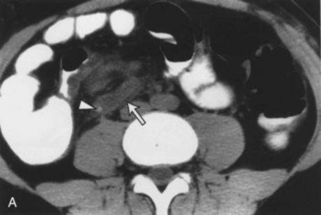

Figure 21-8 Abdominal computed tomographic scans. A, This image shows an inflamed tubular structure consistent with appendicitis (arrow) with a fecalith (arrowhead) at its base. B, This image shows a large pelvic abscess (arrow) containing fluid and gas. BL, Bladder; UT, uterus.
Ref.: 1, 2
Comments
A ruptured appendix may result in a localized periappendiceal abscess, diffuse peritonitis, or abscesses at other abdominal sites, notably in the pelvis, in the right subhepatic region, or between loops of bowel. Unrelenting obstruction of the appendix leads to gangrene and rupture of the organ. Because the patient is ill, the abdominal pain is more severe and diffuse and evidence of sepsis is apparent. Physical signs are more obvious after rupture, depending on the position of the appendix. With a periappendiceal abscess or phlegmon, a mass is usually felt, and its nature can be further clarified by ultrasound or CT.
A patient with a late manifestation of appendicitis (>48 hours) who is found to have an appendiceal abscess may benefit from nonoperative management. Nonoperative management of an appendiceal abscess reduces complications and overall hospital stay and can be accomplished through a percutaneous, transrectal, or transvaginal approach. Patients with abscesses larger than 4 to 6 cm benefit from abscess drainage, and those with a smaller abscess or phlegmon may be treated with antibiotics alone (Figure 21-9). Patients who are not improving with nonoperative treatment should be considered for operative drainage and appendectomy. After the acute inflammation has subsided, adults should undergo colonoscopy or barium enema to evaluate for cancer. Elective performance of interval appendectomy in 6 to 8 weeks is controversial in adults and more commonly accepted in children.
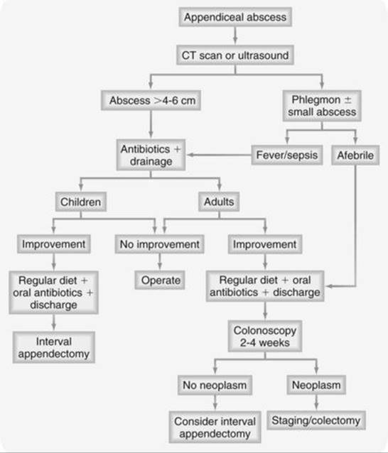
Figure 21-9 Treatment options for appendiceal abscess.
Answer
E
13 For which of the following patients would nonoperative treatment of appendicitis be appropriate?
A A pregnant woman during the third trimester
B A 35-year-old patient with subsiding symptoms and a right lower quadrant mass
C An elderly patient with concomitant cardiac disease
D A 20-year-old woman with Crohn’s disease
E A 20-year-old woman with a thickened appendiceal wall and right ovarian cyst noted on ultrasound
Ref.: 1, 2
Comments
When the diagnosis of appendicitis is a strong consideration but not certain, in most instances surgery should be undertaken because delay involves a risk for rupture with its accompanying increased morbidity and mortality. Surgery should not be delayed during pregnancy because doing so increases the risk to both the mother and fetus. Nor should it be delayed for elderly patients because they face an increased risk for appendiceal rupture and death.
The optimal timing of surgery for a ruptured appendix with an established periappendiceal abscess has been controversial. However, initial nonoperative therapy followed by interval appendectomy in 6 to 8 weeks may be considered for select patients whose symptoms are clearly subsiding and in whom a discrete right lower quadrant mass is palpable. Such expectant treatment consists of intravenous fluids and appropriate antibiotics. Vital signs, WBC count, and the size of the mass are watched closely. With these measures, most abscesses resolve, but prolonged hospitalization and antibiotic therapy are needed. Should progression occur, the abscess is drained. The manifestations of acute regional enteritis often mimic those of appendicitis. Acute ileitis should be distinguished from Crohn’s disease because progression of the former to the latter occurs in only 10% of cases. If exploration reveals an acutely inflamed ileum and a normal appendix, an appendectomy may be performed, but only if the cecum is normal. Performance of an appendectomy in the face of cecal inflammation risks the formation of a fecal fistula.
Answer
B
14 A 20-year-old woman is operated on through a right lower quadrant incision for presumed appendicitis, but the appendix is normal. At this point, which of the following would be appropriate treatment?
A Proceeding with appendectomy if no other pathology is found
B Exploration and treatment of any associated pathologic condition, as indicated, without appendectomy
C Exploration and diverticulectomy if a Meckel diverticulum is present and is normal by inspection and palpation
D Exploration and, if no pathology is found, closure without appendectomy
E Exploration and ileal resection if the terminal ileum appears acutely inflamed
Ref.: 1, 2
Comments
If appendicitis is not found at the time of surgery, careful exploration for other pathologic conditions must be carried out. The accuracy of the preoperative diagnosis should be 85%. In general, appendectomy is performed, except in some cases of Crohn’s disease with extensive involvement of the ileum and cecum. The pelvic organs, gallbladder, colon, and gastroduodenal areas should be inspected to the extent possible. A laparoscopic approach may allow better evaluation of other areas than can be accomplished through a limited right lower quadrant incision.
The differential diagnosis of appendicitis is basically that of an acute abdomen. The surgeon must be prepared to treat other pathologic entities should they be found on exploration for appendicitis. Such differential diagnoses include acute mesenteric adenitis, gastroenteritis, diverticulitis, epiploic appendagitis, and cancer.
Acute mesenteric adenitis is most often confused with appendicitis in children. Frequently, an upper respiratory tract infection precedes or is present at the onset of diffuse abdominal pain. Generalized lymphadenopathy or relative lymphocytosis, when present, can be of help. At surgery, the mesenteric lymph nodes are assessed. If they are enlarged, a biopsy is performed. The lymph nodes are examined histologically for granulomas (including Crohn’s disease), and tissue is cultured for mycobacteria and Yersinia. Infection with Yersinia causes mesenteric adenitis, ileitis, colitis, and acute appendicitis.
Acute gastroenteritis is characterized by cramping pain followed by watery stools, nausea, and vomiting. Laboratory results are usually normal. Diagnosis of a specific bacterial infection (e.g., Salmonella or typhoid fever) is made by stool culture.
The small intestine is inspected in a retrograde manner for evidence of inflammatory bowel disease or an inflamed Meckel diverticulum. The incidence of perforation or peritonitis with Meckel diverticulitis is about 50%. Resection of a Meckel diverticulum is indicated if diverticulitis is present. An asymptomatic Meckel diverticulum found incidentally during laparotomy in adults should not necessarily be removed. Diverticulitis of the cecum may be impossible to distinguish from acute appendicitis or cancer clinically. Both may be manifested as a right lower quadrant mass with evidence of infection and peritonitis. Sigmoid diverticulitis may also mimic appendicitis if a mobile, inflamed sigmoid colon is located in the right lower quadrant.
Epiploic appendagitis results from infarction of the appendage secondary to torsion. The pain is short-lived and well localized, recovery is fairly rapid, and patients do not appear ill.
If no pathology is found and a right lower quadrant incision is made, an appendectomy should be performed to eliminate potential confusion in the management of right lower quadrant abdominal pain in the future.
Answer
A
15 A patient suspected of having appendicitis underwent exploration. Crohn’s disease was found. Which of the following is true?
A The normal appendix should always be removed.
B All grossly involved bowel, including the appendix, should be resected.
C An inflamed appendix, cecum, and terminal ileum should be resected.
D Perforated bowel and advanced Crohn’s disease with obstruction should be resected.
E Only the tip of the appendix should be resected if the base is found to be involved.
Ref.: 1, 2
Comments
If a normal appendix is found at the time of laparotomy, other causes should be sought. If Crohn’s disease is encountered and the cecum and base of the appendix are normal, an appendectomy should be performed. If the base is involved with Crohn’s disease, appendectomy should be avoided. If the areas involved with Crohn’s disease are not complicated by perforation or obstruction, bowel resection is not indicated and medical therapy should be instituted. However, in the case of perforation or high-grade obstruction from a fibrotic segment, the involved bowel should be resected.
Answer
D
16 With regard to carcinoid of the appendix, which of the following statements is true?
A The ileum is the most common location for GI carcinoids.
B An appendectomy is sufficient for incidental carcinoid tumors less than 1 cm with positive lymph nodes.
C Most carcinoid tumors occur at the tip of the appendix.
D Right hemicolectomy is routinely indicated, regardless of nodal status.
E Carcinoid tumors of the appendix are usually manifested as acute appendicitis.
Ref.: 1, 2
Comments
The most common locations for GI carcinoids are the rectum, ileum, and appendix, in order of increasing frequency. Appendiceal carcinoids are usually solitary. About 75% of them occur at the distal tip and less than 10% occur at the base. They are usually an incidental finding and only rarely cause appendicitis. Carcinoids of the small bowel are multiple in approximately 30% of patients. Carcinoid syndrome usually occurs in patients with small bowel tumors that have metastasized to the liver. Appendiceal carcinoids less than 1 cm are generally considered biologically benign lesions, and only 3% metastasize. If the lesion is found at the tip of the appendix and is smaller than 1 cm, appendectomy is considered adequate treatment because these lesions have a lower risk for metastasis. Right hemicolectomy is indicated if tumor is present at the surgical margins, if there is nodal involvement, and if the lesion is larger than 2 cm. For tumors 1 to 2 cm in size, the decision to perform right hemicolectomy must be individualized because these patients have a substantial risk for metastasis. Chemotherapy with a combination of 5-fluorouracil and streptozocin has provided some palliation of carcinoid syndrome in patients with unresectable disease. Up to 25% of patients who undergo palliative resection survive for 5 years.
Answer
C
17 When a mucocele of the appendix is found at the time of surgery, which of the following is appropriate initial therapy?
A Incisional biopsy with subsequent appendectomy if malignancy is confirmed by frozen section
B Routine right hemicolectomy with lymph node dissection
C Needle aspiration of cystic fluid for cytologic examination
D Appendectomy
E Closure and observation
Ref.: 1, 2
Comments
Appendectomy is adequate treatment of a mucocele, but care must be taken to avoid rupture, because pseudomyxoma peritonei has been reported following rupture and peritoneal dissemination of the appendiceal contents even if the appendix was free of cancer. Histologically, mucoceles can be categorized as a benign type, which is the result of occlusion of the proximal lumen of the appendix, or a malignant type, which is a variant of a mucous papillary adenocarcinoma. Treatment of an appendiceal adenocarcinoma is right hemicolectomy.
Answer
D
18 Which of the following statements is true regarding laparoscopic versus open appendectomy?
A Laparoscopic appendectomy is associated with less postoperative pain, shorter hospital stay, faster recovery, and lower wound infection rates.
B Surgical costs for laparoscopic appendectomy are less.
C The laparoscopic approach is contraindicated in morbidly obese patients.
D Conversion from laparoscopic to open appendectomy occurs in approximately 50% of cases.
E Laparoscopic appendectomy results in a lower intra-abdominal abscess rate in patients with advanced appendicitis.
Ref.: 1, 2, 6, 7
Comments
Laparoscopic appendectomy has several advantages over the open technique, including a shortened hospital stay, faster recovery, and lower wound infection rates. The wound infection rate for laparoscopic appendectomy is less than one half that for open appendectomy. The major disadvantage of laparoscopic appendectomy is the use of disposable instruments, which can increase surgical cost; however, this may be offset by shorter length of stay. A laparoscopic approach may be preferable in the morbidly obese because of fewer wound-related difficulties. Rates of conversion are variable but occur approximately 10% of the time. For patients with advanced appendicitis at initial evaluation, there is a greater tendency for the formation of intra-abdominal abscesses with laparoscopic appendectomy than with an open technique.
Answer
A
References
1 Maa J, Kirkwood KS. The appendix. In Townsend CM, Beauchamp RD, Evers BM, et al, editors: Sabiston textbook of surgery: the biological basis of modern surgical practice, ed 18, Philadelphia: WB Saunders, 2008.
2 Matthews JB, Hodin RA. Acute abdomen and appendix. In Mulholland MW, Lillemoe KD, Doherty GM, et al, editors: Greenfield’s surgery: scientific principles and practice, ed 4, Philadelphia: Lippincott Williams & Wilkins, 2006.
3 Galindo GM, Fadrique B, Nieto MA, et al. Evaluation of ultrasonography and clinical diagnostic scoring in suspected appendicitis. Br J Surg. 1998;85:37-40.
4 Roa PM, Rhea JT, Novelline RA, et al. Effect of computed tomography of the appendix on treatment of patients and use of hospital resources. N Engl J Med. 1998;338:141-146.
5 Raptopoulos V, Katsou G, Rosen MP, et al. Acute appendicitis: effect of increased use of CT on selecting patients earlier. Radiology. 2003;226:521-526.
6 Golub R, Siddiqui F, Pohl D. Laparoscopic versus open appendectomy: a metaanalysis. J Am Coll Surg. 1998;186:545-553.
7 Liu SI, Siewert B, Raptopoulos V, et al. Factors associated with conversion to laparotomy in patients undergoing laparoscopic appendectomy. J Am Coll Surg. 2002;194:298-302.