José M. Velasco, M.D., Chad E. Jacobs, M.D.
A
Fluid and Electrolytes
David D. Shersher, M.D.
1 Which of the following statements regarding total body water is false?
A In males, approximately 60% of total body weight is water
B The percentage of total body weight that is water is higher in males than in females
C Lean individuals have a greater proportion of water (relative to body weight) than do obese individuals
D The percentage of total body water decreases with age
E The majority of body water is contained within the interstitial fluid compartment
Ref.: 1-3
Comments
Approximately 50% to 75% of body weight is water. In males, 60% (±15%) of body weight is water, and in females, 50% (±15%) of body weight is water. Age and lean body mass also contribute to differences in the percentage of total body weight that is water. Since fat contains little water, lean individuals have a greater proportion of body water than do obese individuals of the same weight. Because females have more subcutaneous fat in relation to lean mass than do males, they have less body water. Total body water decreases with age as a result of decreasing lean muscle mass. Infants have an unusually high ratio of total body water to body weight: up to 75% to 80%. By 1 year of age, however, the percentage of body water approaches that of adults.
Body water is divided into three functional compartments: the intracellular fluid (ICF) compartment (40% of body weight) and the extracellular fluid (ECF) compartment (20% of body weight), which is further subdivided into the interstitial (15% of body weight) and intravascular (5% of body weight) fluid compartments.
Answer
E
2 Which of the following statements regarding the distribution, composition, and osmolarity of body fluid compartments is not true?
A Most intracellular water resides in skeletal muscle.
B The principal extracellular cation is sodium.
C Nonpermeable proteins determine the effective osmotic pressure between the interstitial and intravascular (plasma) fluid compartments.
D Calcium greatly determines the effective osmotic pressure between the ICF and ECF compartments
E The principal extracellular anions are chloride and bicarbonate.
Ref.: 1
Comments
The ICF compartment (accounting for 40% of total body weight) is contained mostly within skeletal muscle. The principal intracellular cations are potassium and magnesium, whereas the principal intracellular anions are proteins and phosphates. In the ECF compartment (20% of total body weight), which is subdivided into the interstitial (extravascular) and the intravascular (plasma) fluid compartments, the principal cation is sodium, whereas the principal anions are chloride and bicarbonate. The interstitial compartment has a rapidly equilibrating functional component and a slowly equilibrating, relatively nonfunctional component consisting of fluid within connective tissue and cerebrospinal and joint fluid (termed transcellular water). Intravascular fluid (plasma) has a higher concentration of nondiffusible organic proteins than do interstitial fluids. These plasma proteins act as multivalent anions. As a result, the concentration of inorganic anions is lower but the total concentration of cations is higher in intravascular fluid than in interstitial fluid. This relationship is explained in the Gibbs-Donnan equilibrium equation: the product of the concentrations of any pair of diffusible cations and anions on one side of a semipermeable membrane equals the product of the same pair on the other side.
In each body compartment the concentration of osmotically active particles is 290 to 310 mOsm. Although total osmotic pressure represents the sum of osmotically active particles in the fluid compartment, the effective osmotic pressure depends on osmotically active particles that do not freely pass through the semipermeable membranes of the body. The nonpermeable proteins in plasma are responsible for the effective osmotic pressure between plasma and the interstitial fluid compartment (the colloid osmotic pressure). The effective osmotic pressure between the ECF and ICF compartments is due mainly to sodium, the major extracellular cation, which does not freely cross the cell membrane. Because water moves freely between the compartments, the effective oncotic pressure within the various body fluid compartments is considered to be equal after fluid equilibration. An increase in the effective oncotic pressure of the ECF compartment (such as an increase in sodium concentration) causes movement of water from the intracellular space to the extracellular space until the osmotic pressure equalizes. Conversely, loss of sodium (hyponatremia) from the extracellular space results in movement of water into the intracellular space. Thus, the ICF contributes to correcting the changes in concentration and composition in the ECF. Isotonic ECF losses (losses in volume without change in concentration) generally do not cause transfer of water from the intracellular space as long as the osmolarity remains unchanged. Isotonic volume losses result in changes in ECF volume.
Answer
D
3 Which of the following statements regarding changes in volume status of the ECF compartment is true?
A Hyponatremia is diagnostic of excess ECF volume.
B Hypernatremia is diagnostic of depletion of ECF volume
C Excess extracellular volume is usually iatrogenic or due to renal or cardiac failure.
D Central nervous system symptoms appear after tissue signs with acute volume loss.
E The concentration of serum sodium is directly related to extracellular volume.
Ref.: 1, 2
Comments
The serum concentration of sodium is not necessarily related to the volume status of the ECF compartment. Volume deficit or excess can exist with high, low, or normal serum sodium concentrations. Volume deficit is the most frequent volume disorder encountered during surgery. Its most common cause is loss of isotonic fluid (i.e., fluid having the same composition as ECF), for example, through hemorrhage, vomiting, diarrhea, fistulas, or third-spacing. With acute volume loss, central nervous symptoms (e.g., sleepiness and apathy progressing to coma) and cardiovascular signs (e.g., orthostasis, hypotension, tachycardia, and coolness in the extremities) appear first, along with decreasing urine output. Tissue signs (e.g., decreased turgor, softness of the tongue with longitudinal wrinkling, and atonicity of muscles) usually do not appear during the first 24 hours. In response to hypovolemia, body temperature may be slightly decreased. It is therefore important to also monitor the body temperature of hypovolemic patients. Signs and symptoms of sepsis may be depressed in volume-depleted patients. The abdominal pain, fever, and leukocytosis associated with peritonitis may be absent until ECF volume is restored.
Volume overload is generally either iatrogenic or the result of renal insufficiency or heart failure. Both plasma and the interstitial fluid spaces are involved. The signs are those of circulatory overload and include distended veins, bounding pulses, functional murmurs, edema, and basilar rales. These signs may be present in young, healthy patients, but these patients can compensate for moderate to severe volume excess without overt failure or pulmonary edema developing. In elderly patients, however, congestive heart failure (CHF) with pulmonary edema may develop quite rapidly.
Answer
C
4 Which one of the following is not a stimulus for ECF expansion?
A Hemorrhage leading to a reduction in blood volume
B Increased capillary permeability after major surgery
C Peripheral arterial vasoconstriction
D Negative interstitial fluid hydrostatic pressure
E Colloid oncotic pressure
Ref.: 3
Comments
Approximately 85% of the ECF that is within the vascular compartment resides in the venous circulation. Therefore, the remaining 15% resides within the arterial system. The vascular compartment, otherwise known as plasma fluid, constitutes approximately a third of the ECF. Interstitial fluid (i.e., fluid between the cells) makes up approximately two thirds of the ECF. The extracellular fluid constitutes a third of total body water, whereas the ICF represents two thirds. Expansion of ECF is primarily driven by three mechanisms, all of which have the final common stimuli of reduction of intravascular volume. The first mechanism, hemorrhage, is directly responsible for the reduction in blood volume. Through various pathways, this drop in volume signals the retention and sequestration of fluid in the intravascular space. Increased capillary permeability, the second mechanism, occurs following major surgery and is due to the loss of endothelial integrity. This loss of integrity is mediated by several humoral factors that act on the endothelium. The end result of loss of endothelial integrity is extravasation of protein-rich fluid into the interstitium, with a consequent increase in the interstitial fluid space. This constitutes the third mechanism of ECF expansion. Serum albumin is a major determinant of colloid oncotic pressure, and hypoalbuminemia could lead to transudation of fluid from the vascular to the interstitial compartment. This concept is expressed mathematically by the Starling equation: Qf = Kf × (Pv − Pt) − δ × (COP − TOP), where Qf is fluid flux, Kf is the capillary filtration coefficient, Pv is vascular hydrostatic pressure, Pt is interstitial hydrostatic pressure, δ is a reflection coefficient (which defines the effectiveness of the membrane in preventing flow of solutes), COP is colloid osmotic pressure, and TOP is tissue osmotic pressure.
Answer
C
5 Which of the following humoral factors increases arterial vasodilation while not decreasing protein permeability in the capillary membranes?
A Bradykinin
B Nitric oxide (NO)
C Atrial natriuretic factor
D Histamine
E Platelet-activating factor
Ref.: 1
Comments
The protein permeability characteristics of capillary membranes are quantified by a numeric value termed the reflection coefficient. This value ranges from 0 to 1 and is conceptualized as the fraction of plasma protein that “reflects” back from the capillary wall when water crosses. The higher the coefficient, the more impermeable the capillary is to protein. Therefore, the oncotic pressure of the plasma volume declines as the reflection coefficient decreases. Certain intravascular factors can reduce the reflection coefficient and increase arterial vasodilation. Bradykinin, atrial natriuretic factor, histamine, and platelet-activating factor increase microvascular membrane permeability while causing arterial vasodilation. NO, although it causes arterial vasodilation, does not increase microvascular membrane permeability. Membrane permeability causes a shift of fluid and plasma proteins into the interstitium and thereby decreases the intravascular compartment. The protein-rich edema in the interstitium can adversely affect the ability to combat infection.
Answer
B
6 Which of the following statements regarding hypervolemia in postoperative patients is not true?
A Hypervolemia can be produced by the administration of isotonic salt solutions in amounts that exceed the loss of volume.
B Acute overexpansion of the ECF space is usually well tolerated in healthy individuals.
C Avoidance of volume excess requires daily monitoring of intake and output and determinations of serum sodium concentrations to guide accurate fluid administration.
D The most reliable sign of volume excess is peripheral edema.
E The earliest sign of volume excess is weight gain
Ref.: 1, 2
Comments
The earliest sign of volume excess during the postoperative period is weight gain. Normally, during this period the patient is in a catabolic state and is expected to lose weight ( to
to  lb/day). Circulatory and pulmonary signs of overload appear late and usually represent massive overload. Peripheral edema does not necessarily indicate excess volume. In a patient with edema but without additional evidence of volume overload, other causes of peripheral edema should be considered. The most common cause of excess volume in a surgical patient is the administration of isotonic salt solutions in amounts that exceed the loss of volume. In a healthy individual, such overload is usually well tolerated. However, if excess fluid is administered for several days, the ability of the kidneys to secrete sodium may be exceeded, thus resulting in hypernatremia.
Answer
D
7 Which of the following statements regarding loop diuretics is not true?
A Loop diuretics act on the thick ascending limb of the loop of Henle in the nephron.
B Loop diuretics increase blood flow to the kidney.
C Magnesium and calcium are unaffected during diuresis.
D Loop diuretics increase venous capacitance.
E Loop diuretics inhibit the sodium-potassium-chloride cotransporter.
Ref.: 1, 3
Comments
Loop diuretics, most commonly furosemide, are potent inhibitors of the sodium-potassium-chloride cotransporter. They act by competing for the chloride-binding site at the thick ascending limb of the loop of Henle. The effect is inhibition of sodium reabsorption resulting in diuresis. Magnesium, potassium, and calcium will likewise be excreted with the net increase in urine output. Therefore, it is important to monitor their serum levels to prevent depletion while a patient is being treated with a loop diuretic.
Loop diuretics are commonly used for pulmonary edema because of their potency. In addition to inhibition of sodium absorption, they increase blood flow to the kidneys by stimulating vasodilatory prostaglandins and increase venous capacitance, which can quickly relieve pulmonary edema, even before diuresis and natriuresis have occurred. These three mechanisms help decrease ECF volume. Loop diuretics, such as furosemide or bumetanide, are extensively protein bound and must reach their intratubular site of action through active proximal tubular secretion.
Answer
C
8 Which of the following pairing statements regarding daily fluid balance is incorrect?
A Daily water intake, 2000 to 2500 mL
B Average stool loss, 1000 mL
C Average insensible loss, 600 mL
D Average urine volume, 800 to 1500 mL
E Average increase in insensible loss in a febrile patient, 250 mL/day for each degree of fever
Ref.: 2
Comments
The average individual has an intake of 2000 to 2500 mL of water per day—1500 mL is ingested orally and the remainder is acquired in solid food. Daily losses include 250 mL in stool, 800 to 1500 mL in urine, and approximately 600 mL as insensible loss. To excrete the products of normal daily catabolism, an individual must produce at least 500 to 800 mL of urine. In healthy individuals, 75% of insensible loss occurs through the skin and 25% through the lungs. Insensible loss from the skin occurs as loss of water vapor through the skin and not by evaporation of water secreted by the sweat glands. In febrile patients, insensible loss through the skin may increase to 250 mL/day for each degree of fever. Losses from sweating can be as high as 4 L/h. In a patient with a tracheostomy who is being ventilated with unhumidified air, insensible loss from the lungs may increase to 1500 mL/day.
Answer
B
9 Which of the following statements concerning the sodium concentration of various fluids is incorrect?
A Pancreatic secretions, 140 mEq/L
B Sweat, 40 mEq/L
C Gastric secretions, 50 mEq/L
D Saliva, 100 mEq/L
E Ileostomy output, 125 mEq/L
Ref.: 3
Comments
Average daily salt intake ranges from 50 to 90 mEq sodium chloride. Usually, the kidneys excrete excess salt as it is encountered. Under conditions of reduced intake or increased extrarenal fluid loss, renal sodium excretion can be reduced to less than 1 mEq/day. Conversely, in patients with malfunctioning kidneys, sodium loss may be as high as 200 mEq/L of urine. The electrolyte composition of sweat and gastrointestinal secretions varies. Sweat represents a hypotonic loss of fluids. The average sodium concentration in sweat is 15 to 60 mEq/L. Insensible loss from the skin and lungs consists of pure water. Although the various gastrointestinal secretions vary in composition, gastrointestinal losses are usually isotonic or slightly hypotonic. Pancreatic secretions have high bicarbonate concentrations (75 mEq/dL), in contrast to that of bile. Stomach, small intestine, and biliary fluids have relatively high chloride concentrations. Duodenal, ileal, pancreatic, and biliary fluids contain levels of sodium that approximate those seen in plasma. Saliva is relatively high in potassium, a fact that is important to remember when managing a patient with a salivary fistula (Table 4-1). The concentration of sodium varies with gland stimulation and circadian rhythm; it ranges from 3 mmol/L to 70 mmol/L.
TABLE 4-1 Electrolyte Composition
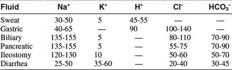
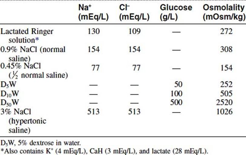
Management of fluid losses should take into account the electrolyte composition of the fluid, as well as that of the solution being used to replace these fluids. Lactated Ringer solution contains 130 mEq/L of sodium and 109 mEq/L of chloride. It also contains 4 mEq/L of potassium, 3 mEq/L of calcium, and 28 mEq/L of lactate. This is in contrast to 0.9% normal saline solution, which contains 154 mEq/L of both sodium and chloride. On the other hand, hypertonic 3% saline solution contains 513 mEq/L of both sodium and chloride.
Answer
D
10 With regard to distributional shifts during an operation, which of the following statements is true?
A The surface area of the peritoneum is not large enough to account for significant third-space loss.
B Approximately 1 to 1.5 L/h of fluid is needed during an operation.
C Blood is replaced as it is lost, without modification of the basal operative fluid replacement rate.
D Sequestered ECF is predominantly hypotonic.
E A major stimulus to ECF expansion is peripheral vasoconstriction.
Ref.: 1-3
Comments
The functional ECF volume decreases during major abdominal operations largely because of sequestration of fluid in the operative site as a consequence of (1) extensive dissection, (2) fluid collection within the lumen and wall of the small bowel, and (3) accumulation of fluid in the peritoneal cavity. The surface area of the peritoneum is 1.8 m2. When irritated, it can account for a functional loss of several liters of fluid that is not readily apparent. It is generally agreed that this lost volume should be replaced during the course of an operation with isotonic saline solution as a “mimic” of sequestered ECF. Although there is no set formula for intraoperative fluid therapy, useful guidelines for replacement include the following. (1) Blood is replaced as it is lost, regardless of additional fluid therapy, provided that the patient meets the criteria for transfusion: hemoglobin concentration less than 7 g/dL. (2) Lost ECF should be replaced during the operative procedure; delay in replacement until after the operation is complicated by adrenal and hypophyseal compensatory mechanisms that respond to operative trauma during the immediate postoperative period. (3) Approximately 0.5 to 1.0 L/h of fluid is needed during the course of an operation, to a maximum of 2 to 3 L during a 4-hour procedure, unless there are measurable losses.
Answer
C
11 With regard to intraoperative management of fluids, which of the following statements is true?
A In a healthy person, up to 500 mL of blood loss may be well tolerated without the need for blood replacement.
B During an operation, functional ECF volume is directly related to the volume lost to suction.
C Functional ECF losses should be replaced with plasma.
D Administration of albumin plays an important role in the replacement of functional ECF volume loss.
E Operative blood loss is usually overestimated by the surgeon.
Ref.: 1, 2
Comments
It is now believed that the routine use of albumin to replace blood and ECF losses intraoperatively is not indicated and may be potentially harmful. Maintenance of cardiac and pulmonary function by replacing blood with blood products and ECF with “mimic” solutions can be achieved without the addition of albumin. In general, it is believed that blood should be replaced as it is lost. However, it is usually unnecessary to replace blood loss of less than 500 mL. Operative blood loss is usually underestimated by the surgeon by 15% to 40% in comparison to the isotopically measured loss, a factor that may contribute to the detection of anemia during the immediate postoperative period.
Answer
A
12 With regard to postoperative fluid management, which of the following statements is not true?
A Insensible loss is approximately 600 mL/day.
B Insensible loss may increase to 1500 mL/day.
C About 800 to 1000 mL of fluid is needed to excrete the catabolic end products of metabolism.
D Lost urine should be replaced milliliter for milliliter.
E Lost gastrointestinal fluids should be replaced milliliter for milliliter.
Ref.: 1-3
Comments
Postoperative fluid management requires assessment of the patient’s volume status and evaluation for possible disorders in concentration or composition. All measured and insensible losses should be treated by replacement with appropriate fluids. In patients with normal renal function, the amount of potassium given is 40 mEq/day for replacement of renal excretion. An additional 20 mEq should be given for each liter of gastrointestinal loss. Insensible water loss is usually constant in the range of 600 mL/day. It can be increased to 1500 mL/day by hypermetabolism, hyperventilation, or fever. Insensible loss is replaced with 5% dextrose in water. Insensible loss may be offset by an insensible gain of water from excessive catabolism in postoperative patients who require prolonged intravenous fluid therapy. Approximately 800 to 1000 mL/day of fluid is needed to excrete the catabolic end products of metabolism. Because the kidneys are able to conserve sodium in a healthy individual, this amount can be replaced with 5% dextrose in water. A small amount of salt is usually added, however, to relieve the kidneys of the stress of sodium resorption. If there is a question regarding urinary sodium loss, measurement of urinary sodium levels helps determine the type of fluid that can best be used. Urine volume should not be replaced milliliter for milliliter because high output may represent diuresis of the fluids given during surgery or the diuresis that takes place to eliminate excessive fluid administration. Sensible or measurable losses such as those from the gastrointestinal tract are usually isotonic and should therefore be treated by replacement in equal volumes with isotonic salt solutions. The type of salt solution selected depends on determination of the patient’s serum sodium, potassium, and chloride levels. In general, replacement fluids are administered at a steady rate over a period of 18 to 24 hours as losses are incurred (Table 4-2).
TABLE 4-2 Composition and Osmolality of Intravenous Solutions
Answer
D
13 With regard to abnormalities in serum sodium concentration, which of the following statements is true?
A Changes in serum sodium concentration usually produce changes in the status of ECF volume.
B The chloride ion is the main determinant of the osmolarity of the ECF space.
C Extracellular hyponatremia leads to depletion of intracellular water.
D Dry, sticky mucous membranes are characteristic of hyponatremia.
E Preservation of normal ECF has higher precedence than does maintenance of normal osmolality.
Ref.: 1, 2
Comments
Although extracellular volume may change without a change in serum sodium concentration (as occurs after isotonic volume losses), changes in serum sodium concentration usually produce changes in ECF volume because the serum sodium concentration is the main determinant of the osmolarity of the ECF space. Alterations in its concentration produce concomitant shifts in water volume. Signs and symptoms of hypernatremia and hyponatremia are not generally present unless the changes are severe or the alteration in sodium concentration occurs rapidly.
Hyponatremia is caused by excessive intake of hypotonic fluids or salt loss that exceeds water loss. With hyponatremia, decreased extracellular osmolarity causes a shift of water into the intracellular compartment. When such a shift occurs, central nervous system symptoms caused by increased intracranial pressure develop, and tissue signs of excess water are noted. Central nervous system symptoms include muscle twitching, hyperactive tendon reflexes, and when the hyponatremia is severe, convulsions and hypertension. Tissue signs include salivation, lacrimation, watery diarrhea, and “fingerprinting” of the skin. When hyponatremia develops rapidly, signs and symptoms may appear at sodium concentrations of less than 130 mEq/L. Acute dilution of osmolality can occur if patients with an ECF deficit are given sodium-free water. The hyponatremia is exacerbated in hypovolemic patients because of secretion of antidiuretic hormone (ADH) as a result of the hypothalamic-pituitary response to both elevated ECF osmolality and a reduction in ECF volume. The normal response of the hypothalamic-pituitary axis to hyponatremia is suppression of ADH release, and as the dilute urine is excreted, there is a corrective increase in serum [Na+]. A moderate or severely hyponatremic patient should have undetectable blood levels of ADH. Preservation of normal ECF has higher precedence than does maintenance of normal osmolality. In symptomatic patients, administration of hypertonic (3%) solutions of sodium may be indicated to correct the problem in those with severe hyponatremia who are at risk for seizures. In less severe cases, restriction of free water and judicious infusion of normal saline solution are usually sufficient. In patients with acute hyponatremia and [Na+] less than 120 mEq/L, the rate of infusion of sodium-containing solutions should not increase serum [Na+] more rapidly than 0.25 mEq/L/h.
Chronic hyponatremia develops slowly, and patients may have sodium levels as low as 120 mEq/L before becoming symptomatic. Severe hyponatremia may be associated with the onset of irreversible oliguric renal failure. Patients with a closed head injury are sensitive to even mild hyponatremia because of increased intracellular water, which exacerbates the increased intracranial pressure associated with the head injury. The syndrome of inappropriate release of antidiuretic hormone (SIADH) and chronic renal failure are frequent causes of hyponatremia. The diagnosis of SIADH can be made only in euvolemic patients who have a serum osmolality of less than 270 mmol/kg H2O along with inappropriately concentrated urine.
Hypernatremia is the result of excessive free water loss or salt intake. Central nervous system signs and symptoms associated with hypernatremia include restlessness, weakness, delirium, and maniacal behavior. The tissue signs are characteristic and include dryness and stickiness of mucous membranes, decreased salivation and tear production, and redness and swelling of the tongue. Body temperature is usually elevated, occasionally to a lethal level. An acute onset of hypernatremia increases ECF osmolality and contracts the size of the ICF compartment. Patients have moderate hypernatremia if their serum [Na+] is 146 to 159 mEq/L. Water loss is the most common explanation for acute hypernatremia. Neurologic damage as a result of contraction of brain cell volume is the primary risk associated with hypernatremia. Patients with diabetes insipidus or nephrogenic diabetes insipidus have a failure to synthesize and release ADH or a failure of the renal tubular cells to respond to ADH, respectively, thus leading to hypernatremia. Treatment of patients with hypernatremia secondary to dehydration involves the administration of water. Hypernatremic patients are frequently hypovolemic, and these patients are treated by the intravenous infusion of isotonic saline solution until the volume deficit has been restored. A rapid decline in ECF osmolality in a severely hypernatremic patient can lead to cerebral injury as a result of cellular swelling. [Na+] should be lowered at a rate not to exceed 8 mEq/day (Table 4-3). Patients with central diabetes insipidus are treated with desmopressin (1-desamino-8-D-arginine vasopressin [DDAVP]). Desmopressin is a synthetic analogue of ADH.
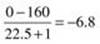
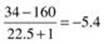
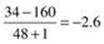
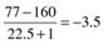
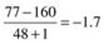
TABLE 4-3 Given a Patient with Hypernatremia (Serum [Na+] = 160 mEq/L), the Estimated Change in [Na+] after Infusion of 1 L
|
|
||
|
Infusate |
Woman Aged 70 Years 50 kg × 0.45 = 22.5 L TBW |
Man Aged 20 Years 80 kg × 0.60 = 48.0 L TBW |
|
D5W |
|
|
|
D5 0.2% NaCl |
|
|
|
D5 0.45% NaCl |
|
|
D5W, 5% dextrose in water; TBW, total body water.
Answer
A
14 Which of the following does not contribute to the development of hypernatremia?
A Excessive sweating
B Hyperlipidemia
C Lactulose
D Glycosuria
E Inadequate maintenance fluids
Ref.: 3
Comments
Hypernatremia is less common than hyponatremia in postoperative patients and is a reflection of elevated serum osmolality and hypertonicity. It is indicative of a deficiency of free water relative to the sodium concentration. Decreased intake of water, increased loss of water, and increased intake of sodium are the main mechanisms responsible for the development of hypernatremia. Loss of the thirst mechanism and an inability to access free water are mechanisms by which hypernatremia secondary to decreased intake of water can develop. Excessive sweating and large evaporative losses are mechanisms of loss of free water. Agents such as lactulose, sorbitol, and carbohydrate malabsorption can cause osmotic diarrhea and result in relative losses of hypotonic fluid. Similarly, hyperglycemia causing glycosuria or diuresis in a catabolic patient excreting excess urea can also cause an osmotic diuresis. Both hyperlipidemia and hyperproteinemia are responsible for an entity known as pseudohyponatremia, which occurs when excess lipids or proteins displace water and create a falsely measured hyponatremia.
Answer
B
15 Which of the following conditions is not associated with hypernatremia?
A Diabetes insipidus
B Tumor lysis syndrome
C Steven-Johnson syndrome
D Primary hypodipsia
E Enterocutaneous fistula
Ref.: 1
Comments
Diabetes insipidus is characterized by the excretion of large volumes of dilute urine, which can lead to hypernatremia. Patients with primary hypodipsia, a rare neurologic deficit of the thirst center, have an impaired or absent thirst response to an increase in extracellular tonicity. Tumor or infection may be responsible for this defect. Dermatologic conditions such as second-degree burns and exfoliative dermatitis can substantially increase transcutaneous water loss and thereby result in the rapid onset of dehydration and hypernatremia. Dehydration from vomiting, diarrhea, or uncompensated loss of hypotonic gastrointestinal fluid, such as occurs with fistulas or endoluminal tubes, may cause hypernatremia. Tumor lysis syndrome, a condition involving cell breakdown and release of their intracellular contents after some chemotherapies, typically develops in patients treated with vinca alkaloid chemotherapy; it causes hyperkalemia, hyperphosphatemia, hyperuricemia, and ultimately, renal failure. Tumor lysis syndrome does not cause hypernatremia.
Answer
B
16 Which of the following clinical situations can be associated with hypovolemic hyponatremia?
A CHF
B SIADH
C Cirrhosis
D Hyperglycemia
E Gastrointestinal losses
Ref.: 1, 2
Comments
Hyponatremia in a surgical patient can be classified into hypervolemic, euvolemic, and hypovolemic categories, which can then be further subclassified according to tonicity (hypertonic, >290 mOsm; isotonic, 280 to 290 mOsm; and hypotonic, <280 mOsm). For simplicity and rapid clinical evaluation, volume status can be used to direct treatment. Hypervolemic hyponatremia may be caused by increased intake of water, postoperative secretion of ADH, and high ECF volume states such as cirrhosis and CHF. Hyponatremia can develop in patients with edema and ascites secondary to CHF, nephrotic syndrome, or cirrhosis despite having an expanded overall volume of extracellular water. These patients have an excess of sodium but an even greater proportional increase in water volume. Their pathophysiologic condition entails an overall contracted intravascular volume, which stimulates the release of vasopressin from the hypothalamus centrally. Peripherally, renal hypoperfusion contributes to water retention. Fluid restriction is crucial to the treatment of this type of hyponatremia. In patients with severe hyponatremia, small volumes of hypertonic saline solution may be administered. Diuresis may be used but is generally unsuccessful. Hemodialysis may be performed in extreme circumstances of fluid excess. Euvolemic hyponatremia may be caused by hyperglycemia, hyperlipidemia or hyperproteinemia (termed pseudohyponatremia because of relative hyperosmolar protein, lipid, or glucose-rich plasma drawing fluid from the interstitial space and diluting plasma sodium), SIADH, water intoxication, and diuretics. SIADH is characterized by functional reabsorption of free water and subsequent dilution of plasma sodium. Hypovolemic hyponatremia may be caused by decreased overall sodium intake, gastrointestinal losses, renal losses associated with the use of diuretics (especially thiazide diuretics), and primary renal disease.
Conversely, hypernatremia can also be subdivided into volume states. Hypervolemic hypernatremia may be caused by iatrogenic sodium administration or mineralocorticoid excess (e.g., aldosteronism, Cushing disease, congenital adrenal hyperplasia). Euvolemic hypernatremia may be associated with renal (renal disease, diuretics, or diabetes insipidus) or nonrenal free water loss through the skin or gastrointestinal tract. Hypovolemic hypernatremia can likewise be subdivided into nonrenal and renal water loss.
Answer
E
17 With regard to diabetes insipidus, which of the following statements is true?
A Diabetes insipidus causes hypervolemic hyponatremia.
B Central diabetes insipidus cannot be corrected by the administration of desmopressin.
C Treatment of diabetes insipidus requires correction of hypernatremia at a rate faster than 12 mEq/day.
D Alcohol intoxication can mimic diabetes insipidus.
E Lithium administration could induce central diabetes insipidus.
Ref.: 1, 3
Comments
Diabetes insipidus is one of the causes of hypovolemic hypernatremia and is marked by continual production of dilute urine of less than 200 mOsm/kg H2O in the context of serum osmolarity of extracellular fluid greater than 300 Osm/L. Patients can have either central (lack of production of ADH by the hypothalamus) or nephrogenic diabetes insipidus (lack of response of the distal tubule of the nephron to ADH). Alcohol causes suppression of vasopressin release and can mimic central diabetes insipidus. Treatment of hypernatremia consists of slow correction of sodium by the administration of free water. Whenever hypernatremia develops, a relative free water deficit exists and must be replaced. The water deficit can be approximated by using the following formula: water deficit = total body water × [(1 − 140 ÷ serum sodium)]. Usually, the rate of correction of hypernatremia should not exceed 12 mEq/L/day. The aim should be to correct approximately half the deficit over the first 24 hours. Too rapid correction of hypernatremia may lead to cerebral edema and seizures.
Desmopressin is a synthetic analogue of ADH that can be used to mimic arginine vasopressin (AVP) and to differentiate between central and nephrogenic diabetes insipidus. It is the agent of choice for treating patients with central diabetes insipidus because the drug increases water movement out of the collecting duct but does not have the vasoconstrictive effects of ADH. Central diabetes insipidus will respond to desmopressin, whereas nephrogenic diabetes insipidus will not. Unlike vasopressin, desmopressin is only renally active and does not have the vasoactive side effects. Lithium and amphotericin B can induce nephrogenic, not central diabetes insipidus.
Answer
D
18 A 30-year-old, 70-kg woman has symptomatic hyponatremia. Her serum sodium level is 120 mEq/L (normal level, 140 mEq/L). Her sodium deficit is:
A 500 mEq/L
B 600 mEq/L
C 700 mEq/L
D 800 mEq/L
E 400 mEq/L
Ref.: 1
Comments
Correction of changes in concentration depends in part on whether the patient is symptomatic. If symptomatic hypernatremia or hyponatremia is present, attention is focused on prompt correction of the abnormal concentration to the point that the symptoms are relieved. Attention is then shifted to correction of the associated abnormality in volume. The sodium deficiency in this patient is estimated by multiplying the sodium deficit (normal sodium concentration minus observed sodium concentration) by total body water in liters (60% of body weight in males and 50% of body weight in females). For the patient in question, the calculation is as follows: total body water = 70 kg × 0.5 = 35 L. Sodium deficit = (140 − 120 mEq/L) × 35 L = 700 mEq sodium chloride.
Initially, half the calculated amount of sodium is infused as 3% sodium chloride. The infusion is given slowly because rapid infusion can cause symptomatic hypovolemia. Rapid correction of hyponatremia can be associated with irreversible central nervous system injury (central pontine and extrapontine myelinolysis). Once the symptoms are alleviated, the patient should be reassessed before additional infusion of sodium is begun. In patients with profound hyponatremia, a correction of no more than 12 mEq/L/24 h should be achieved. If the original problem was associated with a volume deficit, the remainder of the resuscitation can be accomplished with isotonic fluids (sodium chloride in the presence of alkalosis, and sodium lactate in the presence of acidosis). Care must be taken when treating hyponatremia associated with volume excess. In this setting, after the symptoms are alleviated with a small volume of hypertonic saline solution, water restriction is the treatment of choice. Infusion of hypertonic saline solution in this setting has the potential to further expand the extracellular intravascular volume and is contraindicated in patients with severely compromised cardiac reserve. In such a case, peritoneal dialysis or hemodialysis may be preferred for removing excess water.
Answer
C
19 A postoperative patient has a serum sodium concentration of 125 mEq/L and a blood glucose level of 500 mg/dL (normal level, 100 mg/dL). What would the patient’s serum sodium concentration be (assuming normal renal function and appropriate intraoperative fluid therapy) if the blood glucose level were normal?
A 120 mEq/L
B 122 mEq/L
C 137 mEq/L
D 142 mEq/L
E 147 mEq/l
Ref.: 1-3
Comments
Serum osmolality is described as the amount of solutes per unit of water. It can be measured with an osmometer or it can be calculated. It is reported as milliosmoles per liter. Calculation of serum osmolality is performed with the following equation:
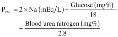
The serum concentrations of sodium, urea, and glucose are required, whereas that of chloride is not required for the calculation. Simply doubling the serum sodium concentration provides an adequate estimate of serum osmolality.
As a general rule, each 100-mg/dL rise in the blood glucose level above normal is equivalent to a 1.6- to 3.0-mEq/L fall in the apparent serum sodium concentration. For example, if the patient has a blood glucose level of 500 mg/dL, or 400 mg/dL above normal, this is equivalent to a 12-mEq/L change in the serum sodium level. If this patient has a measured sodium concentration of 125 mEq/L, the sodium concentration is actually 137 mEq/L once the excess extracellular water has been eliminated.
Answer
C
20 With regard to postoperative hyponatremia, which of the following statements is not true?
A It may easily occur when water is used to replace sodium-containing fluids or when the water given exceeds the water lost.
B In patients with head injury, hyponatremia despite adequate salt administration is usually caused by occult renal dysfunction.
C In oliguric patients, cellular catabolism with resultant metabolic acidosis increases cellular release of water and can contribute to hyponatremia.
D Hyperglycemia may be a cause of hyponatremia.
E Patients with salt-wasting nephropathy could have normal blood urea nitrogen and creatinine values.
Ref.: 1, 2
Comments
Abnormalities in sodium concentration do not usually occur during the postoperative period if the functional ECF volume has been adequately replaced during the operation. The sodium concentration generally remains normal because the kidneys retain the ability to excrete moderate excesses of water and solute administered during the early postoperative period. Hyponatremia does occur when water is given to replace lost sodium-containing fluids or when the amount of water given consistently exceeds the amount of water lost. In patients with head injury, hyponatremia may develop despite adequate salt administration because of excessive secretion of ADH with resultant increased water retention.
Patients with preexisting renal disease and loss of concentrating ability may elaborate urine with a high salt concentration. This salt-wasting phenomenon is commonly encountered in elderly patients and is often not anticipated because the blood urea nitrogen and creatinine levels are within normal limits. When there is doubt, determination of the urine sodium concentration can help clarify the diagnosis. Oliguria reduces the daily water requirement and can lead to hyponatremia if not anticipated. Cellular catabolism in patients without adequate caloric intake can lead to gain of significant quantities of water released from the tissues. Hyperglycemia may produce a depressed serum sodium level by exerting an osmotic force in the extracellular compartment, thus diluting serum sodium levels.
Answer
B
21 An elderly patient with adult-onset diabetes mellitus is admitted to the hospital with severe pneumonia. All of the following conditions can be associated with this patient condition except:
A Hypokalemia
B Hyperkalemia
C Nonketotic hyperosmolar coma
D Hypophosphatemia
E Hyponatremia
Ref.: 1
Comments
Elderly patients with adult-onset diabetes mellitus are at risk for the development of nonketotic hyperosmolar coma during sepsis. As a result of the development of a nonketotic hyperglycemic hyperosmolar state, hypokalemia and hyperglycemia may also occur. Treatment of these patients should include a reduction in the glucose load provided and the administration of isotonic fluid. Patients may also benefit from the administration of insulin. Systemic bacterial sepsis is also often accompanied by a drop in the serum sodium concentration, possibly because of interstitial or intracellular sequestration. It is treated by withholding free water, restoring ECF volume, and treating the source of sepsis.
Answer
B
22 Which one of the following clinical signs or symptoms is not associated with serum sodium concentrations below 125 mEq/L?
A Headache
B Hallucinations
C Bradycardia
D Hypoventilation
E Hyperthermia
Ref.: 2, 3
Comments
In most patients with symptomatic hyponatremia, the serum sodium concentration decreases below 125 mEq/L. When the concentration falls below 125 mEq/L, clinical signs and symptoms may occur, including headache, nausea, lethargy, hallucinations, seizures, bradycardia, hypoventilation, and occasionally coma. Hypothermia, not hyperthermia, occurs.
Answer
E
23 With regard to potassium, which of the following statements is not true?
A Normal dietary intake of potassium is 50 to 100 mEq/day.
B In patients with normal renal function, most ingested potassium is excreted in urine.
C More than 90% of the potassium in the body is located in the extracellular compartment.
D Critical hyperkalemia (>6 mEq/L) is rarely encountered if renal function is normal.
E Administration of sodium bicarbonate shifts potassium from the extracellular space (ECF) to the intracellular space (ICF).
Ref.: 1, 2
Comments
The average daily dietary intake of potassium is 50 to 100 mEq. In patients with normal renal function and normal serum potassium levels, most ingested potassium is excreted in urine. More than 90% of the body’s potassium stores is within the intracellular compartment at a concentration of 150 mEq/L. Although the total extracellular potassium concentration is just 50 to 70 mEq (4.5 mEq/L), this concentration is critical for cardiac and neuromuscular function. Significant quantities of intracellular potassium are released in response to severe injury, surgical stress, acidosis, and a catabolic state. However, dangerous hyperkalemia (>6 mEq/L) is rarely encountered if renal function is normal. The administration of bicarbonate shifts potassium from the ECF across the cell membrane into the ICF.
Answer
C
24 Which of the following electrocardiographic (ECG) findings is not associated with hyperkalemia?
A Peaked T waves
B Prolonged PR interval
C Loss of the P wave
D Narrowing of the QRS complex
E T waves higher than R waves in more than one lead
Ref.: 1, 2
Comments
Hyperkalemia occurs when the serum potassium level exceeds 5 mmol/L. As potassium increases, changes in the resting membrane potential of cells impair depolarization and repolarization and lead to cardiac arrhythmias. The signs of hyperkalemia are generally limited to cardiovascular and gastrointestinal symptoms. Gastrointestinal symptoms include nausea, vomiting, intermittent intestinal colic, and diarrhea. ECG changes could be the first manifestation of hyperkalemia (Figure 4-1) and include peaked T waves and a prolonged PR interval, which are characteristic early findings. These ECG changes may be seen with potassium concentrations greater than 6 mEq/L. Symmetrically peaked T waves indicate dangerous hyperkalemia, particularly if the T waves are higher than the R wave in more than one lead. At higher potassium concentrations (7 mmol/L), loss of P waves, slurring, or widening of the QRS complexes occurs. As [K+] exceeds 8 mmol/L, sudden lethal arrhythmias ensue, such as asystole, ventricular fibrillation, or a wide pulseless idioventricular rhythm.
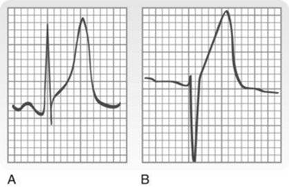
Figure 4-1 A, Electrocardiographic (ECG) changes indicating hyperkalemia. The T wave is tall, narrow, and symmetrical. B, ECG changes indicating acute myocardial infarction. The T wave is tall but broad based and asymmetrical.
(From Somers MP, Brady WJ, Perron AD, et al: The prominent T wave: Electrocardiographic differential diagnosis, Am J Emerg Med 20:243–251, 2002.)
Answer
D
25 Which one of the following is least useful in the immediate treatment of hyperkalemia?
A Calcium salts
B Sodium bicarbonate
C Potassium-binding resins
D Glucose and insulin
E Hemodialysis
Ref.: 1-3
Comments
The most dreaded complication of hyperkalemia is the development of a lethal arrhythmia. Immediate management includes ECG monitoring and cessation of all potassium supplementation and potassium-sparing drugs. Calcium is administered intravenously to stabilize the membrane potential and decrease myocardial excitability. It acts in less than 5 minutes and the effects last for 30 to 60 minutes. Sodium bicarbonate drives potassium into cells, thereby transiently reducing serum potassium levels. Its actions last 15 to 30 minutes. Insulin and glucose also facilitate entry of potassium into cells, with an almost immediate onset of action. In cases of severe hyperkalemia, hemodialysisis the definitive and most rapid method of decreasing extracellular potassium. Potassium-binding resins, such as sodium polystyrene sulfonate (Kayexalate), begin lowering serum potassium within 1 to 2 hours and last 4 to 6 hours. Rectal administration of these binding resins is more effective than oral formulations. However, enemas with sodium polystyrene sulfonate combined with sorbitol have been associated with colon necrosis and perforation. Kaliuresis through the administration of diuretics such as acetazolamide is also effective in reducing serum potassium levels.
Answer
C
26 With regard to hypokalemia, which of the following statements is not true?
A Potassium and hydrogen ions are exchanged for sodium in the renal tubule.
B Respiratory acidosis is associated with increased renal potassium loss.
C Hypokalemia can cause decreased deep tendon reflexes.
D Flattened T waves and a prolonged QT interval are associated with hypokalemia.
E Intravenous potassium administration should not exceed 40 to 60 mEq/h.
Ref.: 1, 2
Comments
Hypokalemia is more common than hyperkalemia in surgical patients. Hypokalemia can result from increased renal excretion, prolonged administration of potassium-free fluids, hyperalimentation with inadequate potassium replacement, or gastrointestinal losses. Respiratory and metabolic alkaloses result in increased renal potassium loss because potassium is preferentially excreted in an attempt to preserve hydrogen ions. Loss of gastrointestinal secretions can also be a significant cause of potassium depletion. This problem is compounded if potassium-free fluids are used for volume replacement. Signs of hypokalemia, including paralytic ileus, diminished or absent tendon reflexes, weakness, and even flaccid paralysis, are related to decreased muscle contractility. ECG changes include flattened or inverted T waves, U waves, and prolongation of the QT interval. The best treatment of hypokalemia is prevention. Gastrointestinal losses should be treated by the administration of fluids containing enough potassium to replace daily obligatory loss (20 mEq/day), as well as the additional losses in gastrointestinal drainage. As a rule, no more than 40 to 60 mEq of potassium should be added to each liter of intravenous fluid, and the rate of potassium administration should never exceed 40 to 60 mEq/h.
Answer
B
27 Which one of the following is not associated with hypocalcemia?
A Shortening of the QT interval
B Painful muscle spasms
C Perioral or fingertip tingling
D Seizures in children
E Prolongation of the QT interval
Ref.: 1-3
Comments
The symptoms of hypocalcemia are generally seen at serum levels of less than 8 mg/dL. Symptoms include numbness and tingling in the circumoral area and in the tips of the fingers and toes. Signs include hyperactive deep tendon reflexes, positive Chvostek sign, positive Trousseau sign, muscle and abdominal cramps, tetany with carpal pedal spasm, or convulsions. The electrocardiogram may show prolongation of the QT interval. Calcium is found in three forms in the body: protein bound (≈50%, mostly to albumin); diffusible calcium combined with anions such as bicarbonate, phosphate, and acetate (5%); and ionized (≈45%). Patients with severe alkalosis may have symptoms of hypocalcemia despite normal serum calcium levels because the ionized calcium is markedly decreased. Conversely, hypocalcemia without signs or symptoms may be present in patients with hypoproteinemia and a normal ionized fraction. Acute symptoms can be relieved by the intravenous administration of calcium gluconate or calcium chloride. Patients requiring prolonged replacement can be treated with oral calcium, often given with vitamin D.
Answer
A
28 Which one of the following clinical scenarios is not associated with acute hypocalcemia?
A Fluid resuscitation from shock
B Rapid infusion of blood products
C Improper administration of phosphates
D Vitamin D–deficient diets
E Acute pancreatitis
Ref.: 1
Comments
Infusion of large volumes of isotonic fluid can cause a modest reduction in serum calcium levels. The concomitant decrease in magnesium also impairs vitamin D activity and makes correction of the hypocalcemia more difficult. Administration of a citrate load during rapid transfusion of blood products can lead to severe hypocalcemia, hypotension, and cardiac failure. In this setting, calcium should be replaced at a dose of 0.2 g/500 mL of blood transfused. Most patients receiving slow, elective blood transfusions do not require calcium supplementation. Acute pancreatitis causes precipitation of calcium salts in the abdomen and may contribute to hypocalcemia. Other common causes include necrotizing fasciitis, renal failure, gastrointestinal fistula, and hypoparathyroidism. In general, calcium replacement should be monitored by measuring the concentration of ionized calcium.
Answer
D
29 Which of the following disturbances is not associated with tumor lysis syndrome?
A Hypocalcemia
B Hyperuricemia
C Hyperkalemia
D Hypermagnesemia
E Hyperphosphatemia
Ref.: 1
Comments
Tumor lysis syndrome is a constellation of electrolyte abnormalities that results from massive tumor cell necrosis secondary to antineoplastic therapy. Hypocalcemia, hyperphosphatemia, hyperuricemia, and hyperkalemia may occur. Hypocalcemia results from the release of intracellular stores of phosphate, which binds with ionized serum calcium to form calcium phosphate salts. Chemotherapy directed against solid tumors, especially lymphomas, is most commonly associated with tumor lysis syndrome. Acute renal failure can occur and prevent spontaneous correction of the electrolyte abnormalities. Hypermagnesemia is not associated with tumor lysis syndrome.
Answer
D
30 An asymptomatic patient is found to have a serum calcium level of 13.5 mg/dL. Which of the following medications should be avoided?
A Bisphosphonates
B Thiazide diuretics
C Mithramycin
D Calcitonin
E Corticosteroids
Ref.: 1
Comments
Hypercalcemia can affect the gastrointestinal, renal, musculoskeletal, and central nervous systems. Early symptoms include fatigability, lassitude, weakness, anorexia, nausea, and vomiting. Central nervous symptoms can progress to stupor and coma. Other symptoms include headaches and the three P’s: pain, polydipsia, and polyuria. The critical serum calcium level for hypercalcemia is 16 to 20 mg/mL. Prompt treatment must be instituted at this level, or the symptoms may progress to death. Two major causes of hypercalcemia are hyperparathyroidism and metastatic disease. Metastatic breast cancer in patients receiving estrogen therapy is the most common cause of hypercalcemia associated with metastases.
Oral or intravenous phosphates are useful for reducing hypercalcemia by inhibiting bone resorption and forming calcium phosphate complexes that are deposited in the soft tissues. Intravenous phosphorus, however, has been associated with the acute development of hypocalcemia, hypotension, and renal failure. For this reason, it should be given slowly over a period of 8 to 12 hours once daily for no more than 2 to 3 days. Intravenous sodium sulfate is effective, but no more so than saline diuresis. Bisphosphonates reduce serum calcium levels by suppressing the function of osteoclasts and thus reducing the bone resorption of calcium. With some malignant conditions such as breast cancer, bisphosphonates may be administered prophylactically to prevent hypercalcemia. Mithramycin lowers serum calcium levels in 24 to 48 hours by inhibiting bone resorption. A single dose may normalize serum calcium levels for several weeks.
Calcitonin is produced by the parafollicular cells of the thyroid gland and functions by inducing renal excretion of calcium and suppressing osteoclast bone resorption. Calcitonin can produce a moderate decrease in serum sodium levels, but the effect is lost with repeated administration. Because corticosteroids decrease resorption of calcium from bone and reduce intestinal absorption, they are useful for treating hypercalcemic patients with sarcoidosis, myeloma, lymphoma, or leukemia. Their effects, however, may not be apparent for 1 to 2 weeks. Chelating agents, such as ethylenediaminetetraacetic acid (EDTA), are not indicated since they can result in metastatic calcification, acute renal failure, and hypocalcemia. Thiazide diuretics are contraindicated because they are calcium sparing (and are often implicated as a cause of iatrogenic hypercalcemia). Acute hypercalcemic crisis from hyperparathyroidism is treated by stabilizing the patient and performing a parathyroidectomy.
Answer
B
31 A 45-year-old alcoholic man is found to have hypomagnesemia. Which of the following statements about magnesium is true?
A The distribution of nonosseous magnesium is similar to that of sodium.
B Calcium deficiency cannot be adequately corrected until the hypomagnesemia is addressed.
C Magnesium depletion is characterized by depression of the neuromuscular and central nervous systems.
D Magnesium supplementation should be stopped as soon as the serum level has normalized.
E The treatment of choice for magnesium deficiency is oral magnesium phosphate.
Ref.: 1, 2
Comments
The body contains 2000 mEq of magnesium, half of which is contained in bone. Most of the remaining magnesium is intracellular (a distribution similar to that of potassium). Plasma levels range between 1.5 and 2.5 mEq/L. Normal dietary intake is 240 mg/day, most of which is excreted in feces. The kidneys excrete some magnesium but can help conserve magnesium when a deficiency is present. Hypomagnesemia (like calcium deficiency) is characterized by neuromuscular and central nervous system hyperactivity. Hypomagnesemia can occur with starvation, malabsorption, protracted loss of gastrointestinal fluid, and prolonged parenteral therapy without proper magnesium supplementation. When there is an accompanying calcium deficiency, the latter cannot be successfully treated until the hypomagnesemia is corrected.
Magnesium deficiency is treated with parenteral administration of magnesium sulfate or magnesium chloride. The extracellular magnesium concentration can be restored rapidly, but therapy must be continued for 1 to 2 weeks to replenish the intracellular component. To avoid magnesium deficiency, patients managed with hyperalimentation should receive 12 to 24 mEq of magnesium daily. Oral supplementation and intramuscular injection are alternative routes for replacement but are not preferred. Magnesium toxicity is rare except in the setting of renal insufficiency. Immediate treatment is infusion of calcium chloride or calcium gluconate; if the symptoms persist, dialysis may be required.
Answer
B
32 Apnea develops in a postoperative patient from narcotics. His PCO2 is 60. With regard to acid-base buffering, which of the following is false?
A The major extracellular buffer is bicarbonate.
B Intracellular pH and extracellular pH are usually the same.
C The major intracellular buffer consists of proteins and phosphate salts.
D Hydrogen ions cannot directly pass through the cell membrane.
E Treating acidosis with bicarbonate infusion can cause cell death.
Ref.: 1
Comments
Two separate physiologic buffering systems exist. Intracellular buffering is mediated mainly by proteins and phosphate, whereas extracellular buffering is mediated by the bicarbonate–carbonic acid system. When serum hydrogen ion concentrations are high (decreased pH), hydrogen ions and sodium bicarbonate form carbonic acid and sodium chloride. Eventually, this reaction yields water and carbon dioxide. The carbon dioxide is expired through alveolar ventilation or crosses cell membranes to contribute to intracellular hydrogen stores. The opposite occurs with an increase in serum pH. This equilibrium can be represented as

Hydrogen ions cannot pass through the cell membrane because of their polarity, so a nonpolar buffering shuttle such as carbon dioxide is needed. Intracellular pH is maintained at 7.1, whereas extracellular pH is normally 7.4 (Table 4-4). The major intracellular buffering system is composed of proteins (which have binding sites for intracellular hydrogen ions) and phosphate salts. When serum pH is low, a bicarbonate infusion can prevent intracellular accumulation of hydrogen ion. However, with excess infusion of bicarbonate, the bicarbonate–carbonic acid equation is pushed to the right to generate more carbon dioxide, which must be expired. If ventilation is inadequate, excess carbon dioxide can pass into cells, oversaturate the intracellular buffering system, and lead to cell death.
TABLE 4-4 Six-Step Sequential Approach to Interpretation of Arterial Blood Gases with Supplemental Information from Serum Sodium, Potassium, and Chloride Concentrations*
|
Observation |
Interpretation |
Intervention |
|
Is pH other than 7.40? |
Acidosis if <7.35 |
Clinical evaluation for causal disease |
|
Is pH <7.20 or >7.55? |
Severe disorder |
Prompt correction required |
|
Is PaCO2 other than 40 mm Hg? |
Ventilation compensates or contributes to the disorder |
Change ventilation so that Paco2 compensates |
|
Is base deficit other than zero? |
Bicarbonate loss/gain compensates or contributes to the disorder |
Infuse NaCO3 or HCl to correct proton concentration |
|
Does urine pH reflect acidosis/alkalosis? |
Acid/alkaline urine indicates that renal function compensates or contributes |
Renally active drugs or electrolyte replacement so that nephrons contribute |
|
Is anion gap† <12 mmol/L? |
Values above 12 mmol/L suggest lactic acidosis or ketoacidosis |
Correct primary metabolic problem |
* The goal is to achieve a normal pH of 7.40.
† Anion gap = [Na+] + [K+] − [Cl−].
Answer
B
33 A 70-year-old man with sepsis has a pH of 7.18. Which of the following statements is true regarding his metabolic acidosis?
A Tissue hypoxia leads to increased oxidative metabolism.
B Acute compensation for metabolic acidosis is primarily renal.
C Metabolic acidosis results from the loss of bicarbonate or the gain of fixed acids.
D The most common cause of excess acid is prolonged nasogastric suction.
E Restoration of blood pressure with vasopressors corrects the metabolic acidosis associated with circulatory failure.
Ref.: 1, 4
Comments
Metabolic acidosis results from the retention or gain of fixed acids (e.g., through diabetic acidosis or lactic acidosis) or the loss of bicarbonate (e.g., through diarrhea, small bowel fistula, or renal tubular dysfunction). Initial compensation is respiratory (by hyperventilation). Renal compensation is slower and occurs through the same means as the renal compensation for respiratory acidosis: excretion of acid salts and retention of bicarbonate. This compensation depends on normal renal function. When kidney damage interferes with the ability to excrete acid and resorb bicarbonate, metabolic acidosis may rapidly progress to profound levels. The most common cause of metabolic acidosis in surgical patients is circulatory failure, with tissue hypoxia and anaerobic metabolism leading to the accumulation of lactic acid. Resuscitation with vasopressors or infusion of bicarbonate does not correct the underlying problem. Replacement of volume with a balanced electrolyte solution, blood, or both results in restoration of the circulation, hepatic clearance of lactate, consumption of the formed bicarbonate, and clearance of carbonic acid by the lung. Excessive use of bicarbonate can cause metabolic alkalosis, which in combination with other sequelae such as hypothermia and low levels of 2,3-diphosphoglycerate (from banked blood), shifts the oxygen-hemoglobin distribution curve to the left and thereby compromises oxygen delivery.
Answer
C
34 A 70-kg man with pyloric obstruction secondary to ulcer disease is admitted to the hospital for resuscitation after 1 week of prolonged vomiting. What metabolic disturbance is expected?
A Hypokalemic, hyperchloremic metabolic acidosis
B Hyperkalemic, hypochloremic metabolic alkalosis
C Hyperkalemic, hyperchloremic metabolic acidosis
D Hypokalemic, hypochloremic metabolic alkalosis
E None of the above
Ref.: 1, 4
Comments
A common problem seen in patients with persistent emesis is hypokalemic, hypochloremic metabolic alkalosis. To compensate for the alkalosis associated with the loss of chloride- and hydrogen ion–rich fluid from the stomach, bicarbonate excretion in urine is increased. The bicarbonate is usually excreted as a sodium salt. However, in an attempt to conserve intravascular volume, aldosterone-mediated sodium absorption occurs and leads to potassium and hydrogen excretion. This compounds the alkalosis and results in a paradoxical aciduria. Management includes resuscitation with isotonic saline solutions and aggressive replacement of lost potassium.
Answer
D
References
1 Mullins RJ. Schock, electrolytes, and fluid. In Townsend CM, Beauchamp RD, Evers BM, et al, editors: Sabiston textbook of surgery: the biological basis of modern surgical practice, ed 18, Philadelphia: WB Saunders, 2008.
2 Shires GT. Fluid and electrolyte management of the surgical patient. In Brunicardi FC, Andersen DK, Billiar TR, et al, editors: Schwartz’s principles of surgery, ed 9, New York: McGraw-Hill, 2010.
3 Fenves AZ, Rao A, Emmett M. Fluids and electrolytes. In O’Leary JP, editor: The physiologic basis of surgery, ed 4, Philadelphia: Lippincott Williams & Wilkins, 2008.
4 Jan BV, Lowry SF. Systemic response to injury and metabolic support. In Brunicardi FC, Andersen DK, Billiar TR, et al, editors: Schwartz’s principles of surgery, ed 9, New York: McGraw-Hill, 2010.
B The Endocrine and Metabolic Response to Stress
Roderick M. Quiros, M.D., F.A.C.S.
1 Which of the following is true with regard to the metabolic response to stress as described by Cuthbertson:
A The flow phase of Cuthbertson’s two-phase model of the metabolic response to injury is characterized by physiologic responses designed to restore tissue perfusion and circulating volume.
B The ebb phase begins once the patient is successfully resuscitated.
C The ebb phase entails both a catabolic and an anabolic period.
D The flow phase occurs initially after traumatic injury.
E The anabolic phase starts after wounds have closed and is characterized by the return of normal homeostasis.
Ref.: 1
Comments
The metabolic response to injury is traditionally broken down into two phases outlined by Cuthbertson. The first part of the response is known as the ebb phase, which is composed of physiologic responses designed to restore tissue perfusion and maintain circulating volume immediately after injury. The flow phase follows once the patient is resuscitated. It can be further broken down into catabolic and anabolic phases. The catabolic phase is characterized by a hyperdynamic response that includes hypermetabolism, hyperglycemia, and water retention. The anabolic phase begins after injuries have started to heal and is characterized by return to normal homeostasis.
Answer
E
2 All of the following activate the sympathoadrenal and hypothalamic-pituitary axes during stress or injury except:
A Pain
B Hypovolemia
C Acidosis
D Hypercapnia
E Acetylcholine
Ref.: 1
Comments
In response to stress or injury, neural afferent signals converge on the brain to activate the sympathetic nervous system and hypothalamic stimulation. Catecholamines are released from the sympathetic nervous system and result in increases in blood pressure, heart rate, cardiac output, and minute ventilation. Hypothalamic release of corticotropin-releasing hormone leads to release of corticotropin from the pituitary gland, which in turn induces the adrenal cortex to synthesize and release cortisol. These responses are designed to compensate for lost circulatory volume, maintain organ perfusion, and provide the energy substrates needed for organ function. Pain is a potent activator of these pathways. Hypovolemia simulates baroreceptors in the aorta and carotid bodies, which stimulates these pathways. Chemoreceptors in the carotid bodies and aorta are activated by hypoxemia, acidosis, and hypercapnia. These receptors also trigger the hypothalamic-pituitary-adrenal axis. Cytokines can likewise affect these pathways, though in a less direct manner since they do not have direct neural input into these axes. Acetylcholine has antiinflammatory effects and is not part of the afferent response to injury.
Answer
E
3 All of the following are a part of the systemic inflammatory response syndrome (SIRS) except:
A Temperature of 36° C or lower
B Pulse lower than 56 beats/min
C Respiratory rate of 20 breaths/min or higher
D White blood cell count of 12,000/µl or greater
E 10% or greater band forms on complete blood count (CBC) with differential
Ref.: 2
Comments
The clinical spectrum of SIRS includes two or more of the following criteria:
• Temperature of 38° C or higher or 36° C or lower
• Pulse of 90 beats/min or greater
• Respiratory rate of 20 breaths/min or greater or a PaCO2 of 32 mm Hg or lower
• White blood cell count of 12,000/µl or greater or 4000/µl or lower or 10% or more band forms on the CBC with differential
SIRS is a sterile response. Sepsis includes an identifiable source of infection in addition to SIRS.
Answer
B
4 Which of the amino acids is critical to the synthesis of catecholamines?
A Tyrosine
B Phenylalanine
C Glutamate
D Aspartic acid
E Methionine
Ref.: 1
Comments
Tyrosine from the diet or from conversion of phen-ylalanine is the prime substrate for the synthesis of catecholamines. Tyrosine is hydroxylated to form dihydroxyphenylalanine (dopa), which undergoes decarboxylation to form dopamine. Dopamine is then hydroxylated to form norepinephrine. Norepinephrine is subsequently methylated in the adrenal medulla to form epinephrine.
Answer
A
5 All of the following are secreted as part of the endocrine response to stress except:
A Corticotropin
B ADH
C Growth hormone
D Thyroid hormone
E None of the above
Ref.: 1
Comments
Trauma induces the release of hormones, which directly affect the metabolism of carbohydrate, fat, and protein. Corticotropin is released from the pituitary gland and stimulates the release of cortisol, which stimulates hepatic gluconeogenesis and increases release of amino acids from skeletal muscles. Release of ADH from the posterior pituitary gland in response to decreases in effective circulating plasma volume leads to increased peripheral vasoconstriction, increased water reabsorption, increased hepatic gluconeogenesis, and glycogenolysis. Growth hormone is released from the anterior pituitary and increases amino acid uptake and hepatic protein synthesis. Release of thyroid hormone increases after injury in response to release of thyroid-stimulating hormone (TSH) from the anterior pituitary after injury. It induces glycolysis and gluconeogenesis and increases the metabolic rate and heat production.
Answer
E
6 Which of the following is true with regard to the renin-angiotensin system?
A It is activated by an increase in the renal tubular sodium concentration.
B Angiotensinogen is found in the renal medulla.
C Angiotensin-converting enzyme in the liver converts angiotensin I to angiotensin II.
D Angiotensin II stimulates the release of aldosterone.
E Angiotensin II decreases splanchnic vasoconstriction.
Ref.: 1
Comments
The renin-angiotensin system is activated by decreases in renal arterial blood flow and renal tubular sodium concentration, as well as increased β-adrenergic stimulation. Renin is secreted from the juxtaglomerular cells of the renal afferent arteriole. Renin converts angiotensinogen in the liver to angiotensin I. Angiotensin-converting enzyme produced by the lung converts angiotensin I to angiotensin II. Angiotensin II simulates the release of aldosterone, increases peripheral and splanchnic vasoconstriction, and decreases the renal excretion of salt and water.
Answer
D
7 Which of the following is not an action of cortisol in a metabolically stressed patient?
A It stimulates release of insulin by the pancreas.
B It induces insulin resistance in muscles and adipose tissue.
C It stimulates release of lactate from skeletal muscle.
D It induces release of glycerol from adipose tissue.
E It leads to immunosuppression.
Ref.: 2
Comments
Cortisol is the major glucocorticoid released during physiologic stress. After injury, levels are elevated in proportion to the degree of stress to the patient. Metabolically, cortisol potentiates the actions of glucagon and epinephrine, which is manifested as hyperglycemia. It also stimulates enzymatic activities favoring hepatic gluconeogenesis. In skeletal muscle, cortisol induces protein degradation and release of lactate, which serves as a substrate for hepatic gluconeogenesis. It also potentiates the release of free fatty acids, triglycerides, and glycerol from adipose tissue to provide additional energy sources. In a stressed patient, cortisol induces insulin resistance in muscles and adipose tissue. All these actions are directed at increasing blood glucose levels in the stressed system. Answer A is therefore incorrect because insulin causes a decrease in blood glucose levels. Additionally, glucocorticoids cause depressed cell-mediated immune responses (decreased killer T-cell and natural killer cell function, as well as T-cell generation) and delayed hypersensitivity responses.
Answer
A
8 Which of the following are effects of epinephrine in response to injury?
A It enhances the adherence of leukocytes to vascular endothelial membranes.
B It stimulates the release of aldosterone.
C It inhibits the secretion of thyroid hormones.
D It increases glucagon secretion.
E It decreases lipolysis in adipose tissue.
Ref.: 2
Comments
The catecholamines norepinephrine and epinephrine are increased up to fourfold in plasma immediately after injury. In the liver, epinephrine promotes glycogenolysis, gluconeogenesis, lipolysis, and ketogenesis. It decreases insulin release and increases glucagon secretion. Epinephrine increases lipolysis in adipose tissue and induces insulin resistance in skeletal muscle. The overall effect of these actions is stress-induced hyperglycemia. Catecholamines also increase the secretion of thyroid and parathyroid hormones as part of the stress response. Epinephrine induces leukocyte demargination from vascular endothelial membranes, which is manifested as leukocytosis.
Answer
D
9 Which of the following substances has been shown to be useful as a measurable marker of the response to injury?
A Tumor necrosis factor-α (TNF-α)
B Interleukin-2 (IL-2)
C IL-6
D IL-10
E C-reactive protein (CRP)
Ref.: 2
Comments
Cytokines released as part of the stress response have a myriad of effects that both drive and inhibit the inflammatory process. TNF-α is among the earliest detectable cytokines after injury. It is secreted by macrophages, Kupffer cells, neutrophils, natural killer cells, T lymphocytes, mast cells, and endothelial cells, among others. It has a half-life of less than 20 minutes. TNF-α induces significant shock and catabolism. IL-2 is secreted by T lymphocytes and has a half-life of less than 10 minutes. It promotes lymphocyte proliferation, immunoglobulin production, and gut barrier integrity. It also regulates lymphocyte apoptosis. IL-6 is released by macrophages, B lymphocytes, neutrophils, basophils, mast cells, and endothelial cells. It has a long half-life and prolongs the survival of activated neutrophils. It is a potent inducer of acute phase proteins in the liver. IL-10 is secreted by B and T lymphocytes, macrophages, basophils, and mast cells. It is an antiinflammatory cytokine and has been shown to reduce mortality in animal models of sepsis and acute respiratory distress syndrome (ARDS). CRP is useful as a marker of the response to injury because it reflects the degree of inflammation fairly accurately. CRP levels are not subject to diurnal variations and do not change with feeding. Consequently, it is used as a biomarker of inflammation and response to treatment.
Answer
E
10 Which of the following is true regarding reactive oxygen metabolites:
A Reactive oxygen metabolites are synthesized and stored within leukocytes before being released in response to injury.
B Reactive oxygen metabolites cause injury by oxidation of unsaturated fatty acids within cell membranes.
C Cells secreting reactive oxygen metabolites are immune to damage after release of these metabolites.
D In ischemic tissue, the mechanisms for production of reactive oxygen metabolites are downregulated.
E Reactive oxygen metabolites are quenched by inhibitory cytokines.
Ref.: 2
CommentS
Reactive oxygen metabolites are short-lived, highly reactive molecules that cause tissue injury by oxidation of fatty acids within cell membranes. They are produced during anaerobic glucose oxidation, with resulting production of superoxide anion from the reduction of oxygen. Superoxide anion is further metabolized to hydrogen peroxide and hydroxyl radicals. Cells are not immune to injury from the reactive oxygen metabolites that they release, but they are usually protected from damage by oxygen scavengers such as glutathione and catalases, not inhibitory cytokines. In ischemic tissues, the mechanisms for production of oxygen metabolites are actually activated, but because of the lack of oxygen supply, production of reactive oxygen metabolites is kept to a minimum. Once blood flow is restored, oxygen is redelivered, thereby allowing large quantities of reactive oxygen metabolites to be produced, which in turn leads to reperfusion injury.
Answer
B
11 Which of the following statements about eicosanoids is true?
A Their synthesis is dependent on enzymatic activation of phospholipase A2.
B They originate from lymphocytes around the site of injury.
C They are stored within inflammatory cells and released on tissue injury.
D The production of leukotrienes is dependent on enzymatic activation of cyclooxygenase.
E The production of prostaglandins is dependent on enzymatic activation of lipoxygenase.
Ref.: 2
Comments
Eicosanoids are a class of mediators that includes prostaglandins, thromboxanes, leukotrienes, hydroxyeicosatetraenoic acids, and lipoxins. They are secreted by all nucleated cells except for lymphocytes. Phospholipids are converted by phospholipase A2 into arachidonic acid. Arachidonic acid is then metabolized by cyclooxygenase to yield cyclic endoperoxides and eventually prostaglandins and thromboxanes. Alternatively, arachidonic acid is metabolized by lipoxygenase to yield hydroperoxyeicosatetraenoic acid and, eventually, hydroxyeicosatetraenoic acid and leukotrienes. Eicosanoids are not stored within cells but are synthesized and released in response to hypoxia or direct tissue injury. Other substances such as endotoxin, norepinephrine, vasopressin, angiotensin II, bradykinin, serotonin, acetylcholine, cytokines, and histamine can also induce the production and release of eicosanoids. Eicosanoids have a variety of deleterious effects, including acute lung injury, pancreatitis, and renal failure. They are extremely potent in promoting capillary leakage, leukocyte adherence, neutrophil activation, bronchoconstriction, and vasoconstriction.
Answer
A
12 Which of the following is true regarding the kallikrein-kinin system?
A Bradykinins are potent vasoconstrictors produced in ischemic tissues.
B Bradykinins are stored in macrophages and released in response to tissue injury.
C Bradykinin release and elevation are proportional to the magnitude of injury.
D Bradykinin antagonists have been shown to improved survival in septic trauma patients.
E Release of bradykinin is actually decreased in sepsis.
Ref.: 2
Comments
Bradykinins are vasodilators produced by kininogen degradation by the protease kallikrein. Kallikrein circulates in blood and tissues in inactive form until is activated by Hageman factor, trypsin, plasmin, factor XI, kaolin, and collagen. Bradykinins increase capillary permeability, which leads to tissue edema. They also increase renal vasodilation, thereby leading to a reduction in renal perfusion pressure, which in turn activates the renin-angiotensin system and culminates in retention of sodium and water. Bradykinins are released during hypoxia and ischemia and after hemorrhage, sepsis, and endotoxemia. Elevations in bradykinins are proportional to the magnitude of the injury present. Studies in which bradykinin antagonists have been used to reduce the effects of sepsis show no improvement in survival.
Answer
C
13 Which of the following is true with regard to the complement cascade in the setting of injury?
A Complement deactivates granulocyte activation.
B Complement induces the release of TNF-α and IL-1.
C Complement induces the relaxation of endothelial smooth muscle.
D The complement components C3b and C5b are strong anaphylotoxins.
E The complement cascade is inhibited by hemorrhage.
Ref.: 3
Comments
Ischemia and endothelial injuries lead to the activation of complement, a series of plasma proteins involved in the inflammatory response. Complement is activated with release of the biologically active anaphylotoxins C3a and C5a during hemorrhage. These components cause granulocyte activation and aggregation, increased vascular permeability, smooth muscle contraction, and release of histamine and arachidonic acid metabolites. They also promote the release of TNF-α and IL-1, both major cytokines in the inflammatory response. Although activation of complement can lead to the destruction and lysis of invading organisms, overactivation may result in tissue destruction and damage, as seen in ARDS.
Answer
B
14 Which of the following is true with regard to the inflammatory response?
A Clot at the site of injury is the primary chemoattractant for neutrophils and monocytes.
B Migration of neutrophils to the site of injury is inhibited by the release of serotonin.
C Mast cells appear at the site of injury after migrating to the injury via chemoattractants such as cytokines.
D Surgical or traumatic injury is associated with upregulation of cell-mediated immunity via type 1 helper T (TH1) cells and downregulation of antibody-mediated immunity via type 2 helper T (TH2) cells.
E Eosinophils involved in the inflammatory response are inactivated by the complement anaphylatoxins C3a and C5a.
Ref.: 2
Comments
Formation of clot at the site of injury serves at the primary chemoattractant for neutrophils and monocytes during the inflammatory response of the body to injury. Migration of neutrophils along with platelets through the vascular endothelium occurs within hours of injury and is facilitated by serotonin, platelet-activating factor, and prostaglandin E2. Mast cells are preexistent in tissues and are therefore the first to be involved in the inflammatory response. They release histamine, cytokines, eicosanoids, proteases, and TNF-α, which results in local vasodilation, capillary leakage, and recruitment of other inflammatory cells to the area. In severe injuries, there is a reduction in cell-mediated immunity and TH1 cytokine production and a shift toward antibody-mediated immunity through the action of TH2 cells. A TH1 response is favored in lesser injuries; with intact cell-mediated opsonizing capability and antibody immunity against microbial infections; and with activation of monocytes, B lymphocytes, and cytotoxic T lymphocytes. A shift to the TH2 response is associated with more severe injuries and includes activation of eosinophil, mast cell, and B-lymphocyte antibody production. Eosinophils involved in the inflammatory response are activated by IL-3, granulocyte-macrophage colony-stimulating factor (GM-CSF), IL-5, platelet-activating factor, and the complement anaphylatoxins C3a and C5a.
Answer
A
15 The initial recruitment of neutrophils to endothelial surfaces is mediated primarily by:
A Immunoglobulins
B Integrins
C Selectins
D All of the above
E None of the above
Ref.: 2
Comments
In endothelial injury, the initial recruitment of inflammatory leukocytes, specifically neutrophils, to the endothelial surfaces is mediated by adhesion molecules known as selectins, which are found on cell surfaces. Neutrophil rolling in the first 20 minutes after injury is mediated by P-selectin, which is stored within endothelial cells. After 20 minutes, P-selectin is degraded and L-selectin becomes the primary mediator of leukocyte rolling. Firm adhesion and transmigration of neutrophils through the endothelium and into the site of injury are mediated by integrins and the immunoglobulin family of adhesion molecules, including intercellular adhesion molecule (ICAM), vascular cell adhesion molecule (VCAM), and platelet–endothelial cell adhesion molecule (PECAM).
Answer
C
16 Which of the following regarding macrophages/monocytes is true?
A Macrophages and monocytes become hyperresponsive to continued injury/insult after trauma.
B Functional impairment in macrophage/monocyte capability may persist for a week and is overcome with the development and growth of newer, more immature monocytes.
C Macrophages present peptides in association with major histocompatibility complex (MHC) class II molecules to prime CD8+ cytotoxic T lymphocytes.
D Human leukocyte antigen/MHC II expression on monocytes increases after major injury.
E Macrophages present peptides in association with MHC class I molecules to prime CD4+ helper T lymphocytes.
Ref.: 4
Comments
After the initial short-lived hyperactivation involving release of TNF and IL-1, macrophages and monocytes actually become hyporesponsive. Deactivation of these cells results in a type of immunologic paralysis. With stress, these cells release prostaglandin E2, which has immunosuppressive effects. It inhibits T-cell mitogenesis, along with IL-1 and TNF-α production. This functional impairment in the patient’s innate cellular immunity lasts for up to 7 days, until newly recruited monocytes are produced to bolster the immune response. Additional mediators such as transforming growth factor-β (TGF-β), IL-10, and IL-4 are also secreted after stress or trauma and inhibit the capability of macrophages and monocytes to present antigen to T cells, thereby contributing to impairment in antigen-specific immunity as well. The overall decrease in the adaptive immune response has been found to be associated with decreased resistance to infection. The functional impairment in macrophage/monocyte capability may persist for up to 7 days and is overcome with the development and growth of newer, more immature monocytes, which may lack the abilities of their predecessor monocytes. HLA-DR/MHC II expression on monocytes decreases after major injury, with prolonged depression being associated with an increased infection rate. Macrophages present peptides in association with MHC class I molecules to prime CD8+cytotoxic T lymphocytes and peptides in association with MHC class II to prime CD4+ helper T lymphocytes.
Answer
B
17 Which of the following is true regarding NO?
A NO is inhibited by acetylcholine stimulation.
B NO is expressed constitutively.
C NO can induce platelet adhesion and thus lead to microthrombosis.
D NO has a half-life of 5 minutes.
E NO is formed from the oxidation of L-alanine.
Ref.: 2
Comments
Nitric oxide is derived from the endothelial surfaces in response to acetylcholine stimulation, hypoxia, endotoxins, and cellular injury. It is expressed constitutively at low levels and helps maintain normal vascular smooth muscle relaxation. It reduces platelet adhesion and aggregation, thus making thrombosis of small vessels less likely. It is diffusible, with a half-life measured in seconds. NO is formed from the oxidation of L-arginine via the enzyme NO synthase.
Answer
B
18 Which of the following regarding TNF-α is true?
A Predominantly a local mediator that induces the classic inflammatory febrile response to injury by stimulating local prostaglandin activity in the anterior hypothalamus.
B Effective in promoting the maturation/recruitment of functional leukocytes needed for a normal cytokine response. Delays apoptosis of macrophages and neutrophils, which may contribute to organ injury.
C Has both a proinflammatory and antiinflammatory role. Is a mediator of the hepatic acute phase response to injury. Induces neutrophil activation, but also delays disposal of neutrophils. Can attenuate TNF-α and IL-1 activity, thereby curbing the inflammatory response.
D An inducer of muscle catabolism and cachexia during stress by shunting available amino acids to the hepatic circulation as fuel substrates. Also activates coagulation and promotes the expression/release of adhesion molecules, prostaglandin E2, platelet-activating factor, glucocorticoids, and eicosanoids.
E Promotes T-cell proliferation, production of immunoglobulins, and gut barrier integrity.
Ref.: 2
Comments
Cytokines are the most potent mediators of the inflammatory response. On a local level, they promote wound healing and proliferation of microorganisms. In excess levels, as sometimes occurs during the response to injury, they may induce hemodynamic instability, which can lead to organ failure or death. There is considerable overlap regarding the effects of cytokines with regard to promoting or attenuating the inflammatory response. Choice A describes IL-1. Choice B describes GM-CSF. Choice C describes IL-6. Choice D describes TNF-α. Choice E describes IL-2.
Answer
D
19 Which of the following is considered an antiinflammatory cytokine?
A IL-1
B IL-4
C IL-6
D IL-8
E Interferon-γ (IFN-γ)
Ref.: 4
Comments
The alterations in the hemodynamic, metabolic, and immune responses evident in stressed patients are orchestrated by endogenous polypeptides known as cytokines. They are produced by immune cells in direct response to injury, with levels correlating with the degree of tissue damage. Despite considerable overlap in bioactivity among cytokines, they are commonly classified by their predominant effect as proinflammatory or antiinflammatory. Those commonly considered proinflammatory include IL-1, IL-6, IL-8, and IFN- γ. Those usually considered antiinflammatory include IL-4, IL-10, IL-13, and TGF-β.
Answer
B
20 Which of the following is true with regard to TNF-α and IL-1?
A Levels of soluble molecules that antagonize the effects of TNF-α and IL-1 have been shown to be predictive of organ failure.
B Secretion of TNF-α and IL-1 is conducive to a hypocoagulable state during acute injury.
C Secretion of TNF-α and IL-1 in response to injury leads to downregulation of the synthesis of NO and subsequent vasoconstriction.
D TNF-α and IL-1 have a long half-life, which makes them effective markers for determining the magnitude and severity of the inflammatory response.
E TNF-α and IL-1 have no natural antagonists; rather, their systemic effects diminish because of natural cytokine degradation.
Ref.: 4
Comments
Tumor necrosis factor-α and Interleukin-1 are overproduced in patients after posttraumatic inflammation. They induce increased synthesis of NO; activation of the cyclooxygenase and lipoxygenasepathways, which leads to the formation of thromboxanes and prostaglandins; and production of platelet-activating factor, intracellular adhesion molecules, and selectins, which is conducive to hypercoagulability. TNF-α and IL-1 have a short half-life, thus making them unreliable predictors of the severity of injury in the clinical setting. Soluble molecules that antagonize their effects are more stable and have been found to be predictive of lethal outcome and end-organ failure. IL-1 receptor antagonist (IL-1Ra) binds to the IL-1 receptor and blocks IL-1 activity. Soluble TNF receptor I and II (sTNF-RI and sTNF-RII) bind biologically active TNF and antagonize its effects.
Answer
A
21 All of the following with regard to IL-6 are true except:
A IL-6 is a sensitive marker for the degree of tissue injury.
B IL-6 induces the synthesis of CRP.
C IL-6 secretion is inhibited by TNF-α and IL-1.
D IL-6 levels peak early after injury.
E IL-6 has antiinflammatory effects.
Ref.: 4
Comments
Interleukin-6 is a very sensitive marker for the degree of tissue injury. It is secreted by monocytes, macrophages, neutrophils, T and B cells, endothelial cells, smooth muscle cells, and fibroblasts. IL-6 expression is induced by bradykinin, TGF-β, platelet-derived growth factor, TNF-α, and IL-1, among others. IL-6 levels peak early after injury, with levels found to be predictive of risk for and mortality from organ failure after trauma. IL-6 induces the synthesis of acute phase proteins such as fibrinogen, complement factors, α1-antitrypsin, and CRP. CRP itself is a marker for states with increased inflammation and in addition is predictive of adverse outcomes following secondary surgery. IL-6 also has some antiinflammatory effects, including inhibition of proteases and reduction of TNF-α and IL-1 synthesis; furthermore, it can cause the release of immunosuppressive glucocorticoids.
Answer
C
22 All of the following with regard to IL-8 are true except:
A IL-8 levels after injury have been shown to correlate with the onset of multiorgan failure.
B IL-8 exerts important inhibitory effects on polymorphonuclear cells.
C IL-8 is associated with ARDS.
D Local hypoxia induces production of IL-8 from macrophage.
E IL-8 does not produce the hemodynamic instability characteristic of TNF-α and IL-1.
Ref.: 4
Comments
Like IL-6, Interleukin-8 levels peak within the first 24 hours after injury. Prolonged elevation of IL-8 is predictive of the onset of multiorgan failure and even mortality. IL-8 is secreted by monocytes, macrophages, neutrophils, and endothelial cells. It is a potent chemoattractant for polymorphonuclear cells, particularly in the lung, where it is thought to have a role in initiating ARDS. Local hypoxia is thought to play a role in stimulating IL-8 production by pulmonary macrophages. Circulating polymorphonuclear cells migrate in response to IL-8 production, thereby leading to massive infiltration into the lungs, which in turn can progress to full-blown ARDS. Interestingly, IL-8 does not produce the hemodynamic instability characteristic of TNF-α and IL-1.
Answer
B
23 Which of the following with regard to IL-10 is true?
A IL-10 is a strong proinflammatory cytokine.
B IL-10 is secreted primarily by platelets in response to injury.
C IL-10 inhibits some proinflammatory cytokines such as IL-1.
D IL-10 has a short half-life and is therefore not a useful marker for assessing the severity of injury.
E IL-10 secretion is inhibited by the stress of surgical procedures.
Ref.: 4
Comments
Interleukin-10 originates from T cells and monocytes. It has strong antiinflammatory properties and is capable of inhibiting the synthesis of proinflammatory cytokines such as IL-1 and TNF-α. IL-10 also induces a reduction in class II MHC molecules on monocytes, thereby leading to downregulation of the immune response. IL-10 levels in trauma patients have been shown to reflect the severity of injury and are predictive of patients in whom sepsis or multiorgan dysfunction syndrome will develop. Release of IL-10 is increased in direct proportion to tissue damage, thus suggesting that more invasive surgical procedures augment release of IL-10.
Answer
C
24 Which of the following with regard to metabolism during fasting is true?
A The main source of fuel in short-term fasting (<5 days) is derived from hepatic glycogen stores.
B Norepinephrine, vasopressin, and angiotensin II promote the assembly of glycogen chains during fasting.
C In prolonged starvation, ketone bodies become the primary fuel source for the brain.
D Lipid stores in adipose tissue provide 80% of the caloric expenditure during starvation.
E Release of free fatty acids is stimulated by an increase in serum insulin levels.
Ref.: 2
Comments
The normal adult contains up to 400 g of carbohydrates in the form of glycogen. Of this, approximately 100 g is stored in the liver and up to 250 g is stored in skeletal, cardiac, and smooth muscle cells. Glycogen stores within muscle are not readily available for systemic use but are available for the energy needs of muscle cells. In the fasting state, hepatic glycogen stores are therefore depleted rapidly, with a fall in serum glucose concentration in less than 16 hours. Glucagon, norepinephrine, vasopressin, and angiotensin II promote the utilization of glycogen stores. After that time, glucose must come from gluconeogenesis in the liver, with lactate from skeletal muscle serving as a substrate. The result, in simple starvation and fasting, is protein degradation in skeletal muscle. In prolonged starvation, systemic proteolysis is reduced as vital organs (myocardium, brain, renal cortex, skeletal muscle) start to use ketone bodies as a primary fuel source. In continued fasting, lipid stores provide an additional source of glucose and supply up to 40% of the caloric expenditure during starvation. Up to 160 g of free fatty acids and glycerol can be mobilized from adipose tissue in a fasting 70-kg patient. Release of free fatty acids is stimulated in part by a reduction in serum insulin levels and in part by an increase in circulating glucagon and catecholamine levels.
Answer
C
25 All of the following regarding insulin therapy are true except:
A Hyperglycemia increases the morbidity of critically ill patients in the surgical intensive care unit (ICU) setting without significantly affecting mortality rates.
B Maintaining blood glucose levels of 80 to 110 mg/dL is beneficial in surgical ICU patients.
C Hyperglycemia impairs macrophage ability.
D Insulin has antiinflammatory effects.
E Hyperglycemia promotes coagulation.
Ref.: 4
Comments
Prospective, randomized data from Van den Berge and colleagues show that hyperglycemia increases mortality rates in critically ill surgical ICU patients. Hyperglycemia promotes oxidative stress, coagulation, and phagocyte dysfunction. Advanced glycation end products resulting from hyperglycemia are themselves proinflammatory. Insulin has anabolic, antiinflammatory, and antiapoptotic effects. For all these reasons, insulin therapy for tight blood glucose control has been shown to improve outcomes in ICU patients.
Answer
A
26 Which of the following provides the main energy source during critical illness/injury?
A Skeletal muscle
B Liver
C Adipose tissue
D Kidney
E Gut
Ref.: 2
Comments
Lipids are nonprotein, noncarbohydrate fuel sources that minimize protein breakdown in injured patients. In response to catecholamines released during stress, triglyceride lipase induces fat mobilization/lipolysis from adipose stores. Glycerol is released and provides a substrate for hepatic gluconeogenesis. Fatty acids are released and processed into ketone bodies by the liver to provide an additional fuel source. Free fatty acids can also serve as a direct source of energy for such tissues as cardiac, kidney, liver, and muscle cells.
Answer
C
27 Which of the following is correct with respect to the respiratory quotient (RQ)?
A RQ = 1: greater oxidation of protein for fuel
B RQ > 1: overfeeding/greater carbohydrate oxidation
C RQ = 0.7: greater oxidation of carbohydrate for fuel
D RQ = 0.85: greater oxidation of fatty acid for fuel
E RQ < 1: excess breakdown of proteins for fuel
Ref.: 2
Comments
The respiratory quotient is a unitless number used for the calculation of basal metabolic rate when estimated from carbon dioxide production. It is calculated from the ratio of CO2 produced and O2consumed. The RQ in patients in metabolic balance usually ranges from 1.0, the value expected for pure carbohydrate oxidation, to 0.7, the value expected for pure fat oxidation. A mixed diet of fat and carbohydrate results in an average value between these numbers. The RQ may rise above 1.0 in an organism oxidizing carbohydrate to produce fat, as in overfeeding. In summary,
RQ = 1: greater oxidation of carbohydrate for fuel
RQ > 1: overfeeding/greater carbohydrate oxidation
RQ = 0.7: greater oxidation of fatty acid for fuel
RQ = 0.85: oxidation of equal amounts of fatty acids and glucose
Answer
B
References
1 Zukerbraun BS, Harbrecht BG. The physiologic response to injury. In Peitzman AB, Rhoes M, Schwab CW, et al, editors: The trauma manual: trauma & acute care surgery, ed 3, New York: Lippincott Williams & Wilkins, 2008.
2 Jan BV, Lowry SF. Systemic response to injury and metabolic support. In Brunicardi FC, Andersen DK, Billiar TR, et al, editors: Schwartz’s principles of surgery, ed 9, New York: McGraw-Hill, 2010.
3 Phelan HA, Esatman AL, Frotan A, Gonzales RP. Shock and hypoperfusion states. In O’Leary JP, Tabuenca A, Capote LR, editors: The physiologic basis of surgery, ed 4, New York: Lippincott Williams & Wilkins, 2008.
4 Faist E, Trentzsch H. The Immune Response. In Feliciano DV, Mattox KL, Moore EE, editors: Trauma, ed 6, New York: McGraw-Hill Medical, 2007.
C Nutrition
Janet Deselich Millikan, M.S., R.D., L.D.N.
1 For an adult patient consuming a normal diet, which of the following is the most calorically dense energy source?
A Fat
B Alcohol
C Protein
D Carbohydrate
E Water
Ref.: 1
Comments
See Question 2.
Answer
A
2 The gastrointestinal tract can secrete and reabsorb how much water in the form of gastric juices per day (in a 70-kg adult male)?
A 1 to 2 L/day
B 4 to 5 L/day
C 6 to 7 L/day
D 8 to 10 L/day
E 50 L/day
Ref.: 2
Comments
Understanding body composition is important for comprehending the metabolic changes that occur in various clinical settings. The science of nutrition is primarily the study of nutrient metabolism at the cellular level. The digestive tract allows the utilization of nutrients via various mechanisms of digestion, including ingestion of food, separation of nutrients from food (digestion), movement of nutrients into the body for use (absorption), and release of by-products. Interruption of any of these stages of intake, digestion, and absorption creates a deviation from normal nutrition and can lead to unfavorable nutritional status. For a 70-kg man, body composition can be generalized as follows in terms of percentage of body weight: 40% ICF; 20% ECF, composed of 13% interstitial fluid, 2% transcellular fluid, and 5% plasma; 7% minerals; 18% protein; and 15% lipid. Fat stores can equal 160,000 kcal, with higher stores present in obese individuals. Lean body mass proteins can supply 30,000 kcal of the body’s energy stores. Although energy in the diet is provided entirely by carbohydrates (4 kcal/g), fats (9 kcal/g), proteins (4 kcal/g), and alcohol (7 kcal/g), maintenance of fluid status is essential for nutrient use and nutritional equilibrium. The end products of protein, carbohydrate, and fat oxidation include water, with 1 g of carbohydrate yielding 0.6 mL of water, 1 g of protein yielding 0.42 mL; and 1 g of fat yielding 1.07 mL. In addition, the gastrointestinal tract may secrete and reabsorb as much as 8 to 10 L/day of water as digestive juices in the following estimated amounts: saliva, 1500 mL; gastric juice, 2500 mL; bile, 500 mL; pancreatic juices, 700 mL; intestinal juices, 3000 mL; and water intake, 2000 mL. Regulation of fluid via the thirst mechanism and ADH allows stable fluid status.
Answer
D
3 Glucagon mobilizes which of the following:
A Glycogen from muscle tissue
B Liver glycogen
C Insulin to improve cellular uptake of glucose
D Glucose to the liver for storage
E None of the above
Ref.: 3
Comments
See Question 4.
Answer
A
4 The protein-sparing effect of glucose administration begins to be manifested after the administration of how much glucose?
A 1 L of 5% dextrose in water (D5W)
B 2 L of D5W
C 3 L of D5W
D 4 L of D5W
E 5 L of D5W
Ref.: 1
Comments
Dietary carbohydrates provide 4 kcal/g and can be classified as complex (polymeric) or simple (monomeric or dimeric). The major role of carbohydrates in the body is to provide energy for body tissues to use for metabolic processes. Approximately 30% to 60% of the calories consumed are in the form of carbohydrates. Digestion of starches begins orally via salivary amylase, followed by pancreatic and intestinal enzymes (amylase and disaccharidases) to reduce complex carbohydrates to disaccharides (maltose, sucrose, and lactose), which can then be hydrolyzed to primary derivatives of carbohydrates—the monosaccharides or hexoses (glucose, fructose, and galactose)—via specific disaccharidases. Glucose is the preferred fuel in humans, with all metabolism beginning or ending with this hexose. The monosaccharides are transported to the liver via the portal circulation. They form pyruvate or glycogen, or they are used by red blood cells or the brain or in the formation of fat in adipose tissue.
In a 70-kg man, the liver can store as much as 70 g of glycogen (10% of the liver’s wet weight), thereby allowing a 12- to 24-hour nutritional reservoir during fasting, and 120 g (1% to 2%) of the wet weight of the muscle mass can be attributed to glycogen. Release of muscle glycogen to the bloodstream, as seen with liver glycogen, cannot occur because muscle tissue lacks glucose-6-phosphatase. Thus, liver glycogen is the glucose reserve used to maintain blood glucose levels as needed.
Blood glucose levels are regulated by hormones in response to carbohydrate intake. Insulin secretion increases with intake of glucose, and glucagon secretion declines, thus allowing increased uptake of glucose by liver, muscle, and adipose tissue. Conversely, glucagon mobilizes liver glycogen via the cyclic adenosine monophosphate (cAMP) protein kinase system when blood glucose levels decrease because of decreased intake. Glucose tolerance is determined by the rate at which mechanisms of glucose removal can operate. Administration of 100 g of glucose (or 1 mg/kg/min) has a protein-sparing effect that suppresses the use of nitrogen (from amino acids) for gluconeogenesis.
All major pathways of carbohydrate metabolism start or end with glucose. The three major types of glucose metabolism are (1) glycolysis, a process by which all cells can oxidize glucose to pyruvate (aerobic conditions), lactate (anaerobic conditions), and adenosine triphosphate (ATP); (2) oxidation of acetyl coenzyme A (CoA) from carbohydrates, fat, or protein for use by the tricarboxylic acid cycle; and (3) the hexose monophosphate shunt (pentose phosphate shunt), which produces reduced nicotinamide adenine dinucleotide phosphate (NADPH), a reducing agent, and enables the degradation of sugars other than hexoses. In addition to glucose from outside sources, gluconeogenesis (formation of glucose from a large variety of noncarbohydrate substrates, including amino acids, lactate, pyruvate, propionate, and glycerol) and glycogenolysis (formation of glucose from glycogen) allow glucose production endogenously when exogenous sources are not available. Endogenous glucose production allows maintenance of plasma glucose levels in the fasting state at a rate of approximately 2 to 3 mg/kg/min. Dietary fiber is a complex carbohydrate that is enzymatically digested and not considered a source of nourishment. Fiber includes cellulose (insoluble) and noncellulose (soluble) forms (including pectins, gums, mucilages, and hemicelluloses), which are broken down by bacterial flora in the gut and degraded primarily in the colon. Soluble fiber is thought to have numerous benefits, including (1) hypocholesterolemic effects; (2) production of short-chain fatty acids, which have trophic effects throughout the intestinal tract; (3) improvement of blood glucose levels by decreasing the rate of glucose absorption; and (4) protection from bacterial translocation.
Answer
B
5 Glutamine is an amino acid that:
A Is categorized as an essential amino acid
B Is found only in muscle tissue
C Has been shown to be conditionally essential during stress
D Maintains stable levels in plasma during stress
E Can be eliminated from the diet during times of stress
Ref.: 4
Comments
See Question 7.
Answer
C
6 Which amino acids can be metabolized outside the liver and are a local source of energy for muscle?
A Leucine, isoleucine, valine
B Alanine, arginine, lysine
C Ethionine, glutamine, lysine
D Phenylalanine, tyrosine, histidine
E None of the above
Ref.: 5
Comments
See Question 7.
Answer
A
7 What are the dietary protein recommendations for a 60-kg woman with intact protein stores?
A 0.7 to 0.8 g/kg/day (30 to 45 g/day)
B 0.8 to 1.0 g/kg/day (48 to 60 g/day)
C 1.2 to 1.5 g/kg/day (72 to 90 g/day)
D 2 to 4 g/kg/day (120 to 240 g/day)
E 5 to 6 g/kg/day (300 to 360 g/day)
Ref.: 6
Comments
Body proteins are made up of 20 different amino acids, each of which has a different metabolic fate and function in the body. There are three categories of amino acids: (1) essential amino acids, which cannot be synthesized by the body; (2) nonessential amino acids, which can be synthesized de novo in the body; and (3) conditionally essential amino acids, which consist of nonessential amino acids that are considered essential during stress or trauma if their use exceeds the body’s capacity for synthesis and an outside source is required. The dietary protein requirement for adults is 0.8 g/kg/day; that is, approximately 20% of the calories consumed should be in the form of protein. One gram of nitrogen equals 6.24 g of protein.
Protein digestion is a result of the sequential hydrolysis of peptide bonds of the protein to form amino acids and peptides by the action of pepsin and pancreatic enzymes (trypsin, chymotrypsin, and carboxypolypeptidase). Protein metabolism depends on numerous endogenous mediators, including endocrine hormones (insulin and glucagon), prostaglandins, cytokines, and lymphokines, with health status and intake determining which substance takes precedence. Endogenous protein production is estimated to be 70 g/day, and approximately 250 g of protein is mobilized daily within the body. Protein breakdown is thought to match protein input, with 60% of protein intake being converted to urea, 25% used to form new amino acids, and 15% used for the synthesis of new protein. Urine contains 90% of all nitrogen lost, with small amounts lost via the skin and stool. Cellular protein and amino acids are thought to be in constant equilibrium in terms of degradation and synthesis. Continuous turnover of protein and amino acids (the amount of synthesis or degradation taking place over time) occurs at the following rates: 30% in muscle, 50% in viscera, and 20% in plasma, without which daily protein requirements would be higher. The liver is the site of urea production, biosynthesis of nonessential amino acids, and degradation of all amino acids. Excess amino acids can be oxidized for energy, stored as fat or glycogen, or excreted. Glutamate dehydrogenase, present in both the cytoplasm and mitochondria of the liver, is the primary enzyme responsible for transamination of amino acids to the end products α-ketoglutarate and ammonia. The branched-chain amino acids, which include leucine, isoleucine, and valine, are the only amino acids metabolized outside the liver. Branched-chain amino acids are extensively oxidized by muscle and adipose tissue and are a local source of energy for muscle.
Preservation of lean body mass is essential during times of stress because synthesis and catabolism are elevated. Providing adequate calorie and protein can help minimize losses to preserve lean body mass, but beneficial amounts vary depending on patient weight and the severity of the illness or trauma. Glutamine, the most abundant amino acid in the body, accounts for 50% of the amino acids in muscle, and concentrations can fall during times of stress because of the body’s inability to meet increases in body requirements for the amino acid. A decrease in muscle and plasma concentrations of glutamine in severe illness has been associated with a worse prognosis.
Answer
A
8 Which of the following forms of fat constitute 95% to 98% of fat in the body?
A Glycerides
B Phospholipids
C Sterols
D Cholesterol
E Linoleic acid
Ref.: 7
Comments
See Question 10.
Answer
A
9 What is the primary substrate for the formation of bile acids?
A Cholesterol
B Triglycerol
C Triglycerides
D Phospholipids
E Insulin
Ref.: 7
Comments
See Question 10.
Answer
A
10 In diabetic patients or those in a fasting state, lipolysis can exceed carbohydrate breakdown and:
A Elevate insulin utilization
B Increase the production of fatty acids, which are then converted to ketones
C Decrease lipase utilization
D Decrease acetoacetate production
E Improve a patient’s response to medical therapies
Ref.: 3
Comments
Fat is considered the most calorie-dense macronutrient in the diet and provides 9 kcal/g. The structure of fat is characterized by its relative lack of oxygen, which necessitates longer oxidative processes than do the less calorie-yielding carbohydrates. Three main forms of fat are found in the body: glycerides, phospholipids, and sterols. Glycerides, principally triglycerides and triglycerol (fatty acid and glycerol), are the storage forms of fat and are the most abundant forms in food; they account for approximately 95% to 98% of ingested fat and the fat in tissues. Essential fatty acids (linoleic, linolenic, and arachidonic acids) cannot be synthesized by humans. Phospholipids are ingested in small amounts and are mainly constituents of cell membranes and myelin sheaths. Sterols consist primarily of cholesterol. Triglycerides store calories, protect organs, and act as insulators. Cholesterol and phospholipids make up cell membranes and are substrates for other essential substances. Cholesterol is the substrate for the formation of bile acids (the primary bile acids are cholate and chenodeoxycholate) and steroid hormones (aldosterone, progesterone, estrogen, and androgens). Phospholipids are substrates for prostaglandins, leukotrienes, and thromboxanes.
Dietary fat is digested in the small intestine. The end products of triglyceride digestion are free fatty acids and monoglycerides. Cholesterol (esters) and phospholipids are hydrolyzed by pancreatic cholesterol ester hydrolase and phospholipase A2, respectively. Once absorbed, the triglycerides, cholesterol esters, and phospholipids are formed and combined with small amounts of protein to generate lipoproteins. Lipoproteins (very low density, low density, and high density) act as transporters for various forms of fat to their ultimate destination (i.e., liver and adipose tissue). It should be noted that only medium-chain fatty acids, which are made up of fewer than 12 carbons, can be directly absorbed via the portal circulation and bypass the lymphatic system. Various hormonal and substrate factors influence rates of lipolysisof adipose tissue. Utilization of fat energy relies on adipose cell lipase, which is regulated by epinephrine, norepinephrine, glucagon, and adrenocorticotropic hormone. Insulin inhibits lipolysis. Lipolysis results in the formation of glycerol and eventually glucose or pyruvate in the liver. If fat breakdown exceeds carbohydrate degradation for energy, which is common in diabetic patients and in the fasting state, fatty acids are converted to ketones (acetoacetate and β-hydroxybutyrate), and oxidation by the tricarboxylic acid cycle is decreased. The ketones are released into the circulation from the liver and converted back to acetyl CoA for use in the citric acid cycle in peripheral tissues. In the heart, muscle, and renal cortex, ketone acetoacetate is the predominant fuel, whereas in the brain and red blood cells, glucose is the predominant fuel. Omega-3 fatty acid (linolenic) is the focus of much research because of its potential benefits in curbing cardiovascular disease. During periods of stress, enteral and parenteral supplementation of omega-3 fatty acid may improve clinical outcomes by curbing the production of highly inflammatory eicosanoids. Sources of omega-3 fatty acids include canola oil, flax seed, and leafy vegetables.
Answer
B
11 Which of the following vitamins is water soluble?
A Vitamin A
B Vitamin D
C Vitamin E
D Vitamin C
E Vitamin K
Ref.: 8
Comments
Vitamins, trace elements, and ultratrace elements are necessary for the release of energy from carbohydrate, fat, and protein; for transfer and delivery of oxygen; and for tissue repair. Vitamins can be either water soluble (vitamin C and B vitamins—thiamin, niacin, riboflavin, folate, vitamin B6, vitamin B12, biotin, and pantothenic acid) or fat soluble (vitamins A, D, E, and K, which dissolve in organic solvents). Trace elements exist as organic ions and include calcium, phosphorus, potassium, sodium, chloride, magnesium, iron, and sulfur. Ultratrace elements are elements that normally constitute less than 1 mcg/g of an organism and include aluminum, arsenic, boron, bromine, cadmium, chromium, fluorine, germanium, iodine, lead, lithium, molybdenum, nickel, rubidium, selenium, silicon, tin, and vanadium. Because early detection of deficiencies may be difficult, patients with malnutrition should be assumed to have inadequate vitamin and mineral intake. Various factors can affect a patient’s micronutrient status, including nutritional intake, medications, availability, and losses via wounds, stool, urine, and metabolic needs.
Answer
D
12 Which of the following equations is thought to be the best predictor of the resting metabolic rate in critically ill, nonobese patients?
A Harris-Benedict equation
B ASPEN equation
C Fick equation
D Penn State 2003 equation
E Arizona State equation
Ref.: 9
Comments
See Question 14.
Answer
D
13 Which of the following visceral proteins has the shortest half-life?
A Retinol-binding prealbumin
B Albumin
C Transferrin
D Thyroxine-binding prealbumin
E Serum globulin
Ref.: 10
Comments
See Question 14.
Answer
A
14 Which of the following information would not be a typical component of the Subjective Global Assessment tool?
A Weight changes
B Serum albumin level
C Changes in muscle mass
D Dietary changes
E Evaluation of gastrointestinal symptoms
Ref.: 11
Comments
Malnutrition is common in hospitalized patients, with as many as 50% having moderate malnutrition, which significantly increases morbidity and mortality, particularly in surgical or highly stressed patients. Nutrition assessmentis done through a review of the patient’s medical history, physical examination, anthropometric characteristics, and laboratory data related to ingestion, digestion, absorption, and excretion. See the American Society for Parenteral and Enteral Nutrition (ASPEN) Chart Reference—Screening and Assessment. Subjective Global Assessment is a tool that uses the patient’s history and physical examination and is less reliant on objective laboratory data and anthropometrics. Additionally, appetite and weight loss, though simple, seem to be as good a predictor of nutritional risk when correlated with global assessment scores. Anthropometric data—height and weight—help relate body size to nutritional needs. A drastic change in weight within a short time (days) is an indication of fluid shifts and should be evaluated appropriately. Adjustments in weight expectations and macronutrient needs should be made for obese patients and those with amputations. Estimates of fat and muscle mass via midarm circumference and triceps skinfold thickness are not widely used for the assessment of hospitalized patients, but they have shown merit in patients monitored long-term.
Visceral protein stores include albumin (half-life, 18 to 21 days), transferrin (half-life, 8 to 10 days), thyroxine-binding prealbumin (half-life, 1 to 2 days), and retinol-binding protein (half-life, 10 hours). Evaluation of proteins with a shorter half-life is most useful in acute care settings. Nitrogen balance is the state when protein intake (nitrogen input) and nitrogen output are equal. Nitrogen output is monitored through 24-hour urine collection and determination of urinary urea nitrogen (UUN) levels. One gram of protein equals 6.24 g of nitrogen. Therefore, protein input (24-hour UUN + 4 for insensible losses) equals the nitrogen balance. The total lymphocyte count and the results of delayed hypersensitivity testing to measure compromised immune function resulting from malnutrition may be affected by many nonnutritional factors. Therefore, the validity of these tests as a measure of malnutrition is debated.
The most common type of malnutrition seen in hospitalized patients is protein-calorie malnutrition as a result of partial or total starvation. Seven days is the absolute maximum period for which a patient should have severely limited nutritional intake. Consequently, early determination of the patient’s daily nutritional requirements is important. Many equations and formulas have been developed and studied to determine energy expenditure, including the Harris-Benedict equation to determine energy needs in hospitalized patients. The Penn State 2003, Swinamer, and Ireton-Jones equations are thought to be the most accurate predictors of the resting metabolic rate in nonobese, critically ill patients. Clinicians have found that a range of 20 to 35 kcal/kg when determining calorie needs tends to work during the initial assessment of patients. The most accurate means of measuring energy expenditure in the hospital setting is to use indirect calorimetric measurements (via a metabolic cart) to measure the resting metabolic rate.
The recommended daily allowance for protein is 0.8 g/kg in healthy adults. An increased need for protein is seen with the catabolic response to injury, with 1.2 to 1.5 g of protein per kilogram or higher necessary for protein synthesis in stressed patients (i.e., postsurgical or sepsis patients). A nonprotein calorie–nitrogen ratio of 150 : 1 in a nonstressed individual or 80 : 1 to 100 : 1 in a stressed individual is typically recommended. Given the availability of endogenous glucose, excessive infusion of parenteral glucose can lead to unwanted side effects, including (1) elevated blood glucose levels; (2) increased rate of fat synthesis leading to fatty liver disease; and (3) increased water and carbon dioxide production, which can result in respiratory compromise and possibly water overload. Glucose administration should be kept below 5 mg/kg/min to prevent these complications. For patients with hepatic disease, protein requirements are based on the stress level. A patient with encephalopathic episodes requires branched-chain amino acids since they do not require metabolism by the liver and can be converted to energy locally in the muscle. Patients with renal disease and acute failure, without dialysis, are typically permitted only 0.4 to 0.6 g/kg of protein. Protein needs increase with dialysis, and such needs should be determined on an individual basis.
Answer
B
15 The decreased insulin-glucagon ratio seen during simple starvation allows:
A Increased lipogenesis
B Increased lipolysis
C Increased protein synthesis
D Increased glycogen production
E Decreased lipolysis
Ref.: 3
Comments
See Question 17.
Answer
B
16 Which amino acid is released in large amounts to be used by the liver during simple starvation?
A Valine
B Serine
C Glutamine
D Cysteine
E Homocysteine
Ref.: 3
Comments
See Question 17.
Answer
C
17 During simple starvation, gluconeogenesis is important for:
A Glycogen storage
B Lipogenesis to continue to allow adequate fat storage
C Protein synthesis to progress to allow muscle health
D Tissues that use only glucose for fuel, such as the brain and blood, which depend on this process for fuel
E None of the above
Ref.: 3
Comments
Surgical patients may be at risk for both simple starvation and stress hypermetabolism, depending on the severity of disease, length of recovery, and the surgical procedure performed and its consequences. Simple starvation results when nutrient intake does not meet energy requirements. Energy expenditure characteristically decreases to help match energy intake, and metabolic responses occur to preserve muscle mass. Initially, during early fasting, the glycogen derived from glycogenolysis supplies glucose for obligatory glucose-using tissues (i.e., red blood cells and brain). Lipogenesis is curtailed since lactate, pyruvate, and amino acids are not diverted to glucose production. The Cori cycle is then activated, which allows the glucose produced by gluconeogenesis in the liver to be converted back to lactate through glycolysis in the peripheral tissues. Skeletal muscle releases amino acids via the alanine cycle, which provides carbon for gluconeogenesis in the liver. The Cori and alanine cycles are important because tissues that use only glucose (i.e., brain, blood, renal medulla, and bone marrow) depend on hepatic gluconeogenesis, primarily from lactate, glycerol, and alanine, during starvation. Glycerol and protein are important substrates for net glucose synthesis. Protein metabolism adapts to starvation as follows: (1) the synthesis of protein decreases because energy sources to generate production are not available, (2) protein catabolism is reduced as other fuels become the primary sources of energy for many tissues, and (3) decreased ureagenesis and urinary nitrogen loss reflect protein sparing (in the initial stages of starvation, the rate of urea nitrogen loss is greater than 10 g/day, with a decline to less than 7 g/day after weeks of starvation). Alanine, glutamine, and glycine are released in large amounts to be used by the liver and kidney for net glucose formation. Glucose synthesis in the liver during simple starvation is linked to the synthesis of urea because of the increased transamination.
During starvation, the insulin-glucagon ratio is decreased, which allows activation of lipolysis and suppression of lipogenesis. Levels of fatty acids are increased during starvation and they are used as alternative fuels by many tissues that prefer fat as a fuel source (i.e., kidney, cardiac muscle, and skeletal muscle). The liver uses fatty acids to meet the energy needs for gluconeogenesis. The acetyl CoA generated by the oxidation of fatty acids in the liver is converted to ketones. As ketone (acetoacetate and β-hydroxybutyrate) levels rise, they can cross the blood-brain barrier to supply fuel, but some glucose is still required. The use of fatty acids as a primary fuel source allows the sparing of body proteins for gluconeogenesis. This sparing effect is important for maintenance of immune functions and liver and respiratory muscle function. An RQ of 0.6 to 0.7 during simple starvation reflects the fact that fat is the body’s primary fuel source during simple starvation.
Answer
D
18 Hyperglycemia during stress hypermetabolism can be attributed to:
A Increased insulin resistance
B Increased glycogen storage
C Decreased lipolysis
D Increased glycogenesis
E Increased insulin uptake
Ref.: 12
Comments
See Question 19.
Answer
A
19 Stress hypermetabolism is characterized by:
A Decreased body temperature
B Hypoglycemia and glycogenesis
C Fluid imbalance and increased resting metabolic rate
D Decreased gluconeogenesis and proteolysis
E Decreased urinary protein retention
Ref.: 12
Comments
In contrast to simple starvation, activation of stress hypermetabolism occurs following surgery, trauma, or sepsis to provide energy and substrates for tissue repair and to activate immune function and the inflammatory response. In the initial period, known as the ebb phase, a decline in oxygen consumption is seen, along with poor circulation, fluid imbalance, and cellular shock lasting 24 to 36 hours. As the body adapts (flow phase), enhanced cellular activity and increased hormonal stimulation take place and lead to an elevated metabolic rate, body temperature, and nitrogen loss. This phase can last days, weeks, or months.
Nutrients are used during hypermetabolism in response to the stress and during the hormonal and inflammatory mediator response to the injury. The earliest stages of response are characterized by increases in gluconeogenesis, resting energy expenditure (REE), proteolysis, ureagenesis, and urinary nitrogen loss. Clinical signs include tachypnea, increased body temperature, and tachycardia, with laboratory results showing increased leukocytosis, hyperlactatemia, azotemia, and hyperglycemia. Liver production of glucose during stress is increased through gluconeogenesis and glycogenolysis (Cori cycle), which are stimulated by endocrine (hormonal) changes: increased cortisol, increased glucagon, increased catecholamines, and decreased insulin. Overall use of protein as an oxidative fuel source by the liver is increased, and typically there is increased turnover of branched-chain amino acids.
Hyperglycemia is characteristic during stress, with (1) increased glycogenolysis occurring initially to elevate the blood glucose level, followed by (2) increased glucose production and (3) reduced peripheral utilization later in response to the stress. Gluconeogenesis in the liver continues despite hyperglycemia. Typically, neither glucose nor insulin infusion can control blood glucose levels (or gluconeogenesis) during times of extreme stress. As a result, protein stores are depleted and insulin resistance continues. Unsuppressed glucose production leads to low rates of glycogen storage, lipolysis, and oxidation of fat. Continuous circulation of insulin, resulting from high plasma glucose levels, prevents extended use of the body’s vast fat stores for energy. Increased fatty acid oxidation occurs with hypermetabolism and results in decreased plasma linoleic and arachidonic acid levels, which can lead to essential fatty acid deficiency in 10 days if exogenous sources are not supplied. Low visceral blood flow rates lead to complications in nutrient utilization and cellular responses. Supplying nutrients intraluminally may help with ischemic injury. Hemodynamic stability must be considered when deciding where feeding tubes should be placed, how quickly feedings should be advanced, and how well the bowel is functioning to achieve the goal of preventing/decreasing gastrointestinal ischemia.
Answer
C
20 Which of the following can lead to errors in information obtained from indirect calorimetry when using the metabolic cart?
A The patient ate breakfast at 7:45 AM and walked to the bathroom before the 8 AM test.
B The patient is ventilator dependent.
C The patient is losing weight after 2 weeks of a nutrition support regimen.
D The patient underwent hemodialysis 2 days before being tested.
E The experience of the personnel administering the test is limited.
Ref.: 13
Comments
Energy expenditure and the resulting caloric needs can be estimated for stressed patients through various equations that have been developed. Indirect calorimetric studies measure a hospitalized patient’s energy released and gas exchange via a portable metabolic cart at the patient’s bedside. In both ventilator-dependent and non–ventilator-dependent patients, energy expenditure can be accurately determined to allow the provision of optimal macronutrient prescription. The use of indirect calorimetry to determine a patient’s nutritional needs has proved useful because certain diagnoses or clinical conditions, such as amputation and sepsis, can alter REE. Indirect calorimetry can also be used to determine whether the nutrition prescription is contributing to metabolic or respiratory problems or whether the nutritional support is accurate. The indirect calorimeter measures O2 consumption ( ) and CO2 production (
 ), which enables the calculation of resting energy expenditure and respiratory quotient. The abbreviated Weir equation is REE = 1.44 [3.9(
 ) + 1.1()], where is O2 consumption in milliliters per minute, is CO2 production in milliliters per minute, and REE is expressed in kilocalories per day.
REE is typically 10% greater than basal energy expenditure (BEE). REE is generally obtained via the metabolic cart for an alert person in a postabsorptive state, and the value reflects 75% to 90% of total energy expenditure(TEE). An additional factor of 1.1 to 1.3 is required to account for the thermodynamic effects of food, shivering, physical activity, illness, and injury in estimations of TEE. When the REE value obtained through indirect calorimetric studies is compared with results predicted with the Harris-Benedict equation, the following assessments can be made regarding a patient’s metabolic state: (1) 110% greater than predicted REE = hypermetabolism, (2) 90% to 100% of predicted REE = normometabolism, and (3) REE measured at 90% less than predicted REE = hypometabolism. Calculation of the RQ allows the clinician to alter the nutrient content of feedings to optimize macronutrient intake. The RQ is the ratio of CO2 expired (VË(tm)CO2) to the amount of O2 inspired (VË(tm)O2): RQ = VË(tm)CO2/VË(tm)O2.
In general, the following nutritional changes can be suggested for the RQ values obtained. An RQ greater than 1 indicates excessive calorie load and necessitates decreased caloric intake. An RQ of 1 indicates a need to decrease carbohydrates, increase lipids, or both. An RQ of less than 0.82 requires an increase in total energy intake. Mixed substrate oxidation with an RQ of 0.85 to 0.95 is considered ideal. Normal deviations in RQ can be seen after eating (RQ = 1.0), in diabetes (RQ = 0.71), and in starvation (RQ = 0.83). Numerous factors can affect the accuracy of indirect calorimetry, including but not limited to positive end-expiratory pressure (PEEP) greater than 12 cm H2O, hyperventilation, leaking chest tube, bronchopleural fistula, errors in calibration or leaking tubes of the indirect calorimeter, and hemodialysis.
Answer
C
21 A 35-year-old man is admitted to the ICU following a diagnosis of acute pancreatitis. After initial resuscitation, the patient’s condition improves and enteral tube feedings are started through a postpyloric tube. Initial intolerance to a tube feeding regimen requires the clinician to:
A Immediately discontinue the tube feeding regimen and start total parenteral nutrition (TPN)
B Add water to feeding regimen to dilute the feedings for better tolerance
C Consider slowing the tube feeding regimen and progress to the goal rate less aggressively
D Immediately change the tube feeding formula
E Increase the tube feeding rate per hour
Ref.: 14
Comments
See Question 22.
Answer
C
22 A 67-year-old woman with a history of atrial fibrillation is admitted to the emergency department with complaints of abdominal pain out of proportion to the physical findings. The patient undergoes diagnostic mesenteric angiography, followed by revascularization of the superior mesenteric artery. At a second-look operation, small bowel resection and right hemicolectomy are performed. The remaining proximal jejunum measures 100 cm. What is the minimum amount of small intestine required for absorption of nutrients before considering the use of enteral feedings?
A 20 cm of small intestine
B 50 cm of small intestine
C 100 cm of small intestine
D 120 cm of small intestine
E 250 cm of small intestine
Ref.: 14
Comments
Enteral nutrition is the provision of a liquid formula diet by mouth or tube into some area of the gastrointestinal tract to maintain or improve nutritional status and to preserve gut integrity. The decision to use enteral nutrition is based on the premise that patients receiving enteral feedings have been found to have fewer septic complications than those receiving TPN, probably because of less bacterial translocation in the gut in the former. Good evidence supports the concept that delivery of early enteral nutrition in critically ill patients improves clinical outcomes even if the initial amounts are suboptimal. The functional capacity of the gut must be considered before prescribing enteral nutrition therapy. There must be (1) at least 100 cm of small intestine for absorption of nutrients, (2) an intact ileocecal valve, and (3) adequate airway protection. Conditions contraindicating use of the gastrointestinal tract include gastroparesis, intestinal obstruction, paralytic ileus, high-output enteric fistula, short bowel syndrome, severe gastrointestinal bleeding, no access to the gastrointestinal tract, aggressive nutrition not wanted by the patient, short-term need for enteral nutrition (<5 to 7 days), severe malabsorption, and hemodynamic instability. Previously contraindicated conditions for enteral feedings have been reviewed extensively, and certain conditions now seem more plausible when considering enteral support with careful clinical review, specific tube placement, and very diligent monitoring of feedings for intolerance. Such conditions include diarrhea/vomiting, gastrointestinal fistula, gastrointestinal bleeding, mechanical obstruction, and situations involving poor digestion and absorption.
Once it has been established that the patient cannot consume adequate nutrition by mouth and that the enteral route can be used, various tubes can be used to deliver the feedings. Nasogastric tubes are preferred for short-term feedings (<4 weeks) and can be inserted in the stomach, duodenum, or jejunum. Long-term feedings (>4 to 6 weeks) require the placement of a more permanent gastrointestinal access device: (1) a percutaneous enteral device (gastric, gastric jejunal, or direct jejunal), (2) a laparoscopically placed tube (gastrostomy or jejunostomy), or (3) a surgically placed tube (gastrostomy or jejunostomy). There are three methods for administering enteral feedings: (1) bolus or gravity, in which 250 to 500 mL of formula is administered quickly several times per day to patients with relatively normal digestion and absorption; (2) intermittent feeding, administered several times per day over a period of at least a half-hour to allow gastric emptying similar to that seen with normal eating; and (3) continuous feeding. In continuous feeding, the formula is typically full strength, initiated at a slow rate (20 to 40 mL/h), and advanced as tolerated until the goal rate is reached.
Most complications associated with tube feeding can be prevented with proper monitoring. Complications can be metabolic (e.g., overhydration or underhydration), gastrointestinal (e.g., diarrhea, nausea, vomiting, delayed gastric emptying, constipation, or abdominal distention), or mechanical (e.g., the wrong tube size or a cracked tube). Note that the typical tube feeding regimen requires additional water to ensure adequate hydration. Diarrhea has been estimated to occur in 2.3% of the enteral population and in 34% to 41% of critically ill patients. Diarrhea may be related to (1) factors not associated with the feeding formula, including medications or antibiotics, fecal impaction, hypoalbuminemia, enteric pathogens, preexisting medical conditions, inflammatory syndromes, or sepsis, or (2) factors related to the tube feeding formula, including too rapid an infusion rate, too rapid initiation or progression, lactose intolerance, microbe contamination, lack of fiber, the osmolality of the formula, or a high fat content in the formula. The diarrhea may be controlled by (1) medications such as diphenoxylate and atropine or Loperamide once Clostridium difficile has been ruled out as a cause of the diarrhea, (2) changing to continuous feeding, or (3) slowing the rate of tube feeding until tolerance is established, before initiation of medication to control the problem.
Answer
C
23 Which type of formula might be appropriate for a patient who has had nothing per mouth for more than a week and has a partially functioning gastrointestinal tract?
A Elemental formula
B Concentrated formula
C Specialty formula
D Modular formula
E Superconcentrated formula
Ref.: 15
Comments
See Question 24.
Answer
A
24 Which of the following is one of the most common food allergies that must be considered when deciding on a tube feeding formula?
A Rice allergy
B Soy allergy
C Nut allergy
D Corn syrup allergy
E Citrus fruit allergy
Ref.: 16
Comments
Many formulas exist for use in tube-fed patients. Carbohydrate is usually provided from intact macronutrient sources, including maltodextrin, hydrolyzed cornstarch, corn syrup solids, and sucrose. Protein sources are casein, soy, whey, lactalbumin, or free amino acids. Fat content may consist of long-chain triglycerides derived from vegetable oil or medium-chain triglycerides derived from coconut or palm kernel oil. Enteral formulas are divided into six product categories. Standard formulas mimic the American diet, with 50% to 60% of calories being derived from carbohydrates, 10% to 15% from protein, and 25% to 40% from fat (e.g., Isocal, Osmolite). These formulas are isosmolar to blood (300 mOsm/kg) and are used for patients with functioning gastrointestinal tracts who have been receiving nothing by mouth for less than 7 days. Concentrated formulas are similar to the standard formula in terms of content to meet the patient’s nutritional requirements, but the density per milliliter is greater than that of standard formulas because of the decreased water content (e.g., TwoCal HN, Isosource 1.5). They are typically used in patients with fluid restrictions or those receiving bolus or nighttime feedings. High–nitrogen-protein formulas contain more than 15% of calories supplied by nitrogen and protein (e.g., Isosource HN, Osmolite HN, and Replete). These formulas are used for patients with higher than normal protein needs (e.g., malnourished, catabolic, or elderly patients with increased protein requirements). Elemental formulas are advocated for patients who have been receiving nothing by mouth for more than 7 days and those with partially functioning gastrointestinal tracts (e.g., Alitraq and Peptamen). They contain hydrolyzed macronutrients, which require less digestion and are potentially better tolerated until the patient is able to transition back to intact nutrients. Generally, the formulas are low in fat or contain fewer long-chain triglycerides and are hyperosmolar (>450 mOsm/kg). Fiber-containing or blenderized formulas contain fiber supplied from added soy polysaccharides or natural food sources, respectively. Fiber formulas are intended to regulate bowel function by eliminating diarrhea and constipation (e.g., Jevity, Fibersource, Replete with Fiber). Fructooligosaccharides are short-chain oligosaccharides that are similar to other dietary fibers but can be digested quickly by colonic bacteria to produce short-chain fatty acids. Short-chain fatty acids help promote intestinal growth and water and sodium absorption and provide an energy source for colonocytes. Blenderized formulas differ from fiber formulas in that they are regular foods and therefore contain all the components of nutrients naturally occurring in foods (e.g., Compleat). Specialty formulas are available for patients with liver, renal, and pulmonary disease and diabetes (e.g., for ARDS/chronic obstructive pulmonary disease (COPD), Oxepa; for pulmonary disease, Nutren pulmonary; for liver disease, Hepatic-Aid II; and for renal failure, Nepro). Their composition varies, depending on the disease state, but they generally have high osmolality and are nutritionally inadequate. The benefit of such formulas remains controversial. Food allergy considerations are important when selecting an enteral formula. Nuts, fruits, and milk are the most common food allergy triggers for 20% of the population of allergy sufferers. Gluten tolerance should also be a consideration. Some enteral products contain lactose, which causes bloating, cramping, and diarrhea in some patients. Because of electrolyte imbalances or the need for varied macronutrient content, some patients have needs that cannot be met by the available commercial products. Enteral feeding modules can be used to create a patient-specific formula (modular formula) that addresses the carbohydrate, protein, and fat requirements. Vitamins and mineral products are available as well.
Answer
C
25 A 65-year-old man is admitted to the hospital because of profuse diarrhea after small bowel resection for an ischemic bowel that resulted in short bowel syndrome. The patient is resuscitated and TPN started. What is the maximum infusion rate for lipids when using TPN?
A 0.5 g/kg/day
B 1.5 g/kg/day
C 2.5 g/kg/day
D 3.0 g/kg/day
E 4.0 g/kg/day
Ref.: 17, 18
Comments
See Question 26.
Answer
C
26 How many calories are provided in one 500-mL bottle of 20% intravenous fat solution?
A 150 kcal
B 550 kcal
C 800 kcal
D 1000 kcal
E 4000 kcal
Ref.: 17
Comments
A patient whose nutritional needs cannot be met via the oral or enteral route requires total parenteral nutrition, the basic goal of which is to meet nutritional needs and maintain or improve metabolic balance safely. Indications for TPN include a nonfunctioning gastrointestinal tract (i.e., short bowel syndrome, intractable vomiting, or diarrhea), the need for bowel rest (e.g., as in severe pancreatitis), and severe malnutrition when the patient has been unable to eat for 5 to 7 days or longer. Two types of solution are available: (1) traditional dextrose and amino acid solutions, in which lipids are piggybacked into the solution, and (2) total nutrient admixture (TNA), which contains all three macronutrients dispensed from a single container. An automated compounding device is needed for accurate mixing, and as with all TPN, mixing should be done under laminar airflow to control bacterial contamination. Crystalline protein and synthetic amino acids are the protein sources for TPN solutions, with standard base solutions ranging from 8.5% to 10.0% amino acids (0.5 L of a 8.5% solution contains 42.5 g of protein). HepatAmine 8% is an amino acid solution sometimes used for patients with encephalopathy since it contains an increased percentage of branched-chain amino acids. Commercially available dextrose solutions contain 5% to 70% glucose (50 to 700 g/L). The final solution (i.e., after all solutions are added) typically contains 15% to 35% dextrose. The monohydrate form of dextrose is used for TPN and provides 3.4 kcal/g on oxidation. Carbohydrates should be administered at a rate no greater than 5 mg/kg/min via TPN, which is the maximum oxidation rate of glucose. Peripheral venous nutrition, at concentrations of less than 10% dextrose and 3% protein (<100 g/day), should be administered only short-term (no less than 5 days and no longer than 2 weeks) since higher concentrations may promote thrombosis because of low peripheral blood flow, thereby preventing the provision of adequate calories or protein (or both) via this method. Fat solutions are considered isotonic and can be administered peripherally or centrally without concern about thrombosis. Fat solutions are available in 10%, 20%, and 30% solutions, which provide 1.1, 2.0, and 3.0 kcal/mL, respectively. Fats can be administered intermittently, continuously, or via TNA. Monitoring clearance by checking triglyceride levels is key to ensuring tolerance to lipid infusions. Patients should also be observed for signs of chills, fever, headaches, or back pain with initiation of fat solutions to rule out intolerance. Fat should be administered at no faster a rate than 2.5 g/kg/day. Lipid solutions should be administered cautiously to patients with ARDS, severe liver disease, or increased metabolic stress because fat may exacerbate these conditions. Patients with hypertriglyceridemia (>250 mg/dL), lipid nephrosis, egg allergy, or acute pancreatitis associated with hyperlipidemia should not be given fat emulsions. Vitamins and minerals should be supplied to meet the recommended daily allowance. Vitamin K must be ordered separately based on coagulation status. Electrolytes are added to TPN to maintain or achieve electrolyte homeostasis, with individual electrolyte needs depending on the patient’s disease state, renal function, drug therapy, hepatic function, and nutritional status.
Answer
D
27 Refeeding syndrome is characterized by which of the following electrolyte abnormalities?
A Hyponatremia, hypokalemia, and hypercalcemia
B Hyperphosphatemia, hypokalemia, and hypocalcemia
C Hypokalemia, hypomagnesemia, and hypophosphatemia
D Hypocalcemia, hyponatremia, and hypomagnesemia
E Hyperkalemia, hypernatremia, and hypercalcemia
Ref.: 18
Comments
See Question 28.
Answer
C
28 Hyperglycemia in a surgical patient receiving TPN may best be managed by:
A Oral hypoglycemics
B Decreasing the dextrose load and doubling the amount of fat
C Adding regular insulin to the TPN
D Discontinuing TPN for 2 weeks and then trying to start TPN again
E Increasing the concentration of protein and carbohydrate calories and decreasing that of lipids
Ref.: 18
Comments
Complication with use of TPN include (1) mechanical, (2) metabolic, and (3) nutritional complications. The incidence of pneumothorax is usually higher in emergency situations and in nutritionally depleted patients. Arterial injury, air embolism, brachial plexus injury, thoracic duct injury, lymphatic injury, catheter embolus, venous thrombosis, and poor catheter position, among other problems, are possible mechanically related complications. Catheter-related sepsis can be expected at a rate of 2% to 5%. The rate of line sepsis is 20% to 30%. Important steps to help prevent line infections include appropriate skin preparation before an operative procedure, ultrasound guidance, appropriate maintenance of the central line, frequent catheter changes, careful use of multiple-lumen catheters, and appropriate antibiotic and thrombolytic treatment if sepsis develops.
A number of metabolic complications may result from TPN. Standard use of vitamins and solutions with trace element has eliminated the problem of deficiency states seen with extended TPN during the early stages of its use. Excess glucose can (1) increase blood glucose levels and induce hyperosmolar nonketotic coma; (2) lead to dehydration; (3) lead to lipogenesis with subsequent hepatic abnormalities (e.g., fatty liver); and (4) increase CO2production, which may compromise respiratory function. Because rebound hypoglycemia can occur with discontinuation of TPN, weaning to 50 mL/h before complete discontinuation is important. Treatment of hyperglycemia in TPN patients typically consists of the addition of regular (not long-acting) insulin to the parenteral solution along with stringent monitoring of glucose levels. Fat deficiency can occur if fat is not provided at least twice weekly to meet essential fatty acid requirements. Clinical signs of essential fatty acid deficiency include dry skin, poor wound healing, and hair loss. Hepatic toxicity and benign transient abnormalities on liver function tests can occur. Gut atrophy and bacterial translocation can occur with gut disuse. Depletion or an excess of vitamins and minerals can cause unwanted deficiencies or elevated levels of nutrients (e.g., hyperkalemia or hypokalemia and hyperphosphatemia or hypophosphatemia). Refeeding syndrome results when glucose is administered quickly to an individual with poor nutrient intake before TPN. Subsequent rapid serum depletion of magnesium, phosphorus, or potassium develops. Immunologic impairment as a result of large doses or rapid lipid administration has been shown to occur. It is thought that once the lipoprotein lipase system becomes overloaded, the reticuloendothelial system helps rid the body of excessive amounts of lipids, which cause neutrophils to become lipid saturated and affects their ability to function. Therefore, careful monitoring of lipid levels by determination of triglyceride concentrations is important.
Nutritional complications of TPN include overfeeding and underfeeding. Careful assessment and monitoring of the patient’s nutritional status help ensure appropriate feeding regimens. Conditions such as acalculous cholecystitis, steatosis, and gut atrophy could occur and lead to significant morbidity.
Answer
C
29 Which of the following is not true regarding nutritional support of hospitalized obese patients?
A Enteral feedings are not a choice for this patient population because of the inability to meet calorie and protein requirements.
B Vascular and enteral access may be difficult in obese patients.
C Critically ill obese patients have an inability to mobilize fat stores to use as an energy source.
D Underfeeding may be advantageous in obese patients to limit metabolic complications.
E Fat sources in feedings should be eliminated.
Ref.: 19
Comments
See Question 30.
Answer
A
30 A 40-year-old man undergoes gastric bypass surgery for morbid obesity. This patient should:
A Eat only high-protein foods
B Begin feeding regimen with small amounts of regular foods
C Begin with small amounts of water
D Eat high-calorie foods six times a day
E Eat only small amounts of high-fat foods
Ref.: 20
Comments
The incidence of overweight or obesity in adults in the United States is on the rise, with 65% of adults being overweight or obese according to the 1999-2002 National Health and Nutrition Examination Survey (NHANES). Obesitycan affect or complicate conditions involving the cardiac, respiratory, gastrointestinal, endocrine, and musculoskeletal systems; psychosocial relationships; and immunity and cancer risk. Other care issues that can affect nutrition management include problems with enteral and vascular access and weight limits for the equipment used for various procedures. Nutrition support regimens for critically ill obese patients must be able to provide appropriate nutrition without giving rise to complications. Challenges in providing nutritional support for critically ill obese patients include altered metabolism and determination of the most appropriate energy requirements. A critically ill overweight/obese patient has an inability to mobilize fat stores during critical illness, which results in accelerated use of lean body mass and endogenous protein stores with increased insulin production. Determining the appropriate energy requirements is most important in the obese population to curb the loss of lean body mass and allow healing and improved patient status. Indirect calorimetry remains the best tool for determining energy needs. The most reliable predictive nutrition equation for this population is debatable and dependent on the primary goal of nutrition intervention: weight maintenance, modest weight reduction, keeping nutritional support complications at bay, or a combination of these goals. Current research seems to support hypocaloric, high-protein feedings to help minimize metabolic abnormalities and decrease the loss of lean body mass. For critically ill obese patients being mechanically ventilated, use of the Ireton-Jones 1992 or Penn State 1998 equations may be the best predictive equations to use for developing feeding regimens.
Bariatric surgery has emerged as a viable weight loss method for patients with medically significant obesity and morbidly obese patients who are unable to achieve or sustain weight loss. Nutritional issues are complex in this population. Patients are expected to lose 50% to 60% of their presurgery weight. Therefore, numerous postoperative nutritional complications are possible. Malnutrition, vitamin and mineral deficiencies, failure of weight loss, dehydration, anemia, and dumping syndrome may occur. Gastrointestinal problems may include nausea, constipation, abdominal pain, marginal ulcers, incisional hernias, vomiting, diarrhea, gallstones, gastritis, and intestinal obstruction. Oral feeding resumes on postoperative day 1 with small volumes of water. A 3-day progression from clear liquids to pureed foods is recommended, with small-volume feedings of 30 to 60 mL at each feeding. The diet eventually returns to regular foods given in small, frequent meals, along with the following general instructions: (1) stop eating when full; (2) chew food well, to a pulplike consistency; (3) avoid high-calorie liquids, especially those with ice cream; and (4) make mealtimes last 30 minutes. Because of bypass of 90% of the stomach, entire duodenum, and a small portion of the jejunum, supplemental nutrient recommendations are necessary. A multivitamin, vitamin B12, calcium, and in some instances iron are typically prescribed.
Answer
C
31 Which of the following is true when considering the nutritional status of a geriatric patient?
A Muscle wasting can be a pathologic process that is mistaken for normal aging.
B Liver function does not affect the selection of nutrition regimens.
C Enteral nutrition is not an option because of slowed gut function.
D Body mass index (BMI) is the best anthropometric measurement for determining nutritional status in an elderly patient.
E Laboratory tests cannot be used to evaluate nutritional status.
Ref.: 21
Comments
Determining the most appropriate nutrition support interventions in elderly surgical patients can be difficult because of the aging process. Use of the BMI and anthropometrics often results in inaccurate measurements because of age-related changes. Many pathologic processes mistaken for normal aging are related to nutrition or affect nutritional status (anthropometrics and biochemical and hematologic aspects) and can include muscle wasting, weight loss, undernutrition, problems with balance and endurance, declining cognition, and depression. Multiple age-related changes in the renal, liver, cardiovascular, and muscular systems can affect overall health and nutrition regimens for surgical patients. Enteral nutrition may be an option for an elderly patient with poor nutritional intake before surgery. Commonly, enteral nutrition has been used in patients with dementia, cancer, dysphagia secondary to stroke, and other neurologic problems. Conditions for which the benefits of enteral support have been demonstrated in the elderly include short-term use in those with stroke and cancer when significant decreases in body mass can probably be prevented, the patient is a candidate for physical therapy, or recovery is probable. Semistarvation, decreased appetite, and decreases in the metabolic rate are seen with changes in cortisol and thyroid metabolism. The use of TPN is indicated in elderly patients with no functional gastrointestinal tract, prolonged ileus, obstruction of the gastrointestinal tract, severe diarrhea/malabsorption, mesenteric ischemia, and peritonitis. Prolonged use of nutrition therapy may have limitations because of financial constraints, lack of home care support systems, and inadequate patient capabilities.
Answer
A
References
1 Blundell JE, Stubbs J. Diet composition and control of food intake in humans. In: Bray GA, Bouchard C, editors. Handbook of obesity: etiology and pathophysiology. New York: Marcel Dekker, 2004.
2 Regulation of gastrointestinal function. In Ganong WF, editor: Review of medical physiology, ed 22, New York: McGraw Hill Lange Medical, 2005.
3 Berg JM, Tymoczko JL, Stryer L, editors. Biochemistry. New York: WH Freeman, 2002.
4 Saito H, Furukawa S, Matsuda T. Glutamine as an immunoenhancing nutrient. JPEN J Parenter Enteral Nutr. 1999;23(Suppl 5):S59-S61.
5 Reeds PJ. Dispensable and indispensable amino acids for humans. J Nutr. 2000;130:S1835-S1840.
6 Ettinger S. Macronutrients: carbohydrates, protein, and lipids. In Mahan JK, Escott-Stumps S, editors: Krause’s food, nutrition, and diet therapy, ed 10, Philadelphia: WB Saunders, 2000.
7 Digestion and absorption. In Ganaong WF, editor: Review of medical physiology, ed 22, New York: McGraw Hill Lange Medical, 2005.
8 Food and Nutrition Board, Institute of Medicine. Dietary reference intakes for vitamin C, vitamin E, selenium, and carotenoids. Washington, DC: National Academy Press; 2004.
9 Frankenfield DC, Rowe WA, Smith JS, et al. Validation of several established equations for resting metabolic rate in obese and nonobese people. J Am Diet Assoc. 2003;103:1152-1159.
10 Fuhrman MP, Charney P, Mueller CM. Hepatic proteins and nutritional assessment. J Am Diet Assoc. 2004;104:1258-1264.
11 Makhija S, Baker J. The Subjective Global Assessment: a review of its use in clinical practice. Nutr Clin Pract. 2008;23:405-409.
12 Chioléro R, Revelly J, Tappy L. Energy metabolism in sepsis and injury. Nutrition. 1997;13(Suppl 9):S45-S51.
13 Holdy KE. Monitoring energy metabolism with indirect calorimetry: Instruments, interpretation, and clinical application. Nutr Clin Pract. 2004;19:447-454.
14 ASPEN Board of Directors and the Clinical Guideline Task Force. Guidelines for the use of parenteral and enteral nutrition in adult and pediatric patients. JPEN J Parenter Enteral Nutr. 2002;26(Suppl 1):SA1-SA138.
15 Parrish CR. Enteral formula selection: a review of selected product categories. Pract Gastroenterol. 2005;29:44.
16 Malone A. Enteral formula selection. In: Charney P, Malone A, editors. ADA pocket guide to enteral nutrition, vol 63. Chicago: American Dietetic Association; 2006.
17 Sacks GS, Mayhew S, Johnson D. Parenteral nutrition implementation and management. In Merritt R, De Legge M, Holcombe B, et al, editors: The A.S.P.E.N. Nutrition support practice manual, ed 2, Silver Spring, Md: American Society for Parenteral and Enteral Nutrition, 2005.
18 Matarese LE. Metabolic complications of parenteral nutrition therapy. In: Gottschlich MM, Furhman MP, Hammond KA, et al, editors. The science and practice of nutrition support: a case-based care curriculum. Dubuque, Iowa: Kendall/Hunt, 2001.
19 Levi D, Goodman ER, Patel M, et al. Critical care of the obese and bariatric surgical patient. Crit Care Clin. 2003;19:11-32.
20 Shikovra S. Techniques and procedures: surgical treatment for severe obesity: the state of the art for the new millennium. Nutr Clin Pract. 2000;15:13-22.
21 Mitchell-Eady C. Nutritional assessment of the elderly. In: Chernoff R, editor. Geriatric nutrition: the health professional’s handbook. Sudbury, Mass: Jones & Barllett, 2006.