
R. Dale Blasier
DEFINITION
 Hip fractures in children may be intra-articular, involving the physis or femoral neck, or extra-articular, involving the intertrochanteric or subtrochanteric regions.
ANATOMY
The femoral head is composed of the capital femoral epiphysis, the subcapital physis, and the most proximal portion of the femoral neck (FIG 1A,B).
The femoral neck forms an angle of about 135 degrees with the femoral shaft. The medial two thirds of the neck is intracapsular; the lateral third is not.
 The important retinacular vessels that supply the capital femoral epiphysis course along the femoral neck.
The intra-articular femoral neck has little if any periosteum.
The lesser trochanter is an apophysis in the child and forms the insertion for the iliopsoas.
Much of the greater trochanter is apophyseal and forms the insertion for the hip abductors.
Fractures may occur through the growth plate (transphyseal), in the intracapsular femoral neck, or in the region distal to the capsule (extracapsular neck), intertrochanteric, and subtrochanteric regions (FIG 1C).
PATHOGENESIS
The hip may be fractured by means of a direct blow to the hip area or an indirect force applied to the limb.
While simple falls are a frequent cause of hip fractures in the elderly, they are less common in children.
An axial load to the femur as in a fall from height or a motor vehicle accident may result in hip fracture.
A direct blow from the side may result in hip fracture.
A violent twisting force to the limb may fracture the hip.
NATURAL HISTORY
Untreated proximal femoral physeal separations that are completely displaced have a poor prognosis because of the high likelihood of avascular necrosis of the capital femoral epiphysis and poor apposition for healing.
Minimally displaced proximal femoral physeal separations have a better prognosis, much like those of an acute slipped capital femoral epiphysis. Untreated they are likely to heal, but there is a possibility of avascular necrosis.
Intra-articular fractures of the femoral neck that are undisplaced may heal but also may displace.
Displaced fractures have a poor prognosis for healing because they are intra-articular and therefore will not generate much subperiosteal new bone.
Furthermore, there usually is not good bony apposition.
Extra-articular fractures of the femur (low neck, intertrochanteric, and subtrochanteric fractures) have a good prognosis for healing but tend to result in shortening, external rotation, and sometimes varus if untreated.
PATIENT HISTORY AND PHYSICAL FINDINGS
A routine history is obtained to elicit the mechanism and energy of injury. Concomitant injury and comorbidities must be recognized.

FIG 1 • A. Diagram of hip from the front. There are growth plates beneath the capital femoral epiphysis, the greater trochanteric apophysis, and the lesser trochanteric apophysis. B. Diagram of hip viewed from medially. The lesser trochanter protrudes posteriorly. C. Femoral regions where hips fracture: intracapsular neck (green), extracapsular neck (blue), and intertrochanteric–subtrochanteric area (red).

FIG 2 • The affected limb appears shortened and externally rotated.
The patient will be unable to bear weight.
Typically the affected limb appears shortened and externally rotated (FIG 2).
IMAGING AND OTHER DIAGNOSTIC STUDIES
Generally, plain films are adequate to diagnose and plan treatment for hip fractures (FIG 3).
Technetium bone scanning or MRI may indicate if the capital femoral epiphysis is perfused, but the accuracy is questionable early on and the avascularity may not be reversible even if diagnosed. Reductive treatment should not be delayed for these imaging studies.
DIFFERENTIAL DIAGNOSIS
Pelvic fracture
Slipped capital femoral epiphysis
Synovitis
Traumatic hemarthrosis of hip

FIG 3 • AP and lateral radiographs of the proximal femur.
NONOPERATIVE MANAGEMENT
Nondisplaced or minimally displaced proximal femoral physeal separations and truly undisplaced neck fractures can be immobilized in a spica cast, but followed closely (in a few days) to watch for displacement. Displacement requires reduction and fixation.
Extra-articular fractures (low neck, intertrochanteric, and subtrochanteric fractures) in children less than 6 years old can be treated by closed manipulation and spica casting. Shortening more than 1.5 cm or varus of more than 15 degrees indicates a need for open reduction and internal fixation (ORIF).
SURGICAL MANAGEMENT
All displaced proximal femoral physeal separations should be reduced and fixed.
All displaced intra-articular femoral neck fractures should be reduced and fixed. It may be prudent to fixate undisplaced fractures to prevent displacement.
Extra-articular hip fractures should be treated by ORIF in children age 6 or older, younger children whose fracture cannot be reduced closed, children with polytrauma, and those with wounds or skin conditions that would preclude casting.
Preoperative Planning
The injured hip should be evaluated under anesthesia using fluoroscopy.
Physeal separations and intra-articular neck fractures that can be reduced anatomically should be treated by percutaneous fixation: pins in infants and toddlers (FIG 4A,B), screws in older children (FIG 4C,D).


FIG 4 • Fixation of an intra-articular femoral neck fracture. A. Displaced fracture. B. Pins are drilled from the lateral femoral cortex retrograde across the fracture. For toddlers, pins are sufficient. Fractures below the physis in older children need screws. C. Displaced basilar fracture of neck. D. After reduction, the fracture is fixed with screws.
Extra-articular fractures that are stable after reduction should be immobilized in a spica cast.
Positioning
Positioning should be supine on a radiolucent table. A fracture table is generally not necessary (FIG 5A).

FIG 5 • Patient positioning to achieve fluoroscopic views.
The C-arm should be brought in from the opposite side.
Lateral views of the hip can be obtained by “frogging” the leg (FIG 5B).
Approach
Many neck fractures can be reduced closed and fixed percutaneously from laterally.
Neck fractures that cannot be reduced closed will need to be openly reduced by a Watson-Jones approach.
Extracapsular fractures that need fixation are approached from direct laterally.
TECHNIQUES
CLOSED REDUCTION AND PERCUTANEOUS FIXATION
 This technique is suitable for proximal femoral physeal separations and intra-articular neck fractures.
With fluoroscopy engaged, the hip is examined under anesthesia. If displaced, it should be reduced, usually by traction and internal rotation and abduction (TECH FIG 1A).
If the fracture can be anatomically reduced, the surgeon should proceed with percutaneous fixation; if not, open reduction should be undertaken.
Percutaneous fixation is inserted from laterally, just below the greater trochanteric apophysis and up the femoral neck as visualized under the C-arm (TECH FIG 1B).
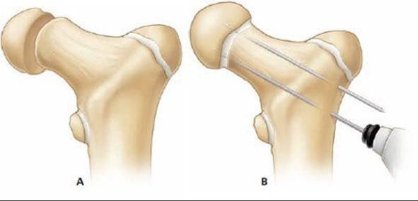

TECH FIG 1 • Fixation of an intraarticular femoral neck fracture. A. Displaced fracture. B. After reduction, pins are drilled from the lateral femoral cortex retrograde across the fracture. C. Drilling is performed over the guidewires with a cannulated drill. D. Cannulated screws are placed and the guidewires are removed.
It is customary to use smooth pins for physeal separations or neck fractures in very young children. In older or larger children, cannulated screws are necessary. Screws should stop short of the physis unless there is so little room between the physis and fracture that adequate fixation is precluded.
A lateral view, usually by frogging the hip, is necessary to confirm pin placement.
Two pins are usually sufficient to provide safe fixation. A total of three is even better, but three are often difficult to insert.
Once reduction and pin placement are confirmed by fluoroscopy is two planes, pins are cut and left protruding from the lateral femoral cortex for later removal, or drilling is performed over pins (TECH FIG 1C) and cannulated screws are placed (TECH FIG 1D).
Because there may be a tense hemarthrosis that tamponades flow in the retinacular vessels of the neck, it may be wise to aspirate the joint capsule to evacuate or decompress the hip joint.
OPEN REDUCTION AND INTERNAL FIXATION WITH PINS OR SCREWS
If the fracture is found to be irreducible by closed means, open reduction must be performed.
The goal is anatomic reduction to maintain perfusion to the capital femoral epiphysis, optimize bony apposition for healing, and prevent deformity, especially varus and external rotation.
The hip is approached laterally (Watson-Jones approach).
A direct lateral incision is made (TECH FIG 2A
The fascial lata is incised longitudinally (TECH FIG 2B).
The vastus lateralis is incised longitudinally, and the muscles overlying the anterior hip capsule are elevated anteriorly.
The anterior hip capsule is exposed (TECH FIG 2C).
The hip capsule is incised longitudinally and “T”-ed if necessary to visualize within (TECH FIG 2D).
The hematoma is evacuated.
The fracture is reduced under direct vision (TECH FIG 2E). It is often necessary to pull the neck “up” with a bone hook to get it reduced. Reduction is confirmed by palpation and then fluoroscopy.
Fixation is then performed from the lateral femur as described above.


TECH FIG 2 • Open reduction and internal fixation of intra-articular femoral neck fracture. A. Incision is made for Watson-Jones approach. B. The fascia lata is split longitudinally. C. The anterior capsule is exposed. D. A “T” incision is made in the capsule. E. The fracture is reduced under direct vision. Pins or screws are placed as described above.
OPEN REDUCTION AND INTERNAL FIXATION WITH FIXED-ANGLE
PLATE AND SCREWS
Exposure and Fracture Reduction
It is contemplated that the fracture will be fixed with a pediatric-sized lag-screw side-plate device.
The hip is approached from laterally. An incision is made from just proximal to the greater trochanter, extending about 4 to 5 inches distally (TECH FIG 3A). The fascia lata is split in line with its fibers (TECH FIG 3B).
The vastus fascia is cut with a “hockey stick” incision, starting anteriorly just distal to the apophysis of the greater trochanter (TECH FIG 3C). The incision is curved posteriorly and then extends distally in the posterior third of the vastus fascia. The muscle is elevated anteriorly, exposing the lateral femoral cortex.
The fracture is reduced by direct manipulation.
Fracture Fixation
A guidewire is then drilled up the center of the femoral neck (TECH FIG 4A). The angle the guidewire makes with the femoral shaft is dictated by the fixation device to be used. Generally a fixed-angle plate–lag screw combination will be used.
 The guidewire should be confirmed to be centered in the neck on anteroposterior (AP) and lateral C-arm views. The guidewire need not cross the capital femoral physis.
The proper length of lag screw is measured (TECH FIG 4B).
The channel for the lag screw is reamed (TECH FIG 4C) and tapped (TECH FIG 4D). The lag screw is inserted (TECH FIG 4E).
A side plate is selected; usually three or four holes is sufficient. It is passed over the lag screw (TECH FIG 4F).
The plate is secured to the femur with appropriate screws and fixed with a compression screw to the lag screw (TECH FIG 4G).
Reduction and fixation are checked by fluoroscopy. Closure is routine.

TECH FIG 3 • Open reduction of extraarticular hip fracture. A. Lateral incision on the thigh. B. The fascia lata is split longitudinally. C. The fascia of vastus lateralis is “hockey sticked” and vastus muscle is retracted anteriorly, exposing the lateral femoral cortex.

TECH FIG 4 • Internal fixation of extra-articular hip fracture. A. After fracture reduction, a guidewire is inserted from the lateral femoral cortex up the femoral neck. The angle the wire makes with the lateral cortex should match the angle of the fixation device (usually 135 degrees). B. The length of the intended lag screw is measured from the protruding guidewire. The lag screw should stop short of the physis. C.Reaming is accomplished over the guidewire to accommodate The lag screw and the barrel of the side plate. D. The channel is tapped because the child's bone is usually quite hard. E. The lag screw is inserted. F. The side plate is placed. G. The plate is secured to the femur with cortical screws and the compression screw locks the lag screw in the side plate.

POSTOPERATIVE CARE
Hip fractures fixed with pins, fractures in small children, and fractures in patients who cannot refrain from weight bearing should be immobilized for 4 to 6 weeks in a spica cast.
Controlled children can be maintained non-weight bearing with crutches.
Pins should be removed after fracture healing. Screws and hardware should be removed before they are covered by new bone growth.
OUTCOMES
Satisfactory healing is to be expected in most cases.
Stiffness after cast removal usually resolves without treatment.
Avascular necrosis may have a benign course similar to that of Perthes disease or may result in relentless disintegration of the femoral head, for which there is no satisfactory treatment in a child.
COMPLICATIONS
Avascular necrosis
Varus malunion
Nonunion
Leg-length discrepancy
REFERENCES
1. Canale ST. Fractures of the hip in children and adolescents. Orthop Clin North Am 1990;21:341–352.
2. Cheng JC, Tang N. Decompression and stable internal fixation of femoral neck fractures in children can affect the outcome. J Pediatr Orthop 1999;19:338–343.
3. Davison BL, Weinstein SL. Hip fractures in children: a long-term follow-up study. J Pediatr Orthop 1992;12:355–358.
4. Hughes LO, Beatty JH. Current concepts review: fractures of the head and neck of the femur in children. J Bone Joint Surg Am 1994;76A:283–292.
5. Jerre R, Karlsson J. Outcome after transphyseal hip fractures: 4 children followed 34–48 years. Acta Orthop Scand 1997;68:235–238.