
J. Richard Bowen
DEFINITION
 Epiphysiodesis (epiphysio–diaphyseal fusion) is an established method to treat a mild limb-length discrepancy (2 to 5 cm) in children.
With epiphysiodesis, growth of a longer extremity is inhibited by prematurely arresting a selected physis so that the remaining growth of the shorter extremity may approximate or equalize limb lengths at maturity.
The open epiphysiodesis technique was first described by Phemister in 193320 and modified by White in 1944.26 “Percutaneous epiphysiodesis” was reported first by Bowen and Johnson in 1984.4,25
 Subsequently, other variations of percutaneous epiphysiodesis with their outcomes have been reported.
When equalizing limb-length discrepancy by epiphysiodesis, caution should be used in terms of the patient's age at operation, the physis selected for premature closure, and the number of physes necessary to correct the discrepancy.
Useful data in decision making include.
Body length from head to foot (to determine percentile of height)
Length of the bones of the lower extremity (to determine degree and source of discrepancies)
Skeletal maturation age (to determine potential remaining growth), and the disease course that caused the limb inequality (to determine the predictability of remaining growth)
Proper patient age for timing of the epiphysiodesis may be determined by several methods, including the Green and Anderson method,10 the Mosley straight-line method,16 the “rule of thumb” method,15 and the multiplier method.1,18
Physeal stapling is an alternative operative procedure for correcting limb inequality by retarding growth in a longer extremity. The goal of physeal stapling is to retard growth of a physis with staples until the desired correction is obtained, after which the staples can be removed, with physeal growth resuming until maturity.
ANATOMY
An epiphysiodesis can be achieved by ablation of the medial and lateral peripheral margins of the physis. These subsequently form bony bars that restrict growth, and then the central aspect of the physis closes spontaneously, resulting in a total epiphysio–diaphyseal fusion.
Caution is required in the femur distally because the epiphysis is narrow at the central area of the physis.
Posteriorly the neurovascular structures are deeply positioned within the condyles, and anteriorly the patella–femoral joint is close.
 Injury to the neurovascular structures may be catastrophic. Therefore, I prefer the peripheral margin ablation technique as described here, in which the central area of the physis remains undisturbed.
The common peroneal nerve at the knee runs obliquely along the lateral side of the popliteal fossa, close to the medial border of the biceps femoris muscle and the lateral head of the gastrocnemius muscle, toward the head of the fibula.
The nerve winds posteriorly around the neck of the proximal fibula and passes deep to the peroneus longus muscle, where it divides into the superficial and deep peroneal nerves.
PATHOGENESIS
The etiology of a limb-length discrepancy may be important in determining the appropriate patient age at which the percutaneous epiphysiodesis is performed.
Shapiro reported different patterns of growth inhibition that may cause shortening of a limb.22
Predictable growth of the shorter extremity is required to achieve optimal results with a percutaneous epiphysiodesis.
NATURAL HISTORY
After a percutaneous epiphysiodesis, bony bridges form at the peripheral margins of the physis both medially and laterally. These bony bars prevent further physeal growth.
After peripheral bony bar formation following a percutaneous epiphysiodesis, the central area of the physis (unoperated area) will spontaneously close within 6 to 8 months.23,24
The scientific reason for this spontaneous closure of the central area is unclear. Confusion exists in that staples do not cause physeal closure, whereas peripheral bony bars from an epiphysiodesis result in progressive physeal closure.
I have studied this phenomenon but found no definitive causes.23,24
Reversal of limb-length discrepancy could occur if the shorter limb (contralateral) were to overgrow the epiphysiodesis limb (ipsilateral) before maturity.
I have not had a patient have this complication, but follow-up until skeletal maturity is advised.
By following growth until maturity, this potential problem may be detected and a contralateral epiphysiodesis may prevent a limb-length discrepancy at maturity.
A percutaneous epiphysiodesis can be used in combination with contralateral limb lengthening in patients with severe shortening.
In major limb-length discrepancies, lengthening may not be able to correct the full discrepancy, and remaining small discrepancies of 2 to 5 cm may be more easily corrected by a contralateral percutaneous epiphysiodesis than by a secondary ipsilateral lengthening.
After leg-lengthening procedures, growth of the lengthened limb may be retarded or occasionally stimulated.
PATIENT HISTORY AND PHYSICAL FINDINGS
The patient history and physical examination should include the following:
Etiology of the limb-length discrepancy19,22
The patient's height (percentile of height)8,10.
The total height and the leg lengths (in centimeters) are measured. The percentile of height is then determined and used to plot limb-length predictive charts.
The level of maturation, based on the appearance of secondary sexual characteristics and the Tanner scale.11 Caution should be used if the clinical maturation scale differs significantly from the Greulich and Pyle skeletal developmental age.
Foot height (foot height is not typically considered in radiographs that measure limb length)
The patient's “bone age” (expressed in years) using the Greulich-Pyle method. Caution should be used if the chronological age differs from the “bone age.”
Radiographs of the limbs are obtained to measure the tibia and femur (expressed in centimeters). These measurements are plotted on growth charts to predict the discrepancy at maturity and to determine the age for epiphysiodesis.
Blocks of differing thicknesses are placed under the foot of the shorter limb until the pelvis is level to determine the length discrepancy (expressed in centimeters). This method also helps evaluate discrepancies in the foot that are not reported by radiographs.
After length measurements of both limbs are obtained, the ratio of femur to tibial discrepancy of the normal to abnormal limb is determined.
A disease diagnosis can be made with Shapiro developmental patterns.22 However, proportional inhibition of growth is more predictable, while disproportional growth is difficult to predict.
IMAGING AND OTHER DIAGNOSTIC STUDIES
Skeletal age determination by the Greulich and Pyle method11
Green-Anderson method of predicting limb-length discrepancy (my preference)2,3
Mosley graphic method of predicting limb-length discrepancy16
Multiplier method of predicting limb-length discrepancy1,18
Menelaus “rule of thumb” method of predicting limb-length discrepancy15
NONOPERATIVE MANAGEMENT
No treatment is required if the limb-length discrepancy is less than 2.5 cm at maturity.
A shoe lift can be used to treat mild limb-length discrepancies of more than 2.5 cm.
Often a lift is used in children until an appropriate skeletal age is reached to perform an equalization procedure.
A prosthesis may be necessary if deformities are so severe that adequate length or ambulatory ability cannot be achieved by operative methods.
Surgery may be necessary to provide an appropriate stump for the prosthesis, and a percutaneous epiphysiodesis is used occasionally to achieve correct stump length.
SURGICAL MANAGEMENT
Epiphysiodesis is most commonly performed at the distal femoral physis or the proximal tibia–fibula physis.
Ablation of the peripheral margins both medially and laterally in a physis causes bony bridges to form between the epiphysis and metaphysis that accomplish growth inhibition in that physis (epiphysiodesis).
This can be accomplished by different techniques and instruments, which include a curette, a drill, a burr, a reamer, and a circular tube saw.
I prefer a curette because surgeon control is easy and the curette can be passed percutaneously. I have used various instruments, but drills and burrs tend to burn and occasionally grab tissue, and reamers and circular saws require a larger incision (really not percutaneous, almost the size of a typical open epiphysiodesis).
A bony bridge needs to form only at the peripheral margins of the physis both medially and laterally to accomplish an epiphysiodesis. The central part of the physis does not require treatment because it will close spontaneously.
The stability of the bone is maintained postoperatively and the patient may continue to ambulate.
I restrict sports for 6 weeks to reduce the possibility of a fracture.
Preoperative Planning
Anticipated remaining growth is determined by one of the following methods:
Green-Anderson method10
Mosley graphic method16
Multiplier method18
Menelaus “rule of thumb” method15
I prefer the Mosley straight-line method because growth inhibition is expressed graphically, multiple data entries can be charted to help predict growth more accurately, and the method is easy to calculate in a brief outpatient visit.
I perform a proximal fibular epiphysiodesis in addition to the proximal tibial epiphysiodesis if the final discrepancy between the tibia and fibula is anticipated to be more than 1 cm.
If the discrepancy is anticipated to be less than 1 cm, I do not perform a proximal fibular epiphysiodesis and have not detected a clinical problem with such a mild discrepancy.
Positioning
Image intensification
Supine position
The limb is prepared and draped up to the proximal thigh into a sterile field. A tourniquet is placed on the proximal thigh but is not inflated unless bleeding occurs.
Approach
Femoral epiphysiodesis at distal physis
Longitudinal incisions of 3 mm (stab incisions) medially and laterally in the skin at the level of the physis at its peripheral area
Tibial epiphysiodesis at proximal physis
Longitudinal incisions of 3 mm
Medially the incision is 3 mm at the level of the physis at its peripheral area.
Laterally the incision is 3 mm at the level of the physis and at the anterior border of the fibula.
Fibular epiphysiodesis at the proximal physis
The same incision is used for the fibular epiphysiodesis as for the lateral physeal area of the tibia; however, in the epiphysiodesis of the fibula the curette is directed differently to avoid injury to the common peroneal nerve (described below). (See above section on anatomy.)
TECHNIQUES
EPIPHYSIODESIS OF THE FEMORAL PHYSIS DISTALLY OR THE TIBIAL PHYSIS PROXIMALLY
Preparation of the Physis
 A metal marker is placed over the skin and under the image intensifier, and the level of the physis is identified at its peripheral area (either medially or laterally) (TECH FIG 1A).
A 3-mm skin incision is made with a scalpel at the level of the physeal plate on the peripheral side (medially or laterally) (TECH FIG 1B).
Under image intensification control, a 3-mm-wide osteotome is directed through the skin incision to make a longitudinal split in the periosteum–cortex, and the physeal plate is then penetrated to a depth of about 0.5 cm (TECH FIG 1C,D).
The osteotome is rotated to create a hole in the physis and is then withdrawn.
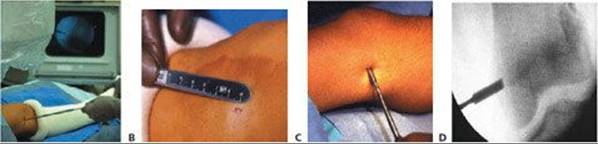
TECH FIG 1 • A. Patient's leg under an image intensifier with a metal pointer identifying the level of the femoral physis distally. B. The incision is only 3 mm. C. A 3-mm-wide osteotome is being directed through the skin incision to make a longitudinal split in the periosteum–cortex and the physeal plate. D. A radiographic view of the osteotome penetrating the physeal plate to a depth of about 0.5 cm.
Physeal Plate Ablation
A 3-mm oval curette is advanced through the skin into the hole of the physis (TECH FIG 2A).
Under image intensification control, the curette is rotated and advanced to the level of the inner third of the physis (TECH FIG 2B).
The curette is then swept cephalad and caudad in the physis to ablate the peripheral third of the physis, leaving the middle third intact (TECH FIG 2C).
Specific surgical attention is directed toward adequate ablation of the most peripheral aspect of the physeal plate (TECH FIG 2D–F).
The same operative process is repeated on the opposite side of the physeal plate (medially or laterally), again sparing the middle third of the physeal plate.
The wound is closed by suture (typically one absorbable suture) (TECH FIG 2G).

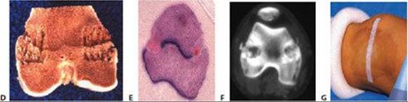
TECH FIG 2 • A. The curette is advanced through the incision into the hole of the femoral cortex. B. The curette is rotated and advanced to the level of the inner third of the physis. C. The anticipated area of ablation of the peripheral third of the physis (both medially and laterally). The curette is swept cephalad and caudad in the physis to ablate the peripheral third of the physis, leaving the middle third of the physis intact. D. A photograph of a specimen demonstrating the ablation of the peripheral third of the physis, leaving the middle third of the physis intact. E. A photomicrograph of the femoral physis distally demonstrating the ablation of the peripheral third of the physis, leaving the middle third intact. F. A computed tomography section through the femoral physis distally demonstrates the ablation of the peripheral third of the physis. Note the narrow space in the middle third of the physis, with the neurovascular bundle posteriorly (between the femoral condyles) and the patellofemoral joint anteriorly. G. The 3-mm incision has been closed with a single suture. The incision is covered by an adhesive strip and bandage.
Epiphysiodesis of the Fibula Proximally
The same incision is used to perform the epiphysiodesis of the fibula as the proximal physis of the tibia laterally.
To avoid injury to the common peroneal nerve with a percutaneous epiphysiodesis of the proximal fibula, direct the curette into the anterosuperior area of the proximal epiphysis and then inferiorly into the physis of the proximal fibula (TECH FIG 3A).
 The most superior extent of the fibular epiphysis proximally is usually at the level of the tibial physis.
The curette is used to ablate the entire central area of the physis of the fibula to achieve the epiphysiodesis (TECH FIG 3B).
The surgeon should not exit the cortex of the fibula as the central area is ablated.
The 3-mm incisions are each closed with a single suture.
The incision is covered by an adhesive strip and bandage.
A knee immobilizer foam brace is applied and maintained postoperatively.
The elastic of the knee immobilizer adds very mild compression to the incision and helps prevent a hematoma or edema.
The knee immobilizer brace should not be applied with a force that impedes circulation.
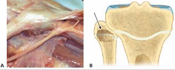
TECH FIG 3 • A. Dissection of the common peroneal nerve at the level of the fibula head. The nerve passes posteriorly and inferiorly to the fibula epiphysis. B. Technique of percutaneous epiphysiodesis of the fibula proximally. To avoid injury of the common peroneal nerve, the curette is directed into the anterosuperior area of the proximal epiphysis and then inferiorly into the physis of the fibula. (The most superior extent of the fibular epiphysis proximally is usually at the level of the tibial physis.) The curette is used to ablate the entire central area of the physis of the fibula to achieve the epiphysiodesis. The surgeon should not exit the cortex of the fibula as the central area is ablated.

POSTOPERATIVE CARE
Postoperatively the leg is protected by a knee immobilizer brace, and full weight bearing with crutch support is allowed.
The patient may usually be discharged within the same operative day (outpatient surgery).
The knee immobilizer is used for 3 to 4 weeks to protect the extremity from fracture.
The scars of the percutaneous epiphysiodesis are small and have an acceptable appearance (FIG 1).
Radiographs are performed about 4 to 6 months postoperatively to ensure adequate healing.
Bony bars at the peripheral margins of the physis are usually observed radiographically by 6 months postoperatively.
Total closure of the physis occurs 8 to 12 months postoperatively.
Correction of the leg-length discrepancy may be determined by periodic radiographs and clinical examinations.
During postoperative evaluations the extremity should be evaluated to ensure appropriate length correction and to watch for an angular deformity.
Lack of appropriate closure may result in undercorrection and asymmetric closure causes angulation.
OUTCOMES
Recently I reviewed the outcome of percutaneous epiphysiodesis as described above in a consecutive series of 97 patients (56 girls and 41 boys) with a mean skeletal age of 12.6 years (range 10 to 16 years) at surgery. All patients were followed until skeletal maturity, a mean of 3.8 years. The mean residual limb-length discrepancy in 88 patients at maturity was 1.3 cm (I consider normal to be a lower-limb discrepancy of less than 2.5 cm). In nine patients, the epiphysiodesis was combined with a femoral lengthening or femoral shortening.

FIG 1 • Very small leg scar after a percutaneous epiphysiodesis of the femoral physis distally.
The Moseley straight-line method accurately and efficaciously predicted the timing for percutaneous epiphysiodesis in all but one patient, who had unpredictable growth from hemihypertrophy secondary to a hemangiomatosis.
Horton and Olney12 reported the results of 42 percutaneous epiphysiodeses in 26 patients. Physeal arrest developed in all cases and no angular deformities occurred. They considered the percutaneous epiphysiodesis to be reliable and safe. Stated advantages include a cosmetic scar, short hospital stay, low incidence of complications, and reliability.
Canale and coworkers6 reported on 13 children treated by a percutaneous epiphysiodesis; growth plate fusion occurred in all cases. They used a pneumatic burr under image intensification to perform the procedure.
Brax and Gille5 performed a percutaneous epiphysiodesis using a drill and radiographic control in 10 children. They had good results in all but one case and considered the procedure to be minimally traumatic, cosmetically preferable, and safe.
Craviari and colleagues7 reported the results of 60 cases followed to skeletal maturity who were treated by a percutaneous epiphysiodesis. They concluded that the procedure was satisfactory and complications were rare. Complications comprised hematoma in 2 cases, need for surgical revision in 10 cases, limb deviation in 4 cases, and inverted discrepancy in 2 cases.
Gabriel and associates9 reported the results of 29 patients who underwent 56 physeal procedures with a percutaneous epiphysiodesis. The procedure lasted a mean of 36 minutes, and in all patients a physeal closure developed. No unplanned angular growth, no deep infections, and no cases of joint stiffness were reported.
Kemnitz and coworkers13 performed a retrospective review of 57 patients who underwent percutaneous epiphysiodesis. They reported no significant operative problems. A final limblength discrepancy greater than 2 cm was seen in 17.5% of the cases, and they concluded the timing of the procedure remains the main problem.
Macnicol and Gupta14 reported 35 cases of epiphysiodesis in which a cannulated tube saw was used to ablate the physis. The mean anticipated discrepancy was 3.3 cm, and at maturity the discrepancy averaged 0.7 cm. One patient had slight overgrowth of the fibula and another had an unsightly scar; otherwise the results were favorable.
Ogilvie and King17 used a low-speed, high-torque drill to create an epiphysiodesis in seven children. This technique required a 1-cm incision. There were no cases of failure of physeal fusion, no infections, no angular deformity, and no restriction of joint motion.
Porat and coworkers21 performed epiphysiodesis in 20 children, with good results in 90% of the patients. They recommended the Moseley straight-line graph and percutaneous epiphysiodesis.
COMPLICATIONS
Errors in predicting growth
Infection
Angulation
Lack of physeal closure
Temporary exostosis formation (believed to develop as bone forms in the elevated periosteum)
Hematoma
Neurovascular injury
REFERENCES
1. Aguilar JA, Paley D, Paley J, et al. Clinical validation of the multiplier method for predicting limb length at maturity, part I. J Pediatr Orthop 2005;25:186–191.
2. Anderson M, Green WT. Growth and predictions of growth in the lower extremities. J Bone Joint Surg Am 1963;45A:1–8.
3. Anderson M, Messner M, Green WT. Distribution of lengths of the normal femur and tibia in children from one to eighteen years of age. J Bone Joint Surg Am 1964;46A:1197–1202.
4. Bowen JR, Johnson WJ. Percutaneous epiphysiodesis. Clin Orthop Relat Res 1984;190:170–173.
5. Brax P, Gille P. Percutaneous epiphysiodesis: first trials with the long bones of the lower limbs. Chir Pediatr 1989;30:263–265.
6. Canale ST, Russell TA, Holcomb RL. Percutaneous epiphysiodesis: experimental study and preliminary clinical results. J Pediatr Orthop 1986;6:150–156.
7. Craviari T, Berard J, Willemen L, et al. Percutaneous epiphysiodesis: analysis of a series of 60 full-grown patients. Rev Chir Orthop Reparatrice Appar Mot 1998;84:172–179.
8. Dimeglio A. Growth in pediatric orthopaedics. J Pediatr Orthop 2001;21:540–555.
9. Gabriel KR, Crawford AH, Roy DR, et al. Percutaneous epiphysiodesis. J Pediatr Orthop 1994;14:358–362.
10. Green WT, Anderson ML. Skeletal growth and the control of bone growth. AAOS Instr Course Lect 1960;17A:199–217.
11. Greulich WW, Pyle S. Radiographic atlas skeletal development of the hand and wrist, 2nd ed. Stanford: Stanford University Press, 1959.
12. Horton GA, Olney BW. Epiphysiodesis of the lower extremity: results of the percutaneous technique. J Pediatr Orthop 1996;16:180–182.
13. Kemnitz S, Moens P, Fabry G. Percutaneous epiphysiodesis for leg length discrepancy. J Pediatr Orthop B 2003;12:69–71.
14. Macnicol MF, Gupta MS. Epiphysiodesis using a cannulated tubesaw. J Bone Joint Surg Br 1997;79B:307–309.
15. Menelaus M. Correction of leg length discrepancy by epiphyseal arrest. J Bone Joint Surg Br 1966;48B:336–339.
16. Moseley C. A straight-line graph for leg-length discrepancies. J Bone Joint Surg Am 1977;59A:174–179.
17. Ogilvie JW, King K. Epiphysiodesis: two-year clinical results using a new technique. J Pediatr Orthop 1990;10:809–811.
18. Paley D, Bhave A, Herzenberg JE, et al. Multiplier method for predicting limb-length discrepancy. J Bone Joint Surg Am 2000; 82A:1432–1446.
19. Pappas AM, Nehme AM. Leg length discrepancy associated with hypertrophy. Clin Orthop Relat Res 1979;144:198–211.
20. Phemister DB. Epiphysiodesis for equalizing the length of the lower extremities and for correcting other deformities of the skeleton. Mem Acad Chir (Paris) 1950;76:758–763.
21. Porat S, Peyser A, Robin GC. Equalization of lower limbs by epiphysiodesis: results of treatment. J Pediatr Orthop 1991;11:442–448.
22. Shapiro F. Developmental patterns in lower-extremity length discrepancies. J Bone Joint Surg Am 1982:64:639–651.
23. Synder M, Harcke HT, Bowen JR, et al. Evaluation of physeal behavior in response to epiphysiodesis with the use of serial magnetic resonance imaging. J Bone Joint Surg Am 1994;76A:224–229.
24. Synder M, Harcke HT, Conard K, et al. Experimental epiphysiodesis: magnetic resonance imaging evaluation with histopathologic correlation. Int Orthop 2001;25:337–342.
25. Timperlake RW, Bowen JR, Guille JT, et al. Prospective evaluation of fifty-three consecutive percutaneous epiphysiodesis of the distal femur and proximal tibia and fibula. J Pediatr Orthop 1991;11:350–357.
26. White JW SS. Growth arrest for equalizing leg lengths. JAMA 1944;126:1144.