
Anthony A. Scaduto and Robert M. Bernstein
DEFINITION
 Fibular deficiency, previously known as fibular hemimelia, is a longitudinal deficiency of the fibula. It is the most common long bone deficiency and may be either partial or complete.
A wide spectrum of associated anomalies also may be seen on the affected limb. The extent of limb shortening and the degree of foot deformity are the most important components that determine treatment. Treatment options include use of a shoe lift, amputation, and limb lengthening.
Delayed amputation should be avoided whenever possible. Ideally, amputation is performed at 10–18 months of age when the child is beginning to pull to stand.3 Psychosocial adjustment to amputation and the adjustment to prosthetic wear are rapid at this age.
 A common dilemma for parents and consulting physicians is an unwillingness to commit to a path of either multiple lengthenings or early amputation. It is generally agreed, however, that the least effective approach to fibular deficiency is “let's try lengthening and if it fails, do an amputation.”
The Syme amputation and the Boyd amputation are the two common amputations performed for fibular deficiency.
The Syme amputation is an ankle disarticulation that preserves the heel pad as a weight-bearing surface. This procedure provides better energy efficiency than a transtibial amputation, may be self-suspending, allows weight bearing on the stump without the use of a prosthesis, and is cartilage capped, preventing terminal overgrowth.
The Boyd amputation is a modified ankle disarticulation in which the calcaneus is preserved with the heel pad and fused to the distal tibia.
The best indications for an amputation are a large leg-length discrepancy (ie, a difference of more than 30%) at skeletal maturity and a nonfunctional foot.
The ideal candidate for lengthening has a smaller expected leg-length discrepancy (less than 10%), a stable ankle, and a fully functional foot.
Because both amputation and multiple lengthenings have significant consequences, care must be individualized. This is especially important for patients with leg-length discrepancies between 10% and 30%, for which both amputation and lengthening have been shown to be effective with excellent functional outcomes.
ANATOMY
Fibular deficiency is best considered an abnormality that affects the entire limb, not just the fibula (FIG 1A).
The appearance of the leg can vary from nearly normal to severely deformed (FIG 1B).

FIG 1 • A. Clinical appearance of limb with fibular deficiency. B. Spectrum of fibular deficiency.
Potential ipsilateral deformities associated with fibular deficiency are as follows:
Femur: mild femoral shortening, femoral retroversion, lateral femoral hypoplasia
Knee: cruciate ligament deficiency, valgus alignment, patella-femoral instability
Tibia: shortening, anteromedial diaphyseal bowing
Ankle: ankle valgus, absent lateral malleolus, ball-andsocket ankle
Foot: absent tarsal bones, tarsal coalitions, absence of one or more lateral rays
The amount of fibula present does not aid treatment planning. For example, some patients with complete fibular absence have minimal leg-length inequality and foot deformity.
An understanding of the anatomy of the ankle and heel is necessary to perform either the Syme or Boyd amputation procedure.
The posterior tibial nerve and artery course posterior to the medial malleolus and split into the medial and lateral plantar nerves. These structures must be protected for the heel pad to maintain its sensation and viability.
PATHOGENESIS
Unlike tibial deficiency, fibular deficiency occurs sporadically with no inheritance pattern.
No genetic defect has been identified, and no common teratogen is linked to fibular deficiency.
Major limb malformations associated with fibular deficiency occur by the 7th week of fetal development.
NATURAL HISTORY
Without surgical intervention, the growth of the abnormal limb remains proportional to the normal side. Therefore, a final leg-length discrepancy is predictable.
For example, if the short leg is 85% the length of the long side at age 2 years, the length of the short side at maturity also will be 85% of the estimated length of the long side at maturity.
Tibial bowing is present in most cases of complete absence of the fibula. In some cases this bowing will improve with age.
Unlike anterolateral bowing of the tibia, bowing associated with fibular deficiency does not increase the risk of fracture or pseudarthrosis.
Knee valgus commonly worsens through childhood. It may require surgical treatment when prosthetic modifications are inadequate to compensate for the deformity.
PATIENT HISTORY AND PHYSICAL FINDINGS
Classically, the limb is short, with an equinovalgus foot and skin dimpling over the mid-anterior tibia.
Because presentation varies widely, an examination to assess length, alignment, and function is critical to treatment.
Hip range of motion: a common finding is limited internal rotation (less than 20 to 60 degrees), indicating femoral retroversion.
Leg-length assessment: there should be minimal shortening of the thigh. Otherwise, consider proximal femoral focal deficiency. Small leg-length discrepancies can be corrected with a shoe lift or lengthening.
Lachman's test: severe anterior/posterior laxity increases the risk of subluxation during lengthening.
Valgus alignment and stability: small angulation is accommodated through prosthetic adjustment, but larger angulation requires correction.
Tibial bowing requires prosthetic adjustments or correction.
Ankle alignment and stability: amputation is preferred over lengthening when severe subluxation or instability exists.
Hindfoot mobility: suspect tarsal coalition if subtalar motion is reduced.
Ray deficiency (number of missing rays): amputation is indicated when the foot is nonfunctional.
IMAGING AND OTHER DIAGNOSTIC STUDIES
Anteroposterior (AP) and lateral radiographs of the leg (including the distal femur) should be obtained.
Absence of the anterior cruciate ligament and hypoplasia of the lateral femoral condyle with a valgus joint alignment are common (FIG 2A, B).
The amount of anterior bowing (tibial kyphosis) also can be assessed (FIG 2C).
Additional radiographs of the affected limb (ie, femur, ankle, and foot) are obtained as necessary (FIG 2D).
A full-length standing radiograph from hips to ankles should be obtained to check alignment in those children able to stand (FIG 2E).
A scanogram and bone age should be obtained to determine the expected leg-length discrepancy at maturity.
The desired limb-length difference at maturity should be at least 3.5 cm to accommodate the height of the prosthetic foot. Epiphysiodesis may be necessary to achieve this and should be planned appropriately.
An ankle and foot series should be obtained when abnormal position or motion is present at the ankle or subtalar joint or when lateral rays are absent. These views may reveal a balland-socket ankle (FIG 2F), tarsal coalitions, or absent or hypoplastic tarsal bones (FIG 2G).
DIFFERENTIAL DIAGNOSIS
Proximal femoral focal deficiency
Tibial deficiency
Tibial dysplasia
NONOPERATIVE MANAGEMENT
If the leg-length discrepancy is small, the ankle is stable, and the foot is plantigrade, a shoe insert or lift may be all that is required.
When amputation or lengthening is needed but must be deferred, an atypical prosthesis that accommodates the foot position can be used.
SURGICAL MANAGEMENT
Syme Amputation
Meticulous care is needed to preserve the posterior tibial nerve and vessels to maintain a sensate stump.
Care should be taken not to leave any cartilage remnants of the calcaneus during resection.
The malleoli should not be resected in children.
The heel pad may be proximal to the ankle joint and can be difficult to bring distally, even after sectioning the Achilles tendon.
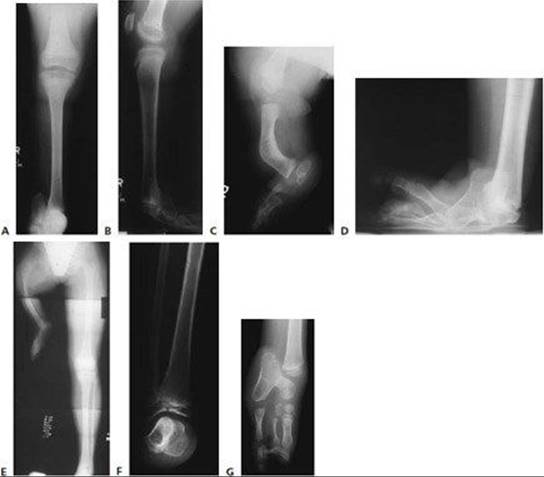
FIG 2 • A,B. AP and lateral radiographs of a child with fibular absence, hypoplasia of the lateral femoral condyle, and anterior cruciate deficiency. C. Anterior bow of the tibia. D. AP radiograph of the same patient's foot, revealing severe equinovarus deformity and talocalcaneal fusion. E. Standing AP radiograph of a child with proximal femoral focal deficiency revealing a substantial leg-length discrepancy and abduction of the affected limb. F. Ball-and-socket ankle. G. Nonfunctional foot with hypoplastic tarsal bones, tarsal coalition, and absent rays. (E: Courtesy of Hugh Watts, MD.)
The Pirogoff modification maintains a portion of the calcaneus, which is fused to the distal tibia to better fix the heel pad.
Because in young children the distal tibial physis must be resected to obtain fusion of the calcaneus to the tibia, this is really a modification of the Boyd amputation, since distal growth of the tibia will be lost.
Advantages
Simple technique
Rapid prosthetic fitting
The stump is shorter and often tapered, which improves cosmesis (but also may inhibit end-bearing)
Disadvantage.
Heel pad migration (FIG 3)
Less end-bearing potential
Boyd Amputation
Advantage.
Maintains maximum length of limb
Eliminates heel pad migration
Flare at the end of the stump improves prosthetic suspension
Maximizes end-bearing potential. This may be especially important if it preserves end-bearing without a prosthesis (eg, not having to put on a prosthesis to go from the bed to the bathroom).
The pin necessary for tibial-calcaneal fixation also can be used to stabilize the mid-tibia osteotomy when bowing of the tibia is corrected simultaneously.
Disadvantages
Delays prosthesis fitting by several weeks while awaiting fusion

FIG 3 • Posterior heel pad migration after Syme amputation.
Excess length may leave less room for energy-storing prosthetic foot options, and the bulbous end may be difficult to hide if it is at the level of the opposite ankle.
Preoperative Planning
Syme Amputation
In patients where the tibial length at skeletal maturity is expected to be equal to that of the opposite side, a Boyd amputation or timed epiphysiodesis should be considered to accommodate the height of the prosthetic foot to achieve equal limb lengths at maturity.
It usually is not necessary to correct mild bowing (less than 30 degrees) of the tibia in a congenital deficiency in a skeletally immature patient. Bowing of more than 30 degrees should be addressed with osteotomy at the time of amputation.
Boyd Amputation
If anterior tibial bowing is present, it is best to correct it at the same time as the Boyd amputation.
The tarsal bones and distal tibia epiphysis are primarily cartilaginous in infancy.
If a Boyd amputation is performed early, it will be necessary to resect a significant portion of the superior calcaneus and distal tibia to achieve bone–bone contact for fusion.
If maximum length of the tibia is a goal of treatment (eg, to allow occasional end-bearing on the stump end without a prosthesis), consider waiting until the distal tibia epiphysis is ossified adequately to avoid resecting the distal physis.
Some authors have suggested that routine resection of the distal tibia physis should be performed.
They observed that most children stop walking around the house without a prosthesis in early adolescence and that ideally the short limb should end in the middle fifth of the shank segment of the prosthesis to optimize cosmesis and allow room for a dynamic-response foot-and-ankle unit.2
Positioning
The patient is positioned supine with a small bump under the greater trochanter. A tourniquet is placed around the upper thigh.
Access to the entire leg from knee to toes is important (FIG 4).

FIG 4 • Intraoperative photos of patient position with tourniquet applied above the thigh. (Courtesy of Hugh Watts, MD.)
TECHNIQUES
AMPUTATIONS FOR FIBULAR DEFICIENCY
Syme Amputation
Incisions
 The dorsal incision is made at the tip of the lateral malleolus (or where it should be) across the ankle joint to end about 1 cm below the tip of the medial malleolus.
 In children with congenital fibular absence, the lateral malleolus is not present, and the end of the first incision must be approximated.
The Achilles tendon (often very tight in patients with congenital fibular absence) can be released through a separate, percutaneous incision posteriorly to improve exposure.
The plantar incision is made at the midportion of the metatarsals and carried proximally up the medial and lateral sides of the foot to meet the anterior incision (TECH FIG 1).
The plantar incision can be cut directly down to bone, with care to be sure that the knife blade remains perpendicular to the skin. Vessels are ligated or cauterized.
Amputation
The foot is now plantarflexed (TECH FIG 2A). The anterior incision is deepened down to bone, again keeping the knife perpendicular to the skin.
The anterior ankle joint is opened, and the deltoid and tibiofibular ligaments are cut sharply, with care not to injure the posterior tibial nerve and artery coursing behind the medial malleolus.
The foot is further plantarflexed to expose the posterior ankle joint, which is released, exposing the posterior calcaneus and the Achilles tendon.
 A bone hook or sharp retractor can be used to pull on the talus distally as the posterior joint is opened (TECH FIG 2B).
The calcaneus is now released from the heel pad extraperiosteally. Care is taken not to separate the calcaneal apophysis from the body of the calcaneus.
The Achilles tendon is now sectioned.
In very tight equinus, the Achilles can be released through a percutaneous incision posteriorly.
Once the tendon is easy to visualize, a 1-cm section of the tendon should be removed to prevent late migration of the heel pad.
The tourniquet is deflated, and perfusion of the heel pad is checked and bleeding is controlled (TECH FIG 2C).
The distal tibial cartilage and malleoli are left intact.
A Steinmann pin or Rush rod may be inserted through the heel pad into the distal tibia to affix the heel pad to the distal tibia (TECH FIG 2D).

TECH FIG 1 • A. Incisions for the Syme amputation. B. Medial incision and identification of the posterior tibial artery and nerve. (B: Courtesy of Hugh Watts, MD.)

TECH FIG 2 • A. The foot is plantarflexed while the dorsal incision is completed. B. A retractor is placed in the talus to expose the posterior capsule and Achilles tendon. C. Intraoperative photograph after deflation of the tourniquet, illustrating a well-perfused heel pad. D. Stump closure with interrupted absorbable sutures after insertion of a Steinmann pin to stabilize the heel pad. (Courtesy of Hugh Watts, MD.)

TECH FIG 3 • A,B. The dorsal and volar parts to the fish-mouth incision meet medially and laterally just distal to the malleoli. C. The plantar incision crosses the foot just distal to the heel pad.

TECH FIG 4 • A. The plantar incision is carried down to the bone. B. Dorsal structures are transected with the foot in plantar flexion. C. Use the forefoot to control the hindfoot. D,E. Release the deltoid and lateral capsule. F,G.Carefully divide the posterior capsule and remove the talus. H,I. Sometimes the talus is small and irregularly shaped, as seen in this case, in which the L-shaped talus hooked around the back of the distal tibia.
Closure is done over a drain using interrupted sutures.
In young children, an absorbable suture is used to avoid later removal.
An antibiotic-impregnated gauze is applied followed by fluffs and Webril.
Boyd Amputation
Incision and Dissection
A fish-mouth incision is made (TECH FIG 3).
The plantar incision crosses the foot where the heel pad ends.
 The dorsal incision crosses the foot at the level of the ankle joint.
The incisions meet medially about 1 cm distal to the medial malleolus and laterally in a similar location (the lateral malleolus often is absent in fibular deficiency).
Midfoot and Forefoot Removal
It is unnecessary to dissect layer by layer on the plantar side. Instead, sharply deepen the plantar incision down to the level of the bone (TECH FIG 4A).
Vessels can be ligated or cauterized (depending on their size) as they are cut or after the tourniquet is deflated before closure.
While maximally plantarflexing the foot, transect the dorsal nerves and extensor tendons, which retract proximal to the incision (TECH FIG 4B).
Do not remove the midfoot and forefoot at this time. They can serve as a handle to control the hindfoot when releasing the tibiotalar capsule and ligaments (TECH FIG 4C).
Expose the tibiotalar joint by releasing the anterior capsule and then release the deltoid ligament medially and the talofibular ligament with the lateral capsule (TECH FIG 4D, E).
Use care to preserve the posterior tibial artery and vein while dividing the posterior ankle capsule.
A bone hook or skin rake on the talar dome will help expose the posterior tibiotalar capsule and Achilles tendon.
Identify the flexor hallucis tendon and protect the neurovascular bundle that lies just medial to the tendon (TECH FIG 4F).
Remove the talus after cutting through the talocalcaneal ligaments (TECH FIG 4G).
Removing the talus may be more difficult in the very young child, in whom the talus is primarily cartilaginous, or when it has an irregular shape (TECH FIG 4D, E).
The midfoot and forefoot are now removed.
Completing the Amputation
Use an oscillating saw (or, in very young children, a knife) to remove the anterior process of the calcaneus and enough of the superior articular surface to expose cancellous bone (TECH FIG 5A–C).
Cut the distal tibia (TECH FIG 5D).
Oppose the cancellous bone surfaces and stabilize with a retrograde K-wire placed through the heel pad and across the tibiocalcaneal surfaces (TECH FIG 5E, F).
Correction of Tibial Bowing
Make a longitudinal anterior incision at the apex of the bowing.
Expose the tibia subperiosteally and place Chandler or Hohmann retractors to protect the soft tissues.
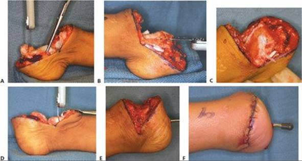
TECH FIG 5 • A–C. Prepare the calcaneus by cutting the anterior and dorsal surfaces. D–F. Stabilize the calcaneus to the end of the tibia with a smooth K-wire.
The superior cut is made perpendicular to the long axis of the proximal tibia, whereas the distal cut is perpendicular to the long axis of the distal tibia, creating a bone wedge with its widest portion located anteromedially (TECH FIG 6A).
The Steinmann pin used to stabilize the calcaneus as part of the Boyd amputation can be extended further into the proximal tibia fragment to simultaneously stabilize the osteotomy site.
Consider using a threaded pin if fixation is inadequate with a smooth pin (TECH FIG 6B).

TECH FIG 6 • A. A saw is used to remove a wedge of bone to correct the anteromedial bow of the tibia. B. A retrograde pin stabilizes calcaneus to distal tibia and the mid-tibia osteotomy.

POSTOPERATIVE CARE
Apply a long-leg cast with the knee flexed 90 degrees to prevent pin migration and keep the cast from slipping off.
Postoperatively, the patient's leg is elevated for 24 hours.
The child should be non-weight bearing.
The cast and pin are removed in the office at 4 to 6 weeks, and, in the Boyd amputation, after radiographic healing is evident (FIG 5A).
A stump wrap or shrinker is then applied.
Once the swelling has resolved, the prosthetist can mold a socket (FIG 5B, C).
OUTCOMES
McCarthy et al4 reported on a comparison of amputation versus lengthening in the treatment of fibular hemimelia.
Patients undergoing amputation were more active, had less pain, were more satisfied, and had fewer complications than those undergoing limb lengthening.
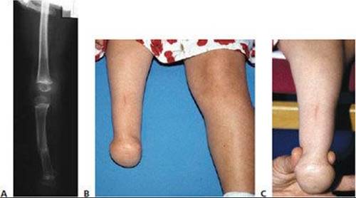
FIG 5 • A. Healed osteotomy and calcaneus fused to tibia. B,C. Postoperative photographs demonstrate a good Syme stump with a bulbous end for possible self-suspending socket. (B,C: Courtesy of Hugh Watts, MD.)
Fulp et al2 reviewed 25 patients (31 extremities) with longitudinal deficiency of the fibula treated with either Syme amputation or Boyd amputation.
Patients undergoing Syme amputation had more problems with prosthetic suspension, reformation of the calcaneus, and migration of the heel pad.
Late progressive genu valgum deformity requiring a stapling or osteotomy of the distal femur occurs in 29% to 58% of cases.
COMPLICATIONS
Wound slough/dehiscence
Migration of the heel pad
Penciling of the distal tibia
Infection
Pin migration
Nonunion
Excess length
REFERENCES
· Eilert RE, Jayakumar SS. Boyd and Syme ankle amputations in children. J Bone Joint Surg Am 1976;58A:1138–1141.
· Fulp T, Davids JR, Meyer LC, et al. Longitudinal deficiency of the fibula. J Bone Joint Surg Am 1996;78A:674–682.
· Krajbich JI. Lower-limb deficiencies and amputations in children. J Am Acad Orthop Surg 1998;6:358–367.
· McCarthy JJ, Glancy GL, Chang FM, et al. Fibular hemimelia: Comparison of outcome measurements after amputation and lengthening. J Bone Joint Surg Am 2000;82A;1732–1735.
· Morrissey RT, Weinstein SL. Boyd amputation with osteotomy of the tibia for fibular deficiency. In Morrissey RT, Weinstein LW, eds. Atlas of Pediatric Orthopaedic Surgery, 4th ed. Philadelphia: Lippincott Williams & Wilkins, 2006:872–876.
· Cummings D. General prosthetic considerations. In: Smith GS, Michael JW, Bowker JH, eds. Atlas of Amputations and Limb Deficiencies: Surgical, Prosthetic, and Rehabilitation Principles, 3rd ed. Rosemont, IL: American Academy of Orthopedic Surgeons, 2004:792–793.