
Scott N. Oishi and Marybeth Ezaki
DEFINITION
 Radial dysplasia represents a spectrum of longitudinal deficiency in radial growth.
This deficiency can be mild or severe based on the deficiency in the radius.
ANATOMY
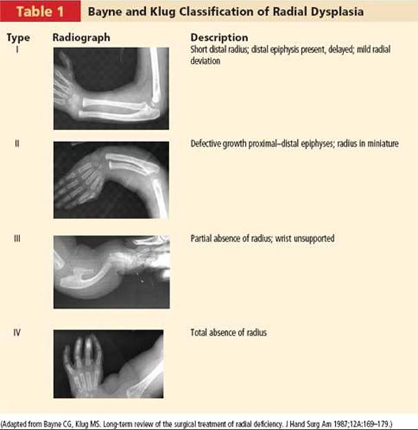
Bayne and Klug1 have provided a classification based on radiographic findings (Table 1).
PATHOGENESIS
Radial dysplasia develops during the period of embryogenesis. During this period, other organ systems are developing and may also be affected, as discussed later in this chapter.
NATURAL HISTORY
The natural history of patients with radial dysplasia clearly depends on the type of dysplasia present and the associated conditions.
 Patients with isolated type I or II radial dysplasia usually do not require surgical intervention.
Patients with more severe dysplasia can frequently benefit from surgical intervention.
Many times radial dysplasia is part of a syndrome, and the associated sequelae clearly affect these patients more than the underlying radial dysplasia. The most common associations are with Holt-Oram syndrome, thrombocytopenia–absent radius (TAR) syndrome, Fanconi anemia, and VACTERL (vertebral anomalies, anal atresia, cardiovascular anomalies, tracheoesophageal fistula, esophageal atresia, renal or radial anomalies, limb anomalies) (in front of or above the central axis of the limb).
An association with several craniofacial syndromes is also common.
No matter what procedure is used for treating the radial dysplasia, the patients all have a high incidence of recurrent deformity as they get older.
PATIENT HISTORY AND PHYSICAL FINDINGS
Clearly the most significant finding is radial deviation at the wrist (FIG 1).
If the patient is older, the affected forearm will also be short.
Frequently there is also associated thumb hypoplasia.
Because of its frequent association with systemic conditions, all patients require careful examination of their cardiac, renal, hematologic, and spinal systems.
IMAGING AND OTHER DIAGNOSTIC STUDIES
Radiographs should be taken of both forearms to assess stage of radial dysplasia (see Table 1).
In addition, all patients warrant a workup for syndromes and associated conditions, such as Holt-Oram syndrome, Fanconi anemia, TAR syndrome, and VACTERL.
This requires cardiac, renal, hematologic, and spinal evaluation.
NONOPERATIVE MANAGEMENT
All patients warrant preoperative stretching and splinting before any surgical intervention.
This may require external fixation techniques to distract the soft tissues in severe cases.
SURGICAL MANAGEMENT
Patients with type I or II radial dysplasia usually do not require surgical intervention.
Surgical treatment has generally ranged from soft tissue rebalancing alone to full centralization of the wrist with or without external fixation.
Before any procedure is contemplated, the surgeon must remember that the patient must maintain the ability to get his or her fingers to the mouth with the wrist in the surgically altered position.
We have had experience with various procedures for the treatment of radial dysplasia, including centralization, free toe transfer for stabilization of the radial wrist, and soft tissue release alone. We do not use formal centralization procedures anymore, as we have found the recurrence rate to be similar to our soft tissue release procedure. In addition, we think that this procedure jeopardizes the ulnar epiphysis, which can lead to an extraordinarily short forearm. Also, loss of mobility can result from a successful centralization procedure.
Vascularized bone transfer can be used in selective cases to provide stabilization of the radial side of the wrist. In the past we have used a vascularized second-toe metatarsophalangeal joint as described by Vilkki.4 In the future we anticipate using the proximal fibula, as its growth potential may be more similar to the ulna than the metatarsophalangeal joint.
The long-term problem for any surgical procedure is the recurrence rate.
For our patients, soft tissue release with a bilobed flap reconstruction has provided the most reliable, effective results.
Preoperative Planning
Before surgery, the patient must have undergone adequate soft tissue stretching.
In the first few months this is accomplished by splinting. In severe cases, serial casting may be necessary.
The splints should be large enough so the child cannot swallow it.
After about 6 months of age, active stretching is started by the parents with use of nighttime splinting.
The bilobed flap design must be drawn appropriately to take advantage of the redundant tissue on the ulnar side of the wrist.
Positioning
The patient is placed in the standard supine position, and a general anesthetic is used in all cases.
We do not use a standard tourniquet as we have found this to be inadequate in young children. Instead, we use the elastic bandage as a tourniquet in the upper arm.
Approach
We use a dorsal surgical approach, although more recently a volar approach has been described that may provide better exposure for soft tissue release.

FIG 1 • Preoperative photo showing radial deviation of the wrist.
TECHNIQUES
RELEASE OF RADIAL DEVIATION OF THE WRIST
 After induction of general anesthesia, the upper extremity is prepared and draped in the usual fashion.
The bilobed flap is then carefully designed using a marking pen (TECH FIG 1A,B).
The Esmarch bandage is used to exsanguinate the limb; it is then wrapped three times around the upper arm for use as a tourniquet. epiphysis, to prevent injury to the vascular supply to this area.
After release is accomplished, the wrist is placed in a neutral position and pinned with a 0.062-inch Kirschner wire. The Kirschner wire is temporary and is put across the joint from either direction (ie, there is no specific location for the exit or entrance site).
The flaps are then rotated and sutured in place (TECH FIG 1D,E).
The tourniquet is removed to ensure perfusion to the fingers, and a long-arm cast is placed.
After careful incision and elevation of the skin flaps (TECH FIG 1C), the finger and extensor tendons as well as the median nerve are carefully identified and preserved.
All other tissues in the radial wrist are released. Care must be taken not to dissect excessively near the ulnar

TECH FIG 1 • A,B. Markings for bilobed flap. C. Initial incision and elevation of flaps. D,E. After release of radial tethering tissue and rotation of flaps, the skin is sutured.


FIG 2 • Postoperative result.
POSTOPERATIVE CARE
The long-arm cast is left on for 3 to 4 weeks.
At that point the pin is removed and the patient is changed to a removable splint.
OUTCOMES
The bilobed flap procedure is an effective procedure for treating radial dysplasia (FIG 2).
Deformity tends to recur, though the incidence of this appears to be similar to that for other procedures used to treat radial dysplasia.
COMPLICATIONS
Very few complications are associated with this procedure.
Partial flap loss can occur, but the risk seems to be minimized by appropriate flap design and immobilization after the procedure.
REFERENCES
· Bayne CG, Klug MS. Long-term review of the surgical treatment of radial deficiency. J Hand Surg Am 1987;12A:169–179.
· Kozin S. Upper-extremity congenital anomalies. J Bone Joint Surg Am 2003;85A:1564–1576.
· McCarroll H. Congenital anomalies: a 25-year overview. J Hand Surg Am 2000;25A:1007–1037.
· Vilkki SK. Distraction and microvascular epiphysis transfer for radial club hand. J Hand Surg Br 1998;23B:445.