
Lori A. Karol and Jeffrey E. Martus
DEFINITION
 Developmental dislocation of the hip (DDH) occurs in 1.5 babies per 1000 live births. When diagnosed in the newborn period, closed treatment with the Pavlik harness is successful in 95% of dysplastic hips and up to 80% of dislocated hips.
Closed reduction is indicated when Pavlik harness treatment has failed, and when presentation for treatment is delayed past 6 months of age.
Open reduction is reserved for babies in whom closed reduction either is unobtainable or lacks stability, or for those who are diagnosed with a dislocated hip later than 18 to 24 months of age.10
The medial approach for open reduction is used most frequently in the young baby less than 12 to 18 months of age in whom an attempted closed reduction under anesthesia has been unsuccessful.
ANATOMY
Reduction of a dislocated hip is impeded by:
 The iliopsoas tendon, which is tautly stretched across the inferior capsule owing to the displacement of the femoral head
The inferomedial hip capsule, which becomes constricted
The transverse acetabular ligament, which spans the inferior aspect of the horseshoe of the acetabulum
The pulvinar, which is fibrofatty tissue occupying the cavity of the true acetabulum
The acetabular labrum, which can be infolded and serve as a doorstop blocking a deep and medial reduction
Anatomic landmarks for the medial approach are as follows:
The adductor longus tendon originating on the pubis
The pectineus, lying anterior to the adductor brevis (which is deep to the adductor longus)
The femoral artery, vein, and nerve, which are located as a bundle anterior to the pectineus muscle
The medial femoral circumflex artery, which lies in the interval between the pectineus and the femoral neurovascular bundle and is important for the circulation of the ossific nucleus of the proximal femur
PATHOGENESIS
DDH is more common in female babies and is linked with breech presentation, oligohydramnios, and firstborn children.
DDH is associated with congenital knee hyperextension and may be more likely in babies with torticollis and metatarsus adductus.
A positive family history is present in a minority of babies with DDH and most likely represents familial hyperlaxity.
Idiopathic DDH must be differentiated from the teratologic hip dislocation seen in infants with such disorders as arthrogryposis and Larsen syndrome.
NATURAL HISTORY
While research has shown that mild dysplasia, seen on ultrasound imaging of the newborn hip, often resolves spontaneously without treatment, the majority of babies with dislocatable or dislocated hips will progress from unstable hips to fixed dislocations in the absence of treatment.
PATIENT HISTORY AND PHYSICAL FINDINGS
The physical examination methods are summarized below. It is very important that the child is relaxed and quiet during the examination.
Limited abduction during range-of-motion testing may signify a dislocation and merits imaging. Abduction can be symmetric in bilateral dislocations.
The patient is examined for the Galeazzi sign. Asymmetry is abnormal and may indicate a hip dislocation or a congenital short femur. The apparent femoral lengths will be equal in bilateral dislocations.
Extra thigh folds may be present in unilateral DDH, but thigh fold asymmetry is often nonspecific.
The child is examined for the Ortolani sign. A positive sign represents the reduction of a dislocated hip. It is usually present in the newborn with DDH but disappears as the dislocation becomes fixed.
A positive Barlow sign represents the ability for a reduced hip to be dislocated because of instability. It disappears as a fixed dislocation develops.
In addition to a complete examination of each hip, the knees, feet, and upper extremities should be examined for contractures to rule out a teratologic dislocation.
The spine should be inspected for signs of dysraphism, which may result in a hip dislocation due to muscle imbalance.
IMAGING AND OTHER DIAGNOSTIC STUDIES
In infants less than 3 months of age, ultrasound of the hip is the preferred imaging study.
The femoral head, acetabulum, and triradiate cartilage can be identified.
The absence of coverage of the femoral head by the bony acetabulum is seen in the dislocated hip, which appears lateralized relative to the pelvis.
The alpha angle, which represents the slope of the bony acetabulum, is decreased (normal is more than 60 degrees), and the beta angle, which represents the cartilaginous acetabulum, is increased (normal is less than 55 degrees) in babies with hip dysplasia and hip dislocation (FIG 1A,B).
In infants 3 months of age and older, an anteroposterior (AP) pelvic radiograph is diagnostic (FIG 1C).
The line of Shenton, drawn along the inferomedial aspect of the proximal femur and the superior aspect of the obturator foramen, is disrupted.

FIG 1 • A,B. Ultrasounds of dislocated right hip with alpha angle of 56 degrees (A) and normal left hip with alpha angle 66 degrees (B). C. AP pelvis radiograph demonstrating dislocated right hip with small ossific nucleus.
The ossific nucleus may be smaller than the contralateral side, or absent altogether.
The medial proximal femoral metaphysis lies lateral to the Perkins line (drawn vertically from the lateral aspect of the bony acetabulum).
The acetabular index, which is the angle subtended by the Hilgenreiner line (a horizontal line drawn through the triradiate cartilages), and a line drawn along the bony acetabulum, is increased.
A pseudoacetabulum proximal and lateral to the true acetabulum may be present.
DIFFERENTIAL DIAGNOSIS
Teratologic hip dislocation (ie, arthrogryposis)
Neuromuscular dislocation (ie, spina bifida)
Congenital short femur
Coxa vara
NONOPERATIVE MANAGEMENT
The Pavlik harness is the treatment of choice for infants with hip dysplasia or instability from birth to age 6 months.
The Pavlik harness is less successful in patients with fixed dislocations but may be attempted for a period not to exceed 4 weeks, following which a reduction must be documented by either sonogram or radiograph.
There is no role for nonoperative management of a baby 6 months of age or older with a fixed hip dislocation.
SURGICAL MANAGEMENT
Surgical management can be delayed in the very young infant until the baby reaches sufficient size for a safe anesthetic and effective cast immobilization. We favor proceeding with surgical reduction (closed or open) at the age of 6 months inthe healthy baby.
The importance of delaying surgery until the presence of ossification within the femoral head remains controversial. While some studies report an increased incidence of avascular necrosis when the ossific nucleus is absent, others report an increased number of surgical procedures in children in whom reduction is delayed. We favor proceeding with reduction rather than waiting for ossification.2
Preoperative Planning
Traction may be used to increase the likelihood of a successful closed reduction, although current trends in the United States show a declining use of preoperative traction.
Positioning
The child is positioned supine on a radiolucent operating table.
The perineum is isolated with adhesive tape.
Surgical drapes are sutured in place to allow free movement of the extremity.
Towel clips should be avoided around the groin as they interfere with fluoroscopic visualization of the hip.
Approach
The medial approach to the dislocated hip is best suited to infants less than 12 months of age, but it has been used by other authors successfully in infants up to 24 months of age.
The medial approach described by Ludloff3 accesses the hip in the interval between the pectineus and the adductor brevis.
We favor the approach described by Weinstein,8,9 which exposes the hip between the femoral neurovascular bundle anteriorly and the pectineus muscle posteriorly. This interval allows for more direct visualization of the hip capsule and the medial femoral circumflex artery.
TECHNIQUES
INCISION AND INITIAL DISSECTION
 With the hip flexed and abducted, a transverse incision is made just lateral to the groin crease, centered over the palpable adductor longus tendon (TECH FIG 1A).
The fascia overlying the adductor musculature is opened (TECH FIG 1B).
The adductor longus tendon is isolated and divided (TECH FIG 1C). The tendon can be dissected free from the underlying adductor brevis with two blunt retractors.
The adductor brevis is identified with its overlying anterior branch of the obturator nerve. The pectineus is located anterior to the adductor brevis (TECH FIG 1D). Blunt retractors are used to identify the superior border of the pectineus.
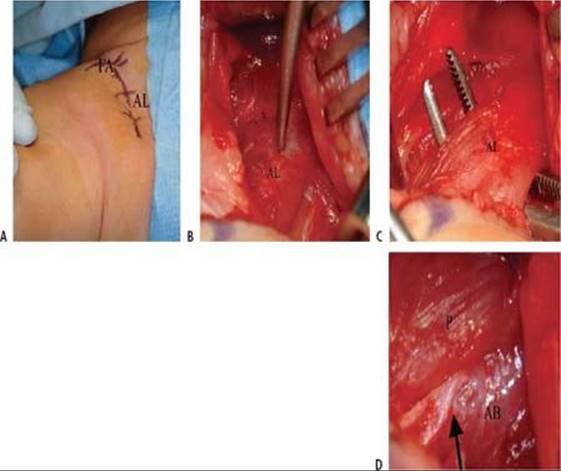
TECH FIG 1 • A. Planned transverse incision over the palpable adductor longus tendon (AL). The femoral arterial pulse is also noted (FA). B. The adductor longus (AL) after longitudinal incision of the overlying fascia. C.The adductor longus (AL) is isolated with a right-angle clamp and divided. D. The adductor longus has been sectioned and retracted distally. Anterior branch of the obturator nerve (arrow), adductor brevis (AB), pectineus (P).
DEEP DISSECTION
The femoral vessels are retracted gently anteriorly, and the pectineus muscle is retracted posteriorly (TECH FIG 2A).
The iliopsoas tendon can be identified. The surgeon can rotate the hip and feel the insertion site on the small prominence of the lesser trochanter. The psoas tendon is isolated and divided (TECH FIG 2B).
The surgeon carefully identifies the medial femoral circumflex artery as it traverses superior to inferior on the capsule of the hip. If possible, a small vessel loop is passed around the vessel to protect it. Otherwise, visualization of the vessel is maintained throughout the procedure.
The femoral head is palpated beneath the medial femoral circumflex artery. Kidners are used to bluntly expose the hip capsule (TECH FIG 2C).

TECH FIG 2 • A. The iliopsoas tendon (IP) is identified distally after retracting the pectineus posteriorly. Femoral vessels (NV), medial femoral circumflex vessels (MFC). B. The iliopsoas tendon (IP) is isolated with a right angle and divided. C. The hip joint capsule (C) has been exposed bluntly. Femoral neurovascular bundle (NV), medial femoral circumflex vessels (MFC).
CAPSULOTOMY AND ACETABULAR EXPOSURE
The capsule is incised along the rim of the acetabulum using a scalpel.
The white glistening cartilaginous femoral head is visualized (TECH FIG 3A). The hip is rotated to identify the ligamentum teres insertion on the superior aspect of the femoral head (TECH FIG 3B), and it is released (TECH FIG 3C).
The stump of the ligamentum is traced into the true acetabulum (TECH FIG 3D).
The incision in the capsule is completed to gain access to the acetabulum.
The ligamentum teres is excised from the floor of the acetabulum using scissors.
With a pituitary rongeur, the fibrofatty pulvinar is removed from the acetabulum. The surgeon should take care to preserve the underlying articular cartilage of the acetabulum (TECH FIG 3E).
The release of the acetabulum is completed by releasing (with scissors) the transverse acetabular ligament at the base of the acetabulum.
The surgeon should not excise or incise the acetabular labrum: it is an important growth center of the acetabulum and should be preserved.

TECH FIG 3 • A. The hip capsule has been incised along the rim of the acetabulum distal to the labrum. Femoral head (H), hip capsule (C), medial femoral circumflex vessels (MFC). B. The ligamentum teres (LT) is isolated with a right-angle clamp. C. The ligamentum teres (LT) is released sharply from the femoral head. D. After release from the femoral head (H), the ligamentum teres (LT) is grasped with a Kocher and traced into the acetabulum. E. The ligamentum teres and the fibrofatty pulvinar have been excised. Femoral head (H), acetabulum (A), hip capsule (C).
HIP REDUCTION, CAPSULORRHAPHY, AND CLOSURE
The hip is reduced under visualization by abducting the hip and lifting the greater trochanter anteriorly (the Ortolani maneuver). The hip should reduce readily and be quite stable in flexion. It will dislocate posteriorly with adduction. The reduced hip will appear relatively superficial, and the femoral head will be visible in the wound.
The reduction should be verified fluoroscopically at this point. If it is difficult to assess the reduction because of absence of ossification of the femoral head, a small drop of contrast can be placed in the acetabulum.
If the hip cannot be reduced, or if the reduction requires undue force to pull the hip into the acetabulum, the surgeon should recheck the capsular release and the transverse acetabular ligament.
In rare instances, the hip cannot be gently reduced and femoral shortening through a separate lateral approach is required. In infants less than 1 year of age who are otherwise normal, this is generally not necessary.
From this point onward, the hip must remain flexed to 90 degrees and gently abducted to maintain reduction. An assistant should be assigned to observe the position of the hip.
The vessel loop is removed from the medial femoral circumflex artery, which has been protected throughout the operation.
The subcutaneous layer and skin are closed. A drain is not necessary.
The wound is dressed and an occlusive bandage applied to deter contamination from diaper contents.
CASTING
The child is transferred to an infant spica table, and a double spica cast is applied with the hips flexed 90 degrees and gently abducted no greater than 60 degrees (TECH FIG 4A).
The surgeon should take care to cover the posterior aspects of the buttocks and mold beneath the greater trochanters to prevent “falling through” the spica posteriorly (TECH FIG 4B).

TECH FIG 4 • A. A double-leg spica cast is applied with careful attention to limb position and the maintenance of hip reduction. B. The limbs are positioned in 90 degrees of flexion and less than 60 degrees of abduction in the spica cast.

POSTOPERATIVE CARE
A radiograph is taken before waking the child from anesthesia to document the reduced hip within the spica cast (FIG 2A).
A limited CT scan may be performed within 24 hours after surgery to visualize the reduced hip after the child moves about in the cast (FIG 2B).
The cast is changed under anesthesia in 6 weeks and the hip is examined fluoroscopically. At this time, a one-and-a-half spica can be applied.
On occasion, a third spica will be needed, to be worn between 12 and 18 weeks after medial open reduction.
Bracing with an abduction orthosis after cast removal is prescribed at the discretion of the surgeon.

FIG 2 • A. Intraoperative AP pelvis radiograph in the cast to confirm reduction. B. Limited CT scan to confirm maintenance of reduction after the child moves about in the cast.
Periodic radiographs are needed to monitor the growth of the ossific nucleus and the resolution of acetabular dysplasia.
OUTCOMES
A satisfactory outcome after medial open reduction of the infant's hip can be expected in about 75% of children.2,5 Reduction can be universally achieved, and redislocation after a medial open reduction is rare. Owing to the limited dissection, prolonged postoperative stiffness is a rare event.
When an adverse outcome occurs after medial open reduction, it usually is related to the diagnosis of postoperative avascular necrosis or residual acetabular dysplasia.
Avascular Necrosis
Avascular necrosis has been documented in 9% to 43% of hips after medial open reduction.1,2,5,7 The large discrepancy in rates between series is due to differing criteria for its diagnosis and variation in the length of follow-up.
The severity of avascular necrosis varies widely between hips that have temporary irregular ossification of the epiphysis (Bucholz-Ogden type I) to complete avascular necrosis with an aspherical femoral head and coxa breva (type IV).
The presence of type I avascular necrosis does not preclude a successful outcome at skeletal maturity, while whole-head avascular necrosis dooms the hip to a poor outcome and leg-length discrepancy despite multiple surgical procedures.
Many series do not include the hips with irregular ossification, so it is imperative when comparing complication rates to ascertain which hips were included in the group with postoperative avascular necrosis. Although the published series of medial open reductions vary in the likelihood of avascular necrosis, it is clear that the proximity of the medial femoral circumflex artery to the medial hip capsule places the hip at greater risk for avascular necrosis during this approach compared to the anterior open reduction.
Because of variation between series in length of follow-up, the rate of type II avascular necrosis ranges widely. Typically, type II avascular necrosis becomes apparent in late childhood and early adolescence, when the proximal femur seems to grow into valgus because of a lateral physeal growth disturbance.1,5
In series with short-term follow-up, the rate of type II avascular necrosis may be lower than in those where skeletal maturity is achieved. Review of the DDH literature shows that type II avascular necrosis is not specific to the medial approach to the hip, however, as it may also be seen after anterior open reduction, and even closed reduction.
Acetabular Dysplasia
Persistent acetabular dysplasia may be seen after medial open reduction of the hip. This approach does not facilitate concomitant pelvic osteotomy, unlike the anterior open reduction, in which the iliac crest is exposed. Yet the optimal age for the medial open reduction (age 1 year or less) is young enough that the surgeon can expect the majority of patients to experience resolution of their dysplasia with growth.
Mankey et al4 found that one third of babies treated with medial approach open reduction required subsequent pelvic osteotomy for residual acetabular dysplasia.
COMPLICATIONS
Redislocation
Avascular necrosis
Infection
Need for future pelvic osteotomy due to persistent acetabular dysplasia
REFERENCES
· Koizumi W, Moriya H, Tsuchiya K, et al. Ludloff's medial approach for open reduction of congenital dislocation of the hip: a 20-year follow-up. J Bone Joint Surg Br 1996;78B:924–929.
· Konigsberg DE, Karol LA, Colby S, et al. Results of medial open reduction of the hip in infants with developmental dislocation of the hip. J Pediatr Orthop 2003;23:1–9.
· Ludloff K. The open reduction of the congenital hip dislocation by an anterior incision. Am J Orthop Surg 1913;10:438–454.
· Mankey MG, Arntz CT, Staheli LT. Open reduction through a medial approach for congenital dislocation of the hip. J Bone Joint Surg Am 1993;75A:1334–1345.
· Morcuende JA, Meyer MD, Dolan LA, et al. Long-term outcome after open reduction through an anteromedial approach for congenital dislocation of the hip. J Bone Joint Surg Am 1997;79A: 810–817.
· O'Hara JN, Bernard AA, Dwyer NStJP. Early results of medial approach open reduction in congenital dislocation of the hip: use before walking age. J Pediatr Orthop 1988;8:288–294.
· Tumer Y, Ward WT, Grudziak J. Medial open reduction in the treatment of developmental dislocation of the hip. J Pediatr Orthop 1997; 17:176–180.
· Weinstein S. Anteromedial approach to reduction for congenital hip dysplasia. Strategies Orthop Surg 1987;6:2.
· Weinstein SL, Ponseti IV. Congenital dislocation of the hip: open reduction through a medial approach. J Bone Joint Surg Am 1979;63A: 110–124.
· Willis RB. Developmental dysplasia of the hip: assessment and treatment before walking age. AAOS Instr Course Lect 2001;50:541–545.