
Anna V. Cuomo, Norman Y. Otsuka, and Richard E. Bowen
DEFINITION
 Shortening of the Achilles tendon, gastrocsoleus complex (triceps surae), or both results in an equinus (plantarflexed) position of the calcaneus relative to the tibia.
An equinus deformity is either congenital or acquired and can be dynamic or rigid.
 A dynamic deformity will correct with passive manipulation.
A rigid, or fixed, deformity does not correct.
Achilles or gastrocsoleus contracture often occurs in combination with other soft tissue contractures.
ANATOMY
The two heads of the gastrocnemius originate on the posterior aspect of the medial and lateral condyles of the distal femur.
The muscle fibers terminate at the muscle–tendon junction at the midcalf.
From here, the Achilles tendon is joined by tendon fibers from the posterior aspect of the soleus as the tendon courses distally.
The tendon is broad proximally and then becomes rounded at the midsection when it undergoes a 90-degree internal rotation before its insertion on the posterosuperior third of the calcaneus.
The rotation causes the medial fibers of the midtendon to insert on the posterior portion of the calcaneus (FIG 1).
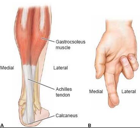
FIG 1 • A. Posterior view of Achilles tendon, demonstrating 90degree rotation of tendon fibers from posterior to medial and anterior to lateral. B. This can be easily remembered since it is a similar alignment to a crossed index and middle finger.
The insertion footprint is delta-shaped, and a small portion of the fibers course distally to meet the origin of the plantar fascia.
The blood supply of the Achilles tendon is limited.
The proximal portion is supplied by branches from within the gastrocnemius muscle.
The distal portion is supplied by branches from the tendon–bone interface.
There is no true synovial sheath. Instead, the surrounding paratenon, comprising loose connective tissue, supplies the rest of the blood supply via branches from the posterior tibial artery and, to a lesser degree, the peroneal artery.2
There are two synovial bursae at the Achilles tendon insertion site.
One is subcutaneous, located between the skin and tendon, and the other is deep, located between the tendon and the calcaneus.
PATHOGENESIS
The pathogenesis of congenital equinus is poorly understood and it is often associated with other limb deformities, such as clubfoot or congenital vertical talus.
Acquired equinus deformity secondary to cerebral palsy results from muscle spasticity or imbalance, leading to subsequent contracture of the Achilles tendon and gastrocsoleus complex.
Muscle imbalance and spasticity in spastic diplegic cerebral palsy often results in equinoplanovalgus deformity.
Muscle imbalance and spasticity in spastic hemiplegic cerebral palsy often results in equinus or equinovarus deformity.
Compensatory balance mechanisms to help maintain ambulation in patients with Duchenne muscular dystrophy also may result in equinus deformity.
Posttraumatic equinus can also be a result of severe burns and posterior scar contracture, postburn positioning, anterior leg muscle loss, or continued tibial growth in a rigid scar.3
Talocrural and subtalar capsular adhesions and an abnormal tibiotalar articulation may also contribute to loss of dorsiflexion and equinus deformity.
NATURAL HISTORY
Fixed equinus deformity will not correct spontaneously and requires prescribed stretching, surgical intervention, or both.4
Equinus associated with cerebral palsy is progressive. Despite both conservative and surgical treatments, the deformity can recur due to persistent spasticity, muscle imbalance, or limb growth.
Equinus deformity results in abnormal gait because of altered ankle range of motion and decreased ankle plantarflexion moment during terminal stance. It can result in chronic pain, poorly fitting footwear, callosities on the plantar forefoot, and possible skin ulceration in patients with altered sensation.
PATIENT HISTORY AND PHYSICAL FINDINGS
Birth history may reveal gestational or perinatal complications, such as traumatic brain injury or global hypoxic events, which are risk factors for cerebral palsy.
Family history may reveal an inherited neuromuscular disease or idiopathic toe walking.
A delay in gross motor milestones may suggest the presence of a static neurologic disorder such as cerebral palsy, while regression of gross motor function may suggest a progressive neuromuscular disease such as muscular dystrophy or Rett syndrome.
The age of equinus deformity onset will depend on the type and severity of the underlying condition.
Posttraumatic equinus, particularly a burn, should prompt questions regarding severity of the soft tissue loss, type of treatment, period of immobilization, and current problems with skin ulceration to assess the severity of scarring and overlying skin quality.
Physical examination should include a thorough examination of the entire lower extremities to look for associated deformities at the hip, knee, hindfoot, and forefoot.
The patient is examined supine on the examination table. It is important that the table has a hard surface so as not to mask any other contractures.
Ankle range of motion: Absence of dorsiflexion beyond neutral is ankle equinus.
Silfverskiöld test: A positive test indicates isolated gastrocnemius contracture. This is present if ankle equinus is present with the knee extended, but improves with knee flexion.
Palpation of Achilles tendon: A tight tendon suggests spasticity of the gastrocsoleus complex or contracture of the Achilles tendon.
Palpation of posterior tibial and peroneal tendons: Taut tendons suggest additional contracture or spasticity of the involved musculotendinous units contributing to the ankle equinus contracture.
Ankle clonus: More than two beats of clonus is abnormal and indicates gastrocsoleus spasticity or an upper motor neuron lesion.
If the child is ambulatory, the clinician should observe the gait in a hallway or large area where the patient can both walk and run.
Socks, shoes, and clothing that extends below the knee are removed.
Hindfoot alignment is best observed from behind.
The foot progression angle (axis of the foot to the axis of progression) and any associated coronal plane abnormalities, such as scissoring (excessive hip adduction during gait), knee progression angle, and pelvic rotation, are best observed from the front.
Ankle equinus and any associated sagittal plane abnormalities, such as a crouch gait (hip and/or knee flexion contracture) or a stiff-knee gait (decreased knee range of motion during swing phase), are best observed from the side.
 In mild equinus, the normal heel-to-toe gait of the plantargrade foot will be replaced with early lift-off during stance. Subtle deformity in patients with cerebral palsy is often unmasked by asking the patient to run. In severe equinus, the heel will not make contact during heel-strike.
Equinovarus or equinoplanovalgus deformity will cause initial contact during gait to occur on either the lateral or medial border of the foot, respectively. There may be a callus or foot pain at the area of initial contact.
The alignment and passive range of motion of the lumbosacral spine, pelvis, hips, and knees must also be tested, since equinus may be a functional compensation for coexistent contractures.
Associated muscle spasticity or contracture in cerebral palsy should be diagnosed with the appropriate physical examination maneuvers described in the relevant chapters.
IMAGING AND OTHER DIAGNOSTIC STUDIES
Anteroposterior (AP) and lateral weight-bearing radiographs of the affected ankle should be obtained.
Ossification centers of the talus, calcaneus, and cuboid are present at infancy. The navicular does not appear until age 3 to 4 years.
Equinus deformity will result in a decreased lateral tibiocalcaneal angle. Normal values range from 25 to 60 degrees. It can be difficult to measure this angle in young children with a partially ossified calcaneus.
Although usually associated with hindfoot varus, equinus deformity can also result in a decreased lateral talocalcaneal angle (intersection of a line through the longitudinal axis of the talus and a line along the plantar surface of the calcaneus; FIG 2). The normal range is 25 to 55 degrees.
Bony abnormalities, such as a flattened talar dome or anterior talar neck and anterior distal tibial osteophytes, can also contribute to ankle equinus contracture.
Arthrography may be useful to compare talocrural and subtalar motion during ankle dorsiflexion and plantarflexion to reveal capsular contractures contributing to restricted dorsiflexion and equinus deformity.6
DIFFERENTIAL DIAGNOSIS
Congenital equinus
Talipes equinovarus (clubfoot)
Planovalgus
Congenital vertical talus
Arthrogryposis
Tibial hemimelia
Acquired equinus
Neuromuscular
Cerebral palsy
Myelomeningocele

FIG 2 • Measurement of the lateral talocalcaneal angle. The normal range of 25 to 55 degrees is decreased in equinus.
Charcot-Marie-Tooth
Spinal muscular atrophy
Sacral agenesis
Rett syndrome or other genetic neuromuscular diseases
Posttraumatic
Posterior scar contracture
Posttrauma positioning
Anterior leg muscle loss
Continued tibial growth in a rigid scar
Other
Idiopathic toe walking
Juvenile arthropathy
NONOPERATIVE MANAGEMENT
Many children with equinus deformity secondary to a contracture of the Achilles tendon or gastrocnemius–soleus complex can be successfully managed with nonoperative treatment.
This is typically performed with serial casting for 3 to 6 weeks or more to achieve neutral sagittal alignment.
The success of nonoperative management depends on the age of the patient, the severity of the deformity, and the cause of the equinus.
In patients with equinus and cerebral palsy, early surgery may have an unpredictable outcome, with high rates of recurrence.
For this reason, surgery is often delayed with nonoperative treatments until after age 6 years.
Physical therapy for Achilles tendon stretching helps correct and maintain correction of equinus deformity.
The efficacy of stretching is likely dependent on the duration and frequency of stretching.
Use of an ankle–foot orthosis (AFO) in patients with cerebral palsy and dynamic equinus is a useful adjunct to nonsurgical management.
Botulinum toxin A (BtA) has been shown to be at least as effective as serial casting, with fewer side effects and more prolonged benefit.1
Serial casting and physical therapy are recommended as an adjunct to BtA injections.
Oral medications for muscle relaxation, such as baclofen, diazepam, dantrolene sodium, and tizanidine, can be helpful in selected patients with cerebral palsy if generalized reduction in tone is desired.
SURGICAL MANAGEMENT
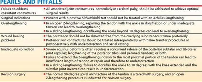
Indications for surgical management include fixed ankle equinus that exists with the knee flexed as well as extended and that also interferes with normal gait.
Clinical difficulties may include pain with weight bearing, toe walking, callosities on the plantar forefoot, poorly fitting orthoses, or plantar midfoot pain.
Surgical management of fixed ankle equinus in knee extension that disappears in knee flexion should consist of surgery to the gastrocnemius fascia alone.
Especially in patients with cerebral palsy, surgical management of ankle equinus should include concurrent treatment of all pelvic and lower extremity deformities, particularly hamstring contractures.5

FIG 3 • Prone positioning of patient on table with appropriate hip and knee padding during anesthesia.
Preoperative Planning
The quality of the overlying skin will be crucial for successful wound healing and should be considered during the preoperative planning phase.
Inadequate skin elasticity may require incomplete correction and staged surgery or staged casting in the postoperative period.
In severe posttraumatic cases, tissue loss and significant scarring may require additional tissue transfer procedures.
In cerebral palsy patients with severe spasticity, examination under anesthesia can help determine if equinus deformity is dynamic or fixed, since paralytic medications during anesthesia eliminate spasticity.
Positioning
The patient can be positioned either prone or supine.
The prone position allows improved access to the tibiotalar and subtalar joint capsules but requires careful padding to the hips and knees (FIG 3).
We prefer the supine position for patients undergoing isolated tendo Achilles lengthening.
A thigh tourniquet can be used.
The leg is prepared sterilely from the tourniquet distally.
If other concurrent soft tissue tendon lengthenings are to be performed, the patient is positioned and prepared according to the additional procedures.
Approach
To avoid postoperative wound complications, a longitudinal incision along the anteromedial border of the Achilles tendon is recommended.
This decreases the risk of wound dehiscence, since the thinnest portion of the overlying skin is directly posterior to the tendon and should remain intact.
For sliding procedures, a modified approach that exposes only the transected portion of the tendon can also be performed.
The open sliding Achilles lengthening technique, first described by White,9 is performed with partial transections at the (1) proximal and medial and (2) distal and anterior portions of the tendon. The 90-degree rotation of the tendon fibers between the transected areas maintains continuity of the tendon fibers as the tendon is lengthened (FIG 4A).
The modified open sliding Achilles lengthening technique decreases the size of the skin incision but uses the same technique of a sliding Achilles lengthening.
The open Achilles Z-lengthening procedure uses the same incision and closure as the modified approach. Here, the entire tendon is divided in a Z-fashion (FIG 4B), and the two sides or ends of the tendon are sutured to each other.
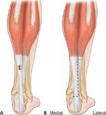
FIG 4 • A. Posterior view of Achilles tendon demonstrating distal anterior and proximal medial transections and subsequent sliding of attached tendon fibers during dorsiflexion. B. Posterior view of planned Achilles tendon sectioning to achieve Z-lengthening.
TECHNIQUES
OPEN SLIDING ACHILLES LENGTHENING
 The incision is made along the anteromedial border of the Achilles tendon.
 The skin incision begins just proximal to the calcaneal insertion (TECH FIG 1A) and continues proximally to the proximal extent of the tendon.
At the proximal portion of the incision, sharply divide the subcutaneous fat in line with the incision.
There are no neurovascular structures at risk here and the incision can be directed deeply until the paratenon sheath surrounding the tendon has been fully incised to form a small opening.
 The sheath comprises multiple thin layers, and full division of these layers is apparent once the gleaming white tissue of the tendon is encountered and the surgeon's forceps can no longer grasp any intervening tissue.
Using round-tipped dissecting scissors directed distally and laterally and aligned longitudinally with the tendon, pass one blade inside the paratenon and cut both the subcutaneous fat and sheath together until the calcaneal insertion is reached.

TECH FIG 1 • A. Planned incision along anteromedial border of tendon. B. Full exposure of the tendon has been achieved and the blade, cutting edge directed inferiorly, is inserted into the tendon in the coronal plane, dividing the anterior two thirds and posterior one third of the tendon. C. The medial two thirds of the proximal Achilles tendon has been transected and the ankle has been dorsiflexed to separate the tendon fibers. D. The ankle is dorsiflexed to the desired position.
This maintains attachment of the sheath to the subcutaneous fat, preserving the blood supply and providing closure of the sheath with approximation of the subcutaneous fat during wound closure.
 The tendon should now be completely exposed.
Insert a new blade, cutting edge inferior, just proximal to the calcaneus in the coronal plane between the anterior two thirds and posterior one third of the distal tendon (TECH FIG 1B).
Rotate the blade so the cutting edge is anterior and divide the anterior two thirds of the tendon transversely.
Identify the most proximal portion of the tendon that has been exposed.
Again, transversely divide the medial two thirds of the tendon, taking care to avoid any underlying muscle fibers of the soleus (TECH FIG 1C).
Slowly dorsiflex the foot with firm pressure to cause the divided portions of the tendon to slide past one another until 10 degrees of dorsiflexion is achieved (TECH FIG 1D).
Maintain the desired correction of alignment while closing the wound to allow even distribution of any new tension of the surrounding tissues.
Avoid overtensioning, which presents as complete blanching of the skin; it may lead to skin necrosis.
Using fine absorbable suture, loosely approximate the subcutaneous fat with several simple interrupted sutures. Then run the suture in the subcuticular tissue to close the skin.
Although a long-leg cast was initially recommended with this surgery, we prefer a short-leg cast with the ankle and subtalar joints in neutral positioning.
MODIFIED OPEN SLIDING ACHILLES LENGTHENING
Identify the anteromedial border of the Achilles tendon as described previously and, with a pen, draw the length of incision as if planning a fully open procedure.
This helps ensure alignment of the two incisions and avoids gaping of the wound after dorsiflexion and subsequent uneven tensioning of the skin.
Using a skin blade, create two short longitudinal incisions at the distal and proximal ends of the drawn line (TECH FIG 2).
As previously described, sharply dissect the overlying tissues to reach the Achilles tendon at the proximal and distal incisions and partially divide the tendon at each area of exposure.
As for the original open lengthening technique, slowly dorsiflex the foot with gentle pressure to cause the divided portions of the tendon to slide past one another until 10 degrees of dorsiflexion is achieved.
Close the subcutaneous tissues and skin and apply a shortor long-leg cast as described previously.

TECH FIG 2 • The incision is marked as if planning for an extensive exposure, except only the most proximal and distal 2 cm are used to expose the medial aspect of the Achilles tendon.
OPEN ACHILLES Z-LENGTHENING
Once the entire tendon has been exposed through the anteromedial approach, gently retract the most distal and medial tissues anteriorly to protect the underlying neurovascular structures.
Align a new no. 15 blade longitudinally with the tendon fibers at the midpoint of the tendon in the sagittal plane. Introduce the blade deeply into the tendon until there is a discernable release in resistance, signifying complete transection through the tendon.
Alternately, a small osteotome may be introduced along the anterior border of the tendon, and the blade can be carried safely through the entire tendon until metal-on-metal contact.
Carry the incision distally until the calcaneal insertion is reached (TECH FIG 3A).
Without removing the blade, rotate the blade medially 90 degrees and with a slight sawing motion, transversely divide the medial portion of the tendon.
Once this is achieved, retraction of the medial structures can be safely released.
Return the blade to the proximal starting point and extend the division proximally until an adequate portion of the tendon is involved.
This is usually about two thirds of the Achilles tendon but will ultimately depend on the desired lengthening and overlap between the two ends.
Take care to stay along the midline of the tendon.
Complete the proximal division of the tendon laterally in this transverse plane (TECH FIG 3B).
The entire tendon should now be divided in a Z-fashion (TECH FIG 3C).
Dorsiflex the ankle to neutral. Under moderate tension, reapproximate the sides of the tendon with a braided nonabsorbable suture.
This can be done with multiple interrupted simple or vertical mattress sutures (TECH FIG 3D).
Alternately, the overlapping ends of the tendon can be excised for approximation of the ends.
Multiple intratendinous and epitendon suture techniques are acceptable for reapproximation.
The intratendinous technique reduces exposed suture and diminishes inflammatory reaction around the suture, and is recommended.
The simplest of these is the modified Kessler suture (locking loops of the core suture; TECH FIG 3E).
Apply either a longor short-leg plaster cast as discussed above.

TECH FIG 3 • A. Full exposure of the Achilles tendon has been achieved and the initial transection in the sagittal plane, dividing the medial and lateral halves of the tendon, has been performed with a blade. B. The entire tendon is exposed and is being transected in a Z-fashion. C. The ankle is dorsiflexed to the appropriate position and the overlapping tendon is noted. D. The desired amount of overlapping tendon has been joined with vertical mattress sutures. E. Alternately, the tendons can be reapproximated end to end. After Z-lengthening, the remaining overlapping tendon has been removed and the ends of the tendon are joined with a nonabsorbable suture and a modified Kessler repair.
POSTOPERATIVE CARE
Adequate pain control in the acute postoperative setting is imperative. This is both to promote the child's comfort and to reduce additional muscle spasms, which may alter the desired surgical correction.
Since children with neuromuscular diseases may have significant communicative barriers, pain should be presumed to be present and should be treated with both morphine derivatives and muscle relaxants.
The limb should be elevated as much as possible for 2 or 3 days until acute swelling resolves.
The child can then become ambulatory and weight bearing as tolerated if a sliding tendon lengthening was performed.
Patients undergoing open Z-lengthening should remain nonweight bearing in a cast until tendon healing is sufficient (6 weeks).
Once the cast is permanently removed, the child needs postoperative physical therapy or use of an AFO, as dictated by the diagnosis.
OUTCOMES
Surgical lengthening results in gains in dorsiflexion, from a preoperative average of 25 degrees of plantarflexion to 8 degrees of dorsiflexion, without significant changes in the arc range of motion.
Correction is maintained in 80% to 90% of patients for at least 7 years postoperatively.4
COMPLICATIONS
Calcaneovalgus deformity occurs in less than 2% of open Achilles tendon lengthenings.
Recurrent deformity is common in neuromuscular diseases owing to continued spasticity and normal longitudinal tibial growth.
After surgical correction, 18% of children with diplegia and 41% of those with hemiplegia will experience recurrence.
Ambulatory patients maintain correction better than nonambulatory patients.
Recurrence is also more frequent in children 4 years or younger.7
Wound dehiscence and necrosis are infrequent and the incidence is not well reported.
These can be devastating, however, and should remain a matter of concern as potential complications.
REFERENCES
1. Ackman JD, Russman BS, Thomas SS, et al. Comparing botulinum toxin A with casting for treatment of dynamic equinus in children with cerebral palsy. Shriners Hospitals BTX-A Study Group. Shriners Hospital for Children, Chicago, USA. Dev Med Child Neurol 2005;47:620–627.
2. Ahmed IM, Lagopoulos M, McConnell P, et al. Blood supply of the Achilles tendon. J Orthop Res 1998;16:591–596.
3. Carmichael KD, Maxwell SC, Calhoun JH. Recurrence rates of burn contracture ankle equinus and other foot deformities in children treated with Ilizarov fixation. J Pediatr Orthop 2005;25:523–528.
4. Damron TA, Greenwald TA, Breed AL. Chronologic outcome of surgical tendoachilles lengthening and natural history of gastroc-soleus contracture in cerebral palsy: a two-part study. Clin Orthop Relat Res 1994;301:249–255.
5. Karol LA. Surgical management of the lower extremity in ambulatory children with cerebral palsy. J Am Acad Orthop Surg 2004; 12:196–203.
6. Mary P, Damsin JP, Carlioz H. Correction of equinus in clubfoot: the contribution of arthrography. J Pediatr Orthop 2004;24:312–316.
7. Rattey TE, Leahey L, Hyndman J, et al. Recurrence after Achilles tendon lengthening in cerebral palsy. J Pediatr Orthop 1993;13:184–187.
8. Rome K. Ankle joint dorsiflexion measurement studies: a review of the literature. J Am Podiatr Med Assoc 1996;86:205–211.
9. White WJ. Torsion of the Achilles tendon: its surgical significance. Arch Surg Am 1943;46:784.