
David Scher
DEFINITION
 A calcaneonavicular coalition is an abnormal connection between the calcaneus and the navicular.
This extra connection between the tarsal bones typically limits subtalar motion.
The major consequence of this condition is a rigid flatfoot that may be painful.
ANATOMY
The coalition typically occurs between the anterior process of the calcaneus and the most lateral aspect of the navicular (FIG 1).
The connection may comprise bone, cartilage, or fibrous tissue (bony, cartilaginous, or fibrous coalitions, respectively).
PATHOGENESIS
The cause of calcaneonavicular coalitions remains unknown.
It has been hypothesized that coalitions may result from failure of segmentation of the individual tarsal bones during fetal development.1
Symptoms typically develop in later childhood, usually between 8 and 12 years old, for calcaneonavicular coalitions.5
It is theorized that the reason for the delayed onset of symptoms, despite presumed presence from birth, is that the coalition ossifies over time, making it more rigid and more likely to limit subtalar motion.5
The pain from a calcaneonavicular coalition may arise from altered kinematics of the foot due to local limitation of motion. It has also been suggested that a fracture through a previously solid coalition could render it painful.
NATURAL HISTORY
Many people with calcaneonavicular coalitions are probably pain-free, although they may have a rigid flatfoot, with loss of the longitudinal arch and valgus alignment of the heel.6
If pain develops in a child with a calcaneonavicular coalition, it usually does so between ages 8 and 12.
PATIENT HISTORY AND PHYSICAL FINDINGS
Patients present with complaints of foot pain exacerbated by activity, typically localized to the lateral aspect of the foot, just distal to the sinus tarsi, in the region of the anterior process of the calcaneus. They may complain of medial foot and ankle pain or pain at the distal tip of the fibula as well.
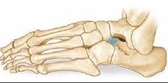
FIG 1 • A complete bony calcaneonavicular coalition.
There may be a history of progressive out-toeing and loss of arch height due to an increase in the planovalgus position of the foot.
Patients may also relate difficulty walking on uneven surfaces, presumably due to decreased subtalar motion.
The physician should observe the patient's gait; he or she may walk with an antalgic gait on the affected side (decreased stance phase) and an out-toeing gait.
The physician should examine the patient's foot alignment. The heel may be in valgus alignment with the forefoot abducted.
The physician should examine the rigidity of the patient's flatfoot. A flexible flatfoot has restoration of the arch upon toe-rise, while a rigid flatfoot has no arch restoration. A rigid flatfoot is a sign of decreased subtalar motion and may indicate a tarsal coalition.
The physician should palpate over the anterior process of the calcaneus and just distal to the anterior process. Point tenderness is suggestive of a painful calcaneonavicular coalition.
IMAGING AND OTHER DIAGNOSTIC STUDIES
Plain radiographs, including anteroposterior (AP), lateral, and oblique views, should be obtained to visualize the coalition.
 A calcaneonavicular coalition is best seen on the oblique view (inversion oblique).
A prominent anterior process of the calcaneus, the “anteater nose” sign, may be seen on the lateral view.4
Standing AP and lateral views can be included to assess foot alignment (FIG 2).
A Harris axial view or Salzman hindfoot alignment view can be obtained to assess heel alignment.
A CT scan should be obtained to rule out a concurrent talocalcaneal coalition or the presence of arthritis in adjacent joints. A CT or MRI may also be useful if the diagnosis is in question.

FIG 2 • Oblique radiograph depicting a cartilaginous calcaneonavicular coalition.
DIFFERENTIAL DIAGNOSIS
Flexible flatfoot
Subtalar arthritis
Other tarsal coalition (talocalcaneal or other less common ones)
Tumor or infection involving the subtalar joint
Idiopathic rigid flatfoot
NONOPERATIVE MANAGEMENT
Nonoperative management is indicated for all patients with calcaneonavicular coalition at first presentation.
Painless coalitions need no treatment.
Initial treatment for painful coalitions consists of activity modification, anti-inflammatory medication, and immobilization in a short-leg walking cast for 4 to 6 weeks.
SURGICAL MANAGEMENT
The indication for surgical management is persistence of pain despite nonoperative management.
The main goals of treatment are, primarily, elimination of pain and restoration of function.
Restoration of subtalar motion is a secondary goal.
Restoration of arch height is unlikely after resection.
Preoperative Planning
All imaging studies are reviewed.
An examination of subtalar motion may be performed under anesthesia to serve as a comparison to the examination immediately after resection.
Positioning
The patient is positioned supine with a bump under the hip of the operative side to slightly internally rotate the leg.
If subcutaneous fat autograft is to be used as an interposition material after resection, the limb should be prepared up to the buttocks and a sterile tourniquet should be used (FIG 3).
Alternatively an eschmarch tourniquet may be used just proximal to the ankle.
Approach
The approach involves exposure and resection of the entire coalition.
A graft material is interposed between the ends of the resected bone consisting of local muscle (peroneus brevis) or autologous fat.
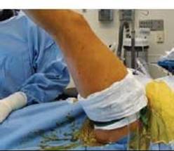
FIG 3 • A sterile tourniquet is used with sufficient room proximal to it for harvesting of fat graft.
TECHNIQUES
INCISION AND DISSECTION
 The procedure can be done under tourniquet control if desired.
An oblique incision is made along the lateral side of the foot between the extensor tendons and the peroneal tendons, directly overlying the anterior process of the calcaneus (TECH FIG 1A).
The skin and subcutaneous tissue are incised sharply, taking care not to undermine the tissues.
The extensor digitorum brevis is exposed and followed proximally to its origin at the sinus tarsi (TECH FIG 1B, C).

TECH FIG 1 • A. The incision lies between the extensor tendons and peroneal tendons. B,C. The extensor digitorum brevis is identified and reflected distally.

TECH FIG 1 • D. The coalition is localized with a needle and confirmed fluoroscopically. E. The coalition is resected with a small osteotome. When performing the medial cut, care is taken to avoid damaging the adjacent articular surface of the talar head.
Fibrofatty tissue within the sinus tarsi is exposed.
This fibrofatty tissue is incised and reflected distally along with the attached origin of the extensor digitorum brevis, exposing the anterior process of the calcaneus and the calcaneonavicular coalition (TECH FIG 1D).
Fluoroscopic confirmation of the coalition is obtained by placing a surgical instrument or needle directly over it (TECH FIG 1E).
RESECTION OF THE CALCANEONAVICULAR COALITION
The extensor digitorum brevis is retracted distally and any remaining fibrofatty tissue from the sinus tarsi is retracted proximally.
A small osteotome is used to remove a trapezoidal piece of bone (TECH FIG 2A, B).
The first cut is made in the region of what would be the middle of the anterior process of the calcaneus. This cut should be inclined about 40 to 60 degrees from the vertical relative to the plantar surface of the foot and directed medially toward the lateral aspect of the navicular, deep within the wound.
The next cut is made at the most lateral aspect of the navicular, directed toward nearly the same point as the first cut.
The ends of these two cuts should not meet, as the goal is to resect a trapezoidal piece and not a triangular piece.
When making these cuts, especially the medial one, care must be taken to avoid injuring the articular cartilage of the talar head, which lies directly medial and proximal to the osteotome.
Attention must also be paid to removing sufficient bone so that there is a visible space between the calcaneus and navicular, which is confirmed fluoroscopically on the inversion view. After resection, the lateral edge of the navicular should line up with the lateral aspect of the talar neck and the medial edge of the anterior process of the calcaneus should line up with the medial edge of the cuboid.
Remaining bone is removed as necessary with rongeurs (TECH FIG 2C).
Bone wax is placed over the exposed cut bone surfaces.

TECH FIG 2 • A,B. The piece of bone removed is trapezoidal in shape, not triangular. C. Remaining bone is removed.
INTERPOSITION OF FAT GRAFT
A piece of subcutaneous fat can be taken from just beneath the buttock crease. Use of this donor site allows for a cosmetic incision with minimal donor site morbidity. There is always abundant fat in this location and there are no neurovascular structures at risk during this dissection.
A transverse incision is made at the base of the buttocks while an assistant elevates the limb.
A piece of subcutaneous fat about 2 cm in diameter is removed and placed directly into the gap that has been created (TECH FIG 3).
Interposition of Peroneus Brevis Muscle (Alternative Technique)
After the coalition has been resected, heavy absorbable sutures are woven through the proximal end of the peroneus brevis that had been detached from its origin.

TECH FIG 3 • A sufficient gap is created and fat is interposed.
The ends of the sutures are passed through Keith needles.
The Keith needles are passed through the space that has been created in the depth of the wound, to exit the medial side of the foot.
The needles are passed through a piece of sterile felt and a button and the sutures are sewn over the button, drawing the muscle into the gap where the calcaneonavicular coalition was previously (TECH FIG 4).
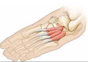
TECH FIG 4 • Absorbable sutures are passed through the proximal edge of the extensor digitorum brevis and the ends are passed into the space created by the resection and out the medial side of the foot. They are then tied over felt and a button.
WOUND CLOSURE
The tourniquet is released and hemostasis is obtained.
If fat was used as graft material, the extensor digitorum brevis is sewn back down anatomically to its origin with absorbable suture.
Subcutaneous tissue and skin are closed in standard fashion.

POSTOPERATIVE CARE
The patient is placed in a cast or splint for 2 to 3 weeks to allow the graft to consolidate and the wound to heal.
Progressive weight bearing is allowed after cast removal, and range-of-motion exercises are performed to address subtalar motion.
OUTCOMES
Greater than 90% good or excellent results have been reported in most series.2
Poor results with persistent pain are attributed to failure to resect adequate bone or the presence of concurrent arthritis in the midfoot or hindfoot.3
COMPLICATIONS
Failure to resect adequate bone
Injury to adjacent articular cartilage
Wound-healing complications
Recurrence of the coalition
REFERENCES
· Harris RI, Beath T. Etiology of peroneal spastic flat foot. J Bone Joint Surg Br 1948;30B:624–634.
· Gonzalez P, Kumar SJ. Calcaneonavicular coalition treated by resection and interposition of the extensor digitorum brevis muscle. J Bone Joint Surg Am 1990;72A:71–77.
· Moyes ST, Crawfurd EJ, Aichroth PM. The interposition of extensor digitorum brevis in the resection of calcaneonavicular bars. J Pediatr Orthop 1994;14:387–388.
· Oestreich AE, Mize WA, Crawford AH, et al. The “anteater nose”: a direct sign of calcaneonavicular coalition on the lateral radiograph. J Pediatr Orthop 1987;7:709–711.
· Stormont DM, Peterson HA. The relative incidence of tarsal coalition. Clin Orthop Relat Res 1983;181:28–36.
· Varner KE, Michelson JD. Tarsal coalition in adults. Foot Ankle Int 2000;21:669–672.