
Karen S. Myung and Kenneth Noonan
DEFINITION
 The incidence of residual deformity in congenital clubfoot ranges from 26.6% to 50%, regardless of the initial treatment provided.2
 The disparity in the reported incidence is due to varying severity of clubfoot deformity, different methods of treatment, and, in part, differing definitions of residual deformity.
Residual deformities include isolated equinus, cavus, metatarsus adductus, forefoot supination, and combinations of the above.
Dynamic forefoot adduction and supination can be observed after clubfoot treatment with or without soft tissue releases.
Dynamic forefoot supination deformity results from residual medial displacement of the navicular on the head of the talus, which results in muscle imbalance. In this case, because its insertion is medially displaced, the anterior tibialis becomes a forefoot supinator instead of a dorsiflexor. In addition, deformity may also occur because the anterior tibialis muscle is relatively strong in comparison to the peroneal muscles (FIG 1).
The aim of treatment is to correct any fixed deformity and to rebalance the muscles of the foot, thereby correcting dynamic deformity and improving foot alignment.

FIG 1 • Normal foot versus supinated foot. Medial subluxation of the navicular, the medial cuneiform, and the first metatarsal results in supination deformity as the line of pull of the tibialis anterior tendon directs the foot into supination instead of dorsiflexion.
ANATOMY
The anterior tibialis muscle originates from the upper two thirds of the tibia.
The anterior tibialis tendon fibers rotate 90 degrees from the musculotendinous junction to its insertion on the medial cuneiform and first metatarsal.
Medial rotation begins proximally, so the most medial muscle fibers proximally rotate to the posterior surface of the tendon near the midpoint and continue to rotate so that their final insertion is as the distal–lateral fibers on the first metatarsal.
Meanwhile, the most lateral muscle fibers proximally rotate to the anterior surface at the midpoint and continue distally to insert on the cuneiform as the proximal–medial fibers (FIG 2).4
The anterior tibialis muscle is active in two important stages of the gait cycle; it concentrically fires during the initiation of swing phase and keeps the foot dorsiflexed during early swing phase and then it relaxes.
The anterior tibialis muscle then fires eccentrically as the foot is lowered to the floor from heel strike to foot flat in stance phase.
As a dorsiflexor, the anterior tibialis muscle opposes gravity and the strong gastrocsoleus complex. Importantly, the anterior tibialis muscle may also be a supinator of the forefoot in the face of peroneal longus weakness or medial displacement of the insertion.
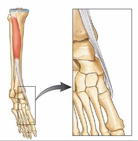
FIG 2 • Anatomy of the tibialis anterior muscle-tendon. The anterior tibialis tendon fibers rotate 90 degrees from their musculotendinous junction to their insertion on the medial cuneiform and the first metatarsal such that the proximal-medial insertional fibers on the cuneiform begin as the lateral fibers at the musculotendinous junction (see window).
There are important bony abnormalities associated with residual clubfoot deformity.
The subtalar joint may have an absent anterior facet and small, narrow medial and posterior facets, resulting in restricted subtalar motion. In this setting, the calcaneus does not slide fully into valgus with casting such that the navicular remains medially displaced.
The navicular itself is wedge-shaped and is medially displaced along with the cuneiforms and metatarsals.10 With
medial displacement of its insertion, the biomechanical advantage favors the action of the anterior tibialis muscle as a strong supinator over its role as a dorsiflexor (FIG 3).
PATHOGENESIS
The cause of residual clubfoot deformity may be incomplete correction or recurrence of deformity as part of the natural history of the resistant clubfoot.
Electromyographic studies have demonstrated that the peroneal muscle group can be relatively weaker, thus increasing the supinator action of the tibialis anterior muscle.1,3
Medial subluxation of the navicular was found to be the most important factor influencing both the appearance of the foot and the lateral rotation of the ankle.9
In addition to the bony abnormalities associated with clubfeet, anatomic variations from the customary insertion of the anterior tibialis muscle into adjacent areas of the first metatarsal and medial cuneiform occur in 10% of pathologic specimens.
In these variants, the distal anterior tibialis muscle inserts more medially than normal, optimizing the force vector for supination.8
NATURAL HISTORY
Residual deformities are usually encountered within the first year after initial treatment and generally before the age of 5 years, even in congenital clubfeet that had been fully corrected since the first month of life.
Residual forefoot adduction and supination are the most common deformities after nonoperative treatment and can also be seen after initial operative repair. They can result from undercorrection at the time of the primary intervention.13
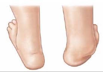
FIG 3 • Bony abnormalities associated with residual clubfoot deformity. The navicular is wedge-shaped and is medially displaced along with the cuneiforms and metatarsals.

FIG 4 • Hindfoot varus. When untreated, residual deformity may become stiff. When fixed inversion deformity is combined with residual equinus deformity, hindfoot varus occurs.
Correction of resistant congenital clubfoot often requires more than one surgery, not because of a “failed initial intervention,” but because of dynamic muscle imbalances that may not be fully manifest at the time of the initial intervention. Thus, the need for an additional operation can be perceived as part of the natural history of congenital clubfoot.12
If left untreated, the dynamic deformity may become stiff and the foot tends to invert.
When inversion deformity is combined with residual equinus deformity, hindfoot varus may recur (FIG 4).
PATIENT HISTORY AND PHYSICAL FINDINGS
Residual deformity is more likely in patients who have clubfoot as a result of myelomeningocele or other neuromuscular syndromes and genetic disorders such as Larsen syndrome. Therefore, it is important to consider neurologic causes, such as tethered cord, when confronted with residual deformity.
Recurrent deformity may be found in children with only four toes on the affected foot, as these individuals may have absence of the peroneal muscle group (similar to that seen in fibular hemimelia), thus leaving them prone to recurrence.
One of the first clinical signs of recurrence is a dynamic inversion of the foot with slight equinus. Equinus may be difficult to quantify as midfoot breech will often accommodate and hide the hindfoot equinus (FIG 5).
Residual deformity most frequently occurs in severe or atypical cases, which are often associated with a small calf size. These children may also have short, fat feet with a deep plantar crease that extends from the medial border to the lateral border of the foot and a shortened first ray. These findings are consistent with severe or atypical clubfeet that have a propensity for residual deformity.
In maximum pronation or maximum supination, the navicular–medial malleolar distance is decreased compared to the normal foot. In fact, the medial malleolus can be difficult to delineate because it is in contact with the navicular. The navicular malleolar distance demonstrates the extent of medial subluxation.
It is important to examine gait when possible.
During examination of gait, the clinician should identify whether the tibialis anterior is a dynamic supinator; this is best observed in swing phase when no antagonist muscles contract.
This finding will confirm the appropriateness of surgery.
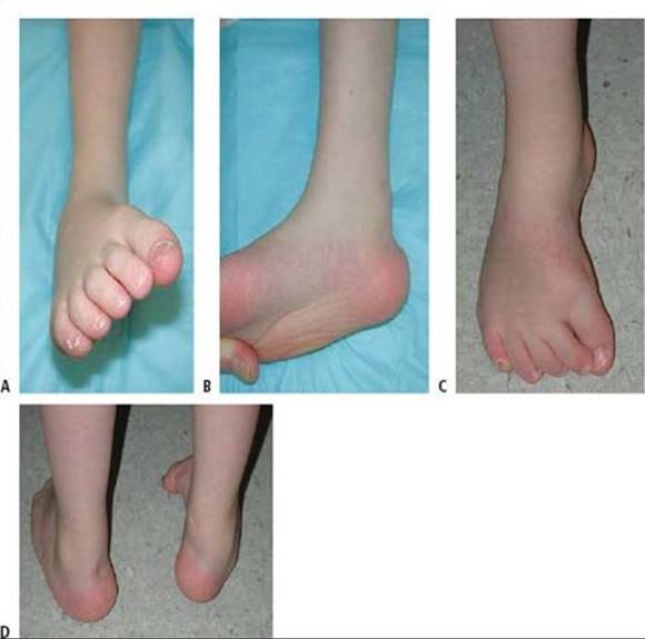
FIG 5 • Examination findings of residual supination and equinus deformity. A. Forefoot supination. B. Hindfoot equinus. C. Anterior view combined forefoot supination and hindfoot equinovarus. D.Posterior view combined forefoot supination and hindfoot equinovarus.
The strength of the tibialis anterior is tested. With dynamic supination deformity, the supinator action of the anterior tibialis muscle will overpower the dorsiflexor action, thus demonstrating the appropriateness of surgery. In addition, good power is needed for a successful transfer.
Range of motion of the ankle is examined. Transfer will work only as long as there is not a fixed contracture.
The clinician should evaluate for other deformities, such as equinus, cavus, varus, adductus, and tibial torsion.
IMAGING AND OTHER DIAGNOSTIC STUDIES
Anteroposterior (AP) and lateral radiographs may be helpful to study and quantify various deformities.
AP radiographs will demonstrate medial deviation of the metatarsals, which can indicate residual medial displacement of navicular, which is yet to ossify (FIG 6).
On an AP radiograph of normal feet, the line drawn through the long axis of the talus should point to the first metatarsal, while the line drawn through the long axis of the calcaneus should point toward the fourth metatarsal.
 In clubfeet, these lines become more parallel, depicting “stacking” of the talus and calcaneus.
Forced maximum dorsiflexion lateral radiographs may reveal hindfoot equinus with midfoot breech.
Stacking of the metatarsals on the lateral radiograph identifies the presence of residual forefoot supination (a decreased talocalcaneal angle).
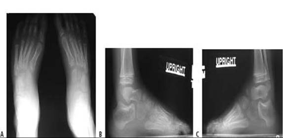
FIG 6 • Weight-bearing AP and lateral radiographs of feet shown in Figure 5. A. The long axes of the talus and calcaneus are somewhat parallel rather than divergent. The metatarsals appear adducted in relation to the talus. B,C. The long axis of the talus and calcaneus appear somewhat parallel rather than divergent on the lateral view of the right foot. The axes of the talus and the first metatarsal do not form a straight line, as opposed to a normal foot. This degree of divergence from this linear alignment represents intrinsic deformity of the clubfoot. The metatarsals are stacked on the weight-bearing lateral views.
Ultrasound evaluation of the foot is not done routinely. However, experimental studies have demonstrated that this technique is capable of documenting the location of the navicular in relationship to the head of the talus. The navicular is subluxated plantarward and medially on the head of the talus.
Similarly, magnetic resonance imaging can be performed to completely identify the relationships of the cartilaginous bones.
Although possible, this technology is rarely clinically used as orthopaedists are aware of the classic deformities that are associated with recurrence.
DIFFERENTIAL DIAGNOSIS
Residual deformities in clubfoot may be due to unrecognized tarsal coalitions or other conditions in severe or atypical feet.
Unexpected and rapid recurrent deformity in children with previously corrected feet and with known myelomeningocele may be a result of continued neurologic involvement, such as tethering of the spinal cord.
NONOPERATIVE MANAGEMENT
Treatment of residual deformity depends on the location, severity, and age of the child.
Recasting may be considered in children less than 18 months of age who have residual deformity.
Most residual deformities at this age can be treated by Ponseti manipulation, followed by application of a toe-togroin plaster cast with the feet in a fully corrected position for 2 weeks.
After 2 weeks, the casts are removed and reapplied.
Usually three casting sessions are required, for a total of 6 weeks.
Thereafter, abduction bracing is reinstituted.
In larger children, ankle–foot orthoses (AFOs) may also be used to prevent recurrence.
Physiotherapy may also be used in patients with residual deformity. The therapist must be familiar with techniques to manipulate residual forefoot adductus and posterior contracture.
In older children and in patients with midfoot breech, it can be difficult to effectively stretch any hindfoot equinus contracture.
With equinus contracture, abduction bracing is difficult.
Unbraceable posterior contracture can then lead to recurrent metatarsus adduction and forefoot supination. Thus, a repeat percutaneous heel cord tenotomy and casting may be required.
SURGICAL MANAGEMENT
In children more than 2 to 3 years of age, it may be preferable to correct any residual deformity using soft tissue lengthenings or transfers with or without bony procedures.
As the anterior tibialis acts as a supinator, lateral transfer of the anterior tibialis tendon is often necessary to correct dynamic supination deformity.
The optimal age for lateral transfer of the anterior tibialis tendon varies from case to case.
Important factors are the rapidity of recurrence, the strength of the anterior tibialis muscle, the presence of fixed forefoot deformity, or the presence of concurrent equinus deformity or cavus.
The surgery should be performed after the lateral cuneiform ossification center appears (2 to 4 years of age).
The split anterior tibialis tendon transfer (SPLATT) of Hoffer6 is rarely used in idiopathic clubfeet but is an excellent method for correcting dynamic supination deformity as a result of spasticity associated with disorders such as cerebral palsy.
This method may have some utility in children with mild, flexible forefoot supination who require surgery for other deformity.
Preoperative Planning
Feet with residual deformity should be extensively evaluated by clinical and radiographic assessment before surgical planning. Each foot should be treated individually, as no single treatment plan is appropriate for all feet.
Associated deformities must be identified. For example, an anterior tibialis transfer will function poorly in the face of a fixed equinus contracture. In this case, it will be necessary to correct equinus deformity with a heel cord tenotomy or lengthening or posterior release.
Residual varus deformity may indicate the need for an opening wedge or sliding calcaneal osteotomy.
Persistent metatarsus adductus may necessitate midfoot osteotomies in order for the lateral border of the foot to be reduced.
Anterior tibialis tendon transfer does not correct restricted subtalar motion.
It is important to confirm that the ossific nucleus of the lateral cuneiform is present in order to place the anterior tibialis tendon into an appropriate anchor site.
Positioning
The patient is placed in the supine position on a standard operating table or a hand table.
Either positioning is done in a way to ensure good fluoroscopic images.
A well-padded thigh-high tourniquet should be placed before preparing and draping the patient.
Approach
A medial incision is based over the insertion of the anterior tibialis tendon.
From this incision the surgeon may be able to perform an opening wedge osteotomy of the medial cuneiform if indicated.
Once the anterior tibialis tendon is detached, a lateral incision is based over the lateral cuneiform.
Fluoroscopic imaging can assist in planning this incision.
The lateral incision may need to be longer and more laterally based should the surgeon decide to perform a cuboid closing wedge osteotomy at the same time.
TECHNIQUES
FULL ANTERIOR TIBIALIS TENDON TRANSFER TO THE LATERAL CUNEIFORM: MODIFIED GARCEAU TECHNIQUE 5,11
Approach
 A 4-cm-long dorsal-medial longitudinal skin incision is made over the course of the anterior tibialis tendon from the inferior margin of the ankle retinaculum (the superior limb of the inferior extensor retinaculum) to its palpable distal insertion based over the medial cuneiform (TECH FIG 1).
Dissection is carried down through subcutaneous tissues and the inferior limb of the inferior extensor retinaculum to expose the tendon sheath.
 The anterior tibialis tendon sheath is incised sharply and opened as far distally as possible and then proximally to just short of the ankle retinaculum.
A hemostat is placed under the anterior tibialis tendon to help expose the insertion.
 This broad extensive insertion is detached as far distally as possible to gain maximum length of tendon for the transfer.
 It is critical to obtain as much length as possible.
Transferring the Tendon
Once the tendon is freed and detached distally, a strong absorbable suture (eg, 1-0 Vicryl) is woven in a Bunnelltype fashion through the anterior tibialis tendon.
Care is taken to weave the suture in a fashion that does not lead to a bulbous end, thus making the tendon difficult to deliver to the lateral wound and subsequently pass into the lateral cuneiform.
Occasionally, the loose ends of the tendon insertion are trimmed or incorporated with a 3-0 absorbable suture to facilitate passage and anchoring.
By pulling on the suture, the tendon is gently pulled distally while the soft tissue attachments to the tendon are freed up to, but not beyond, the ankle retinaculum.
To avoid bowstringing of the tendon, it is important not to release the ankle retinaculum.
A dorsal-lateral longitudinal incision, 1.5 to 2 cm long, is made over the lateral cuneiform.
The lateral cuneiform is identified just proximal to the base of the third metatarsal.
Dissection is carried down through subcutaneous tissues to the toe extensors.
To expose the lateral cuneiform, the toe extensors are retracted medially and the extensor digitorum brevis muscle is retracted laterally.
A cruciate periosteal incision is made directly over the lateral cuneiform, carefully avoiding the adjacent joint articulations.
In young children, a Keith needle is used to fluoroscopically locate the center of the ossific nucleus.
In older children, a small periosteal elevator is used to elevate the periosteal flaps off the lateral cuneiform.
Occasionally, these flaps may be sutured into the transferred tendon, thus supplementing fixation. In young children, however, it may be difficult and futile to elevate perichondrium from the predominantly cartilaginous bone.
A blunt hemostat is then passed from the lateral incision over the lateral cuneiform and under the extensor tendons to the point where the anterior tibialis tendon passes beneath the ankle retinaculum.
Use the hemostat to develop a tract for the transfer of the anterior tibialis tendon.
The hemostat is passed into this same tract into the medial wound to grasp the suture ends and bring the anterior tibialis tendon into the lateral wound (TECH FIG 2).
Ensure that the available length of the tendon will reach the proposed transfer site into the lateral cuneiform.
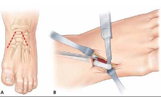
TECH FIG 1 • A. Two incisions are made. B. The medial incision is made over the course of the tibialis anterior tendon. The surgeon frees the tendon from its broad insertion as far distally as possible and proceeds proximally as far as the ankle retinaculum.

TECH FIG 2 • The freed anterior tibialis tendon is brought into the lateral wound.
Attaching the Transferred Tendon
A drill bit is selected to be slightly larger than the diameter of the sutured anterior tibialis tendon end.
Once the bit is selected, make a hole directly in the center of the lateral cuneiform, drilling just through the plantar aspect of the bone (dorsal to plantar while aiming for the arch of the foot).
The suture ends of the tendon are threaded onto Keith needles (TECH FIG 3A).
While the foot is maximally dorsiflexed and everted, the suture needles are passed through the lateral cuneiform drill hole and out through the plantar aspect of the foot, guiding the tendon through the drill hole.
The tendon is confirmed to easily and reproducibly slide into its new insertion.
This is a critical step: be certain that the tendon reliably enters the anchoring hole after the skin is closed when the foot is dorsiflexed and when the suture is tensioned.
The suture needles on the plantar aspect of the foot are passed through a nonadhesive dressing (eg, Adaptic) and a sterile felt pad.
At this time it is advisable to irrigate and close all other associated wounds, leaving the lateral recipient wound for last.
This way the surgeon can ensure that the anterior tibialis is in the intended position just before dressing and cast application.
The periosteum of the lateral cuneiform is sutured with two interrupted absorbable sutures to the transferred anterior tibialis tendon while it is pulled into the recipient site (TECH FIG 3B).
The lateral wound is irrigated and closed in layers while the foot is held in a dorsiflexed position, thus ensuring that the anterior tibialis remains in the hole and the continuity of the periosteal sutures is preserved.
Sterile dressings are applied while an assistant simultaneously maintains the foot dorsiflexed with tension on the suture.
The distal foot and ankle portion of a toe-to-groin cast is applied, while ensuring that the suture ends of the tendon are in tension.
In the past we have tied the button over the felt underneath the cast. However, a high rate of pressure sores has led us to consider alternative fixation.
After the cast is hardened, the suture is tied over a button on the exterior of the plantar aspect of the cast (TECH FIG 3C).
To prevent plantar pressure sores, make sure the plaster is sufficiently hardened.
Some surgeons will perform the exact procedure except transfer the whole tendon into the cuboid. These surgeons choose this insertion site if the foot has a concurrent fixed forefoot deformity and mild hindfoot varus that they choose not to correct.
We prefer to correct the fixed deformity and transfer the anterior tibialis into the lateral cuneiform as we fear overcorrection from the more lateral insertion into the cuboid.
Some surgeons add a third incision at the anterior distal tibia directly over the anterior tibialis tendon and just lateral to the tibial crest. The tendon can be easily palpated. The tendon sheath is incised here and the freed distal tendon end is pulled with a hemostat into this incision. From this incision, the freed distal tendon end is eventually pulled into the lateral incision for attachment.
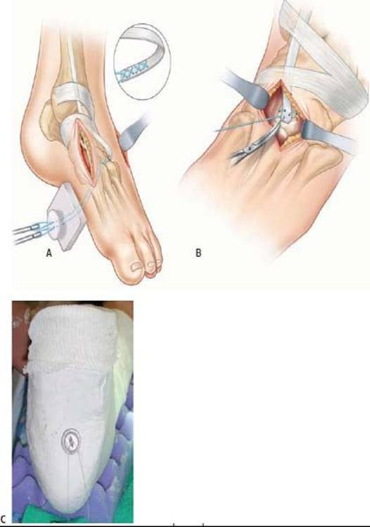
TECH FIG 3 • A. The suture ends of the tibialis anterior tendon are threaded onto Keith needles and passed into the drill hole through the plantar aspect of the foot. The tendon is guided into the drill hole. B.While the foot is maximally dorsiflexed and everted, the tendon is secured. The periosteum of the third cuneiform is sutured with interrupted nonabsorbable sutures into the transferred tibialis anterior tendon. C. The cast is molded and hardened with the foot in dorsiflexion and eversion and with the suture ends under appropriate tension. The suture is tied over a button on the exterior of the hardened cast to prevent plantar pressure sores.
SPLIT ANTERIOR TIBIALIS TENDON TRANSFER (SPLATT)
Approach
Transferring the Tendon
A 4-cm-long dorsal-medial skin incision is made over the course of the anterior tibialis tendon from the inferior margin of the ankle retinaculum (the superior limb of the inferior extensor retinaculum) to its palpable distal insertion based over the medial cuneiform.
Dissection is carried down through subcutaneous tissues and the inferior limb of the inferior extensor retinaculum to expose the tendon sheath.
The anterior tibialis tendon sheath is incised sharply and opened as far distally as possible and then proximally to just short of the ankle retinaculum.
The lateral half of the anterior tibialis tendon insertion is detached as far distally as possible to gain maximum length of tendon for the transfer.
A strong absorbable suture (eg, 1-0 Vicryl) is woven in a Bunnell-type fashion through the lateral half of the anterior tibialis tendon.
Transferring the Tendon
The suture is grasped and pulled, allowing the lateral tendon to be gently dissected proximally but not beyond the ankle retinaculum.
To avoid bowstringing of the tendon, it is important not to release the ankle retinaculum.
A dorsal-lateral longitudinal incision, 1.5 to 2 cm long, is made over the cuboid in line with the fourth metatarsal axis.
Dissection is carried down through subcutaneous tissues to the toe extensors.
To expose the cuboid, the toe extensors are retracted medially.
A cruciate periosteal incision is made directly over the cuboid, carefully avoiding the adjacent joint articulations.
An appropriate drill hole is then made in the cuboid, drilling dorsal to plantar in line with the fourth metatarsal axis and through the plantar aspect of the bone.
A blunt hemostat is then passed from the incision over the cuboid under the extensor tendons to the point where the split anterior tibialis tendon passes beneath the ankle retinaculum.
Use the hemostat to develop a tract for the transfer of the anterior tibialis tendon.
The hemostat is passed into this same tract into the medial wound to grasp the suture ends and bring the split anterior tibialis tendon into the lateral wound.
The suture ends of the tendon are threaded onto Keith needles.
Fixation of the Tendon to Bone
While the foot is maximally dorsiflexed and everted, the suture needles are passed through the cuboid drill hole and out through the plantar aspect of the foot, guiding the tendon through the drill hole.
The tendon is confirmed to easily and reproducibly slide into its new insertion.
The suture needles are passed through a nonadhesive dressing (eg, Adaptic) and a sterile felt pad.
The periosteum of the cuboid is sutured with two interrupted absorbable sutures to the transferred split anterior tibialis tendon.
The wounds are irrigated and closed in layers.
Sterile dressings are applied, while ensuring that the felt pad is flush with the plantar skin and the suture ends of the tendon are at hand.
Alternative fixation may include use of suture anchor into the cuboid or transfer of the lateral half of the tendon into the peroneus tertius tendon prior to its insertion into base of the fifth metatarsal.9
With the standard technique described above, the most medial muscle fibers proximally are the ones attached to the laterally transferred split tendon, resulting in a proximal crossing over as the split tendon is laterally transferred.
Fennell and Phillips4 suggest releasing the proximal medial insertion on the cuneiform instead of the distal lateral insertion on the first metatarsal to avoid this proximal crossing over, allowing for a more direct line of pull of the muscle on the transferred tendon.

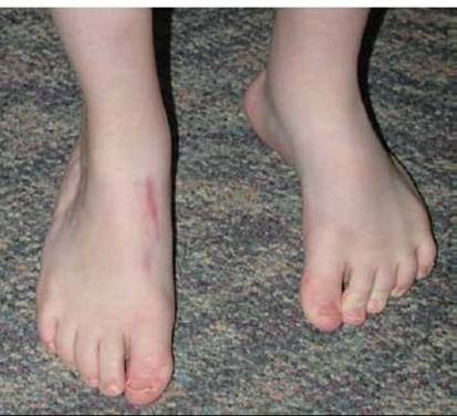
FIG 8 • Postoperative clinical photograph of patient in Figure 5. Foot alignment is restored after full-thickness anterior tibialis tendon transfer in the right foot. The left foot is shown as comparison.
POSTOPERATIVE CARE
In patients under 5 years of age and those who may be noncompliant, a toe-to-groin bent-knee cast is maintained with the patient non-weight bearing for about 6 weeks.
At 6 weeks, the button and suture are removed and the patient is allowed to begin walking.
In older children, a short-leg cast for an initial 6 weeks is maintained.
At 6 weeks, the button is removed and patient is placed in a short-leg walking cast for an additional 3 weeks to ensure healing and to avoid tendon rupture.
Clinical and radiographic assessment of outcomes is performed at the end of healing. Plain radiographs (standing AP and lateral foot radiographs) are usually sufficient. CT examination may be obtained if indicated (FIG 8).
OUTCOMES
Successful surgery will be noted by correction of the supination deformity and conversion of the anterior tibialis into the primary dorsiflexor of the foot. Clinical examination of the foot during active dorsiflexion demonstrates the new insertion site of the anterior tibialis tendon.
Twenty-seven previously treated clubfeet in 25 patients were retrospectively evaluated after tibialis anterior tendon transfer to correct residual dynamic supination deformity.1 All showed active contraction of the transferred tibialis anterior tendon. There was no case of overcorrection.
Clinical and radiographic improvement in both forefoot adduction and supination was demonstrated in 71 cases of residual dynamic congenital clubfoot deformity treated by full and split anterior tendon transfer, with an increase in the eversion strength of the tibialis anterior muscle.8
Farsetti et al2 confirmed the findings of multiple studies, demonstrating that transfer of the anterior tibial tendon to the lateral cuneiform underneath the extensor retinaculum corrects and stabilizes relapsing clubfeet by restoring normal function of foot dorsiflexion–eversion. In their two series of patients reviewed at the end of skeletal growth, none of the operated patients had further relapse.
COMPLICATIONS
Undercorrection
Cast sores
Wound infection
Loosening of the transferred tendon
Rupture of the transferred tendon
Bowstring at the anterior ankle joint resulting in weakness and a cosmetic deformity
Loss of dorsiflexion force
Overcorrection
REFERENCES
· Ezra E, Hayek S, Gilai AN, et al. Tibialis anterior tendon transfer for residual dynamic supination deformity in treated clubfeet. J Pediatr Orthop B 2000;9:207–211.
· Farsetti P, Caterini R, Mancini F, et al. Anterior tibial tendon transfer in relapsing congenital clubfoot. J Pediatr Orthop 2006;26:83–90.
· Feldbrin A, Gilai AN, Ezra E, et al. Muscle imbalances in the etiology of idiopathic clubfoot: an EMG study. J Bone Joint Surg Br 1995;77B: 596–601.
· Fennell CW, Phillips P III. Redefining the anatomy of the anterior tibialis tendon. Foot Ankle Int 1994;15:396–399.
· Garceau GJ. Anterior tendon transposition in recurrent congenital clubfoot. J Bone Joint Surg 1940;22:932–936.
· Hoffer MM, Reiswig JA, Garrett AM, et al. The split anterior tibial tendon transfer in the treatment of spastic varus hindfoot of childhood. Orthop Clin North Am 1974;5:31–38.
· Hui JP, Goh JH, Lee EH. Biomechanical study of tibialis anterior tendon transfer. Clin Orthop Relat Res 1998;349:249–255.
· Kay RM. Lower extremity surgery in children with cerebral palsy. In: Tolo V, Skaggs D, eds. Master Techniques in Orthopaedic Surgery: Pediatrics. Philadelphia: Lippincott Williams & Wilkins, 2008.
· Kuo KN. Anterior tibial tendon transfer. In: Tolo V, Skaggs D, eds. Master Techniques in Orthopaedic Surgery: Pediatrics. Philadelphia: Lippincott Williams & Wilkins, 2008.
· Kuo K, Hennigan S, Hastings M. Anterior tibial tendon transfer in residual dynamic clubfoot deformity. J Pediatr Orthop 2001;21: 35–41.
· Main BJ, Crider RJ. An analysis of residual deformity in clubfeet submitted to early operation. J Bone Joint Surg Br 1978;60:536–543.
· Ponseti I, El-Khoury G, Ippolito E, et al. A radiographic study of skeletal deformities in treated clubfeet. Clin Orthop Relat Res 1981;160: 30–31.
· Ponseti IV, Smoley EN. Congenital clubfoot: the results of treatment. J Bone Joint Surg Am 1963;45A:261–275.
· Sobel E, Giorgini RJ, Michel R, et al. The natural history and longitudinal study of the surgically corrected clubfoot. J Foot Ankle Surg 2000;39:305–320.
· Tarraf YN, Carroll NC. Analysis of the components of residual deformity in clubfeet presenting for reoperation. J Pediatr Orthop 1992; 12:207–216.