
Sami O. Khan and Larry D. Field
DEFINITION
 Valgus extension overload of the elbow is commonly seen in the overhead throwing athlete and is associated with medial compartment distraction, lateral compartment compression, and posterior compartment impingement.4,6
ANATOMY
The bony articulation of the elbow joint provides primary stability to varus and valgus force at angles of less than 20 degrees and greater than 120 degrees of flexion.
 Soft tissues are the chief stabilizers between 20 and 120 degrees, where most athletic activity occurs.
The ulnar collateral ligament (UCL) is the primary restraint to valgus stress.
It is composed of the anterior band, the posterior band, and the transverse ligament.
The anterior band is further divided into anterior and posterior bundle, which perform reciprocal functions (FIG 1).
UCL insufficiency can be subtle, with ligament-sectioning studies showing a 3-degree difference when the anterior band of the UCL is cut.2
PATHOGENESIS
Valgus extension overload typically occurs in repetitive overhead athletes, most commonly with pitchers. The repetitive pitching motion imparts a large valgus force on the elbow. Resulting microtrauma and incomplete recovery can lead to attenuation of the UCL.
Failure of the UCL leads to abnormal valgus rotation of the elbow, affecting the mechanics of the highly constrained articulation of the posterior elbow joint.
This leads to bony impingement of the posteromedial olecranon and its corresponding fossa.
Chronic bony impingement can lead to chondral lesions as well as reactive osteophyte formation of the posterior compartment (FIG 2).

FIG 1 • The ulnar collateral ligament is composed of three bands: anterior, posterior, and transverse. The anterior band is further subdivided into the anterior and posterior bundles.
NATURAL HISTORY
Thus far, no studies have been performed documenting the natural history of the disease process.
It is postulated that chronic impingement and valgus extension overload can lead to posteromedial olecranon osteophyte formation that can cause ulnar nerve irritation and loss of elbow extension as well as posterior compartment elbow arthritis.
PATIENT HISTORY AND PHYSICAL FINDINGS
The patient typically complains of loss of extension with posterior or posteromedial elbow pain.
Pitchers will report pain during the acceleration and followthrough phases of the throwing motion (FIG 3).

FIG 2 • Valgus extension overload. With ulnar collateral ligament insufficiency, medial compartment distraction and lateral compartment compression ultimately lead to posteromedial olecranon impingement.
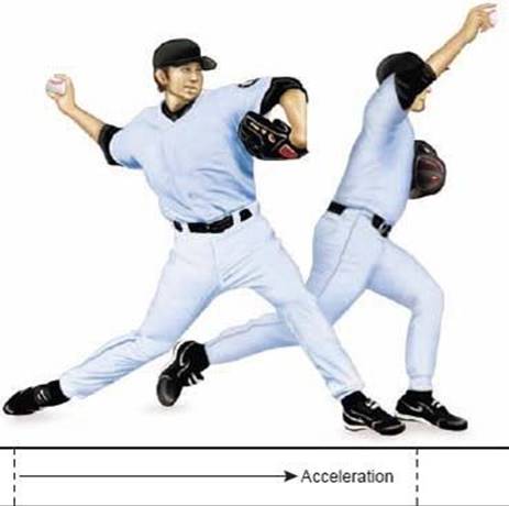
FIG 3 • The pitching athlete with valgus extension overload will often complain of pain during the acceleration and followthrough phases of throwing.
Physical examination maneuvers relevant to valgus extension overload include:
Valgus extension overload test: This maneuver acts to simulate impingement occurring with the throwing motion and to reproduce the symptoms of posterior elbow pain.
Valgus stress test: Increased medial joint space opening, loss of end point, or pain elicited is significant for UCL insufficiency.
Milking maneuver: Maneuver eliciting pain, apprehension, or instability indicates UCL insufficiency.
Posterior olecranon impingement
Range of motion of elbow: may reveal loose bodies, chondromalacia, or osteophyte formation; flexion contracture may signify either osteophyte impingement or anterior capsular contracture
Examination of the elbow should also evaluate for other causes of medial-sided elbow problems, such as isolated UCL insufficiency, ulnar neuropathy, medial epicondylitis, and flexor pronator rupture.
IMAGING AND OTHER DIAGNOSTIC STUDIES
Radiographs frequently reveal a posterior olecranon osteophyte on standard lateral or anteroposterior views (FIG 4A).
Some authors also advocate an olecranon axial view (FIG 4B).
Because radiographs cannot predict chondral lesions and soft tissue injuries and often underestimate loose body formation, magnetic resonance (MRI) and computed tomography are frequently used.
MRI can also be important in investigating a potential UCL tear (FIG 4C).
DIFFERENTIAL DIAGNOSIS
Isolated UCL insufficiency
Medial epicondylitis
Flexor–pronator rupture
Ulnar neuropathy
NONOPERATIVE MANAGEMENT
Before recommending surgery, the physician should usually treat the patient nonoperatively for 3 to 6 months.
Specific goals to be attained during this time are full, nonpainful range of motion; absence of pain and tenderness on physical examination; and satisfactory muscle strength, power, and endurance.
The patient begins treatment with an initial period of rest (1 to 3 weeks), allowing any synovitis and inflammation to resolve.
Next, he or she begins wrist and elbow flexor and extensor muscle stretching and strengthening exercises.
Finally, an interval-throwing program is instituted.
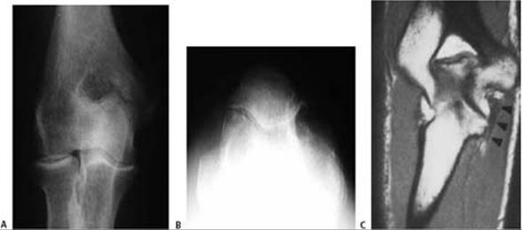
FIG 4 • AP (A) and olecranon axial (B) radiographs showing spur formation along the posteromedial olecranon. C. Coronal section of MRI demonstrating injury to the ulnar collateral ligament.
SURGICAL MANAGEMENT
Relative contraindications to elbow arthroscopy include severe bony or fibrous ankylosis and previous surgery that has distorted the native anatomy, such as a previous ulnar nerve transposition.
Preoperative Planning
A thorough history is paramount to planning for arthroscopic elbow surgery.
The surgeon should confirm that the ulnar nerve is indeed in the groove and cannot be subluxated.
The surgeon must assess for valgus instability when considering valgus extension overload.
Failure to address UCL instability in the setting of valgus extension overload may lead to treatment failure.
Positioning
We prefer the prone position for all elbow arthroscopy because it allows for the elbow to be stabilized as well as giving improved access to the posterior compartment. Posteromedial olecranon spur excision is especially facilitated by the prone position.
We routinely use a pneumatic tourniquet and a prone arm holder.
The elbow should be positioned and draped so that the arm is supported by the holder at the proximal upper arm, the elbow rests at 90 degrees of flexion, and the antecubital fossa is free from contact with the holder (FIG 5).
Approach
Elbow arthroscopy has made open resection of olecranon osteophytes primarily a point of historic interest. However, occasions still exist when an open procedure should be performed.
The determining factor in the decision-making process is whether a contraindication to elbow arthroscopy is present.
A posteromedial approach to the elbow is used when concomitant UCL reconstruction, ulnar nerve transposition, or exploration of a previously transposed ulnar nerve is to be accomplished in conjunction with removal of posteromedial osteophytes.

FIG 5 • Intraoperative photograph showing prone positioning for elbow arthroscopy.
Equipment
General anesthesia is preferred because use of regional anesthesia makes the immediate postoperative motor and sensory examination difficult to interpret.
A standard 4.0-mm arthroscope, power inflow or pump, standard power débrider, and abraders are required.
Hand-held instruments as well as only blunt trocars should be used.
The video monitor is placed on the opposite side of the patient.
Examination Under Anesthesia
Examination under anesthesia is essential to develop a feel for the character and cause of any extension block.
A bony block has a hard, sudden stop and a feeling of bony impingement. Anterior capsular contracture often has a slightly softer feel at terminal extension.
Valgus instability testing throughout the range of motion helps to assess the status of the UCL.
TECHNIQUES
DIAGNOSTIC ARTHROSCOPY
 The medial epicondyle, lateral epicondyle, and ulnar nerve are outlined.
The surgeon confirms that the ulnar nerve is indeed located within the groove and remains so with range of motion of the elbow.
The lateral “soft spot” portal location is identified and 20 cc of saline is injected into the elbow joint. Often a slight elbow extension will be seen as the capsule is insufflated.
Diagnostic arthroscopy must include a complete inspection and evaluation of the elbow.
An arthroscopic valgus instability test should be performed and medial stability should be documented (TECH FIG 1A,B).3
The posterior compartment should be thoroughly evaluated.
 Examination of the olecranon–olecranon fossa articulation may show osteophyte formation of the posteromedial olecranon (TECH FIG 1C,D).
The olecranon fossa of the humerus should be evaluated for hypertrophy, chondromalacia, and spur formation.
A systematic examination of the entire elbow joint is necessary to identify and remove any loose bodies present.
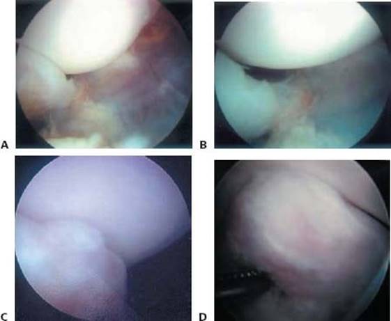
TECH FIG 1 • A. The arthroscopic valgus instability test is performed to assess for significant opening in the ulnohumeral articulation. B. A diastasis in the medial ulnohumeral articulation is noted. C,D.Arthroscopic views of a posteromedial bone spur.
POSTEROMEDIAL OLECRANON SPUR REMOVAL
First, a viewing portal is established through a posterolateral portal.
A direct posterior or triceps-splitting portal is established for access of the motorized resector or burr. The posteromedial spur is then resected (TECH FIG 2A,B).
The ulnar nerve has a close relationship to the medial olecranon. While working medially, the surgeon should minimize the use of suction, use a hooded burr, and always keep the hooded portion oriented toward the ulnar nerve (TECH FIG 2C).

TECH FIG 2 • A,B. Posterior compartment olecranon spur excision. C. Use of a hooded burr for posteromedial spur resection. The instrument is always pointed away from the ulnar nerve.
EVALUATION AND TREATMENT OF ARTICULAR CARTILAGE
After clearing out the olecranon fossa, the articular cartilage is carefully inspected.
Areas of chondromalacia can be treated by the surgeon 's choice of microfracture, abrasion chondroplasty, or benign neglect.
DEEPENING OF OLECRANON FOSSA
Occasionally, hypertrophy of the olecranon fossa necessitates a deepening or fenestration of the olecranon fossa (TECH FIG 3).
This can be performed using the same instruments and positioning.
When resection is complete, the surgeon should assess elbow extension and valgus instability with a repeat arthroscopic valgus instability test.

TECH FIG 3 • Arthroscopic views both during (A) and after (B) olecranon fossa fenestration.

POSTOPERATIVE CARE
Patients with valgus extension overload undergoing isolated spur excision are moved rapidly through the rehabilitation process.5
A sling is used sparingly for comfort for the first 7 to 10 days.
After the first week, patients are encouraged to use the elbow normally for activities of daily living, and they can begin strengthening and range-of-motion exercises.
We include flexor–pronator mass strengthening to improve dynamic valgus instability.
When patients reach a pain-free plateau, they can be advanced through an interval-throwing program.
This throwing program typically begins at 6 weeks.
A target return to competitive pitching is 3 to 4 months.
OUTCOMES
The study by Wilson et al6 included five patients treated with open biplanar spur excision. One reoperation was required in a patient with severe chondromalacia of the olecranon articular surface.
Bartz et al1 reported on a series of 24 baseball pitchers treated with a mini-open technique. Nineteen of the 24 obtained complete relief and were able to equal or exceed their preoperative throwing velocity. However, two patients did require reoperation for UCL reconstruction.
COMPLICATIONS
Thus far, no complications of this diagnosis or procedure have been documented.
Use of a motorized shaver in the medial gutter to débride olecranon osteophytes in close proximity to the ulnar nerve does warrant extreme caution.
In addition, an unrecognized UCL insufficiency has been a documented cause of reoperation.
Often this diagnosis is difficult to make in the immediate postoperative period; it may become apparent only when the athlete cannot regain his or her pitching velocity and control.
REFERENCES
1. Bartz RL, Lowe WR, Bryan WJ. Posterior elbow impingement. Oper Tech Sports Med 2001;9:245–252.
2. Callaway GH, Field LD, Deng XH, et al. Biomechanical evaluation of the medial collateral ligament of the elbow. J Bone Joint Surg Am 1997;79A:1223–1231.
3. Field LD, Altchek DW. Evaluation of the arthroscopic valgus instability test of the elbow. Am J Sports Med 1996;24:177–181.
4. Miller CD, Savoie FH. Valgus extension injuries of the elbow in the throwing athlete. J Am Acad Orthop Surg 1994;2:261–269.
5. Wilk KE, Arrigo C. Current concepts in the rehabilitation of the athletic shoulder. J Orthop Sports Phys Ther 1993;18:365–378.
6. Wilson FD, Andrews JR, Blackburn TA, et al. Valgus extension overload of the elbow. Am J Sports Med 1993;11:83–88.