
Jacob Bickels, Yehuda Kollender, and Martin M. Malawer
BACKGROUND
 Tumors of the upper extremity may cause extensive soft tissue and bone destruction and extend into the main neurovascular bundle. In those extreme situations, limb-sparing may not be feasible, and amputation is required to achieve wide margins of resection and local tumor control.
Above-elbow amputations may be required for advanced soft tissue and bone sarcomas of the forearm and around the elbow (FIG 1A). Below-elbow amputations are performed for such tumors of the forearm and the hand (FIG 1B).
Aboveand below-elbow amputations rarely are done, because (1) the upper arm, elbow, and forearms are rare location for musculoskeletal tumors and (2) when tumors do occur at one of those sites, they are noticed in relatively early stages and in most cases are resectable. Furthermore, administration of preoperative chemotherapy and availability of isolated limb perfusion have made it possible to achieve control in most patients who present with a large tumor.
Nonetheless, aboveand below-elbow amputations retain a definitive role in the management of soft tissue and bone tumors of the upper extremity.
ANATOMY
Above-elbow amputations can be metaphyseal (high), diaphyseal, or supracondylar.
High above-elbow amputations are those proximal to the deltoid tuberosity. Patients who undergo amputation proximal to the insertions of the deltoid and pectoralis major muscles have far greater difficulties adjusting to their prosthesis than do those who have undergone a more distal amputation.
Below-elbow amputations should preserve the maximal length of both radius and ulna. Although tumors of the hand are treated by a standard below-elbow amputation, performed through the distal third of the forearm, tumors of the distal forearm require a higher amputation and warrant special consideration. A minimum of 2.5 to 3 cm of bony stump, measured from the radial tuberosity, is required to preserve function. Additional length in a very short stump can be obtained by releasing the biceps tendon; adequate flexion of the stump will be provided by the brachialis muscle.
INDICATIONS
Extensive soft tissue and bone tumor extension with no option for reconstruction and reasonable function following resection (FIG 2A–E)
Local recurrence formerly was considered a primary indication for amputation, but the mere presence of a recurrent sarcoma no longer is an immediate indication for an amputation. Whether or not it is possible to resect the recurrent tumor without compromising the function of the extremity is the factor on which the decision to amputate is based (FIG 2F).
Major vascular involvement
 The neurovascular bundle within the arm is tightly integrated in a closed anatomic space. The cephalic vein usually provides sufficient collateral flow if the brachial or the axillary vein has to be sacrificed. However, although the tumor mass occasionally can be delicately dissected off the brachial artery, in most cases of vascular involvement the brachial artery is extensively encased and amputation is inevitable.
The compact nature of the vascular supply to the wrist makes involvement of the radial and ulnar arteries likely when a large tumor invades the volar aspect of the distal forearm. In such a case, the incidence of morbidity and failure associated with resection and reconstruction using a vascular graft of one of these vessels is prohibitively high.
Major nerve involvement
In general, one nerve around the arm can be sacrificed, and a two-nerve deficit is tolerated. Sacrifice of the three major nerves leaves the patient with a functionless extremity that is better off amputated.
Techniques for nerve grafting for replacement of a section of the median, radial, or ulnar nerves have not yet developed to a point at which satisfactory function can be obtained.
IMAGING AND OTHER STAGING STUDIES
Patients requiring aboveor below-elbow amputations for a soft tissue or primary bone sarcoma must undergo complete staging to allow the surgeon to determine the level of amputation and extent of soft tissue resection needed. Complete staging allows determination of full tumor extent and, as a result, the site for skin incision, shape of the flaps, and site of osteotomy.
The combined use of plain radiography, CT, and MRI is necessary to determine the proximal extent of the intraosseous and soft tissue components of the tumor. In general, the more proximal of the two levels of involvement (ie, bone or soft tissue) determines the level of amputation.
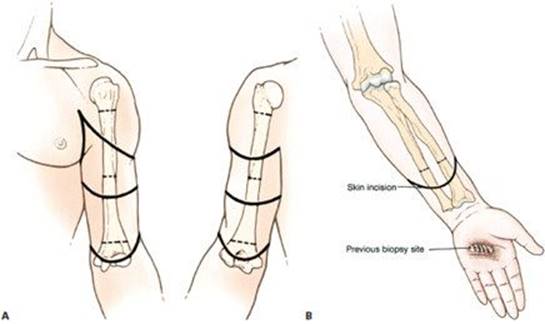
FIG 1 • A. Above-elbow amputations are done for advanced soft tissue and bone sarcomas of the forearm. Skin incisions and osteotomy sites for metaphyseal (high), diaphyseal, and supracondylar above-elbow amputations. B.Below-elbow amputations are done for advanced soft tissue and bone tumors of the forearm and hand. Skin incision and osteotomy site for below-elbow amputation. (Courtesy of Martin M. Malawer.)

FIG 2 • Clinical photograph (A) and plain radiograph (B) showing metastatic carcinoma of lung to the mid-ulna with extensive bone destruction and soft tissue extension, requiring above-elbow amputation to achieve local tumor control and palliate pain. Clinical photograph (C) and plain radiograph (D) showing high-grade sarcoma of the first metacarpus, requiring below-elbow amputation to achieve local tumor control. E. Extensive squamous cell carcinomatosis of the forearm. Above-elbow amputation was done. F. Recurrent high-grade sarcoma of the distal forearm. The recurrent disease is diffused, and wide resection would result in loss of neurovascular structures and all flexor tendons, and would end with an extensive soft tissue defect in a previously irradiated surgical field. Below-elbow amputation was done, therefore following the previously planned incision (outlined).
TECHNIQUES
ABOVEAND BELOW-ELBOW AMPUTATIONS
 The patient is supine with the ipsilateral shoulder slightly elevated. Standard anterior–posterior “fishmouth” flaps usually are used (TECH FIG 1A). However, medial–lateral flaps occasionally may be needed because of local tumor anatomy. Because of the excellent blood supply to the upper extremity, wound healing is rarely a problem, regardless of flap configuration. The skin and superficial fascia are divided perpendicular to the skin surface.
Large blood vessels are ligated in continuity and then suture ligated. The nerves are handled delicately. They are pulled approximately 2 cm from their muscular bed, doubly ligated with nonabsorbable monofilament suture, and cut with a knife.
Muscles are transected according to the flap design, and the humerus or the radius and ulna are cut at the appropriate location, as determined by the preoperative imaging studies (TECH FIG 1B,C). The radius and ulna are transected so that they are the same length.
For optimal function and mobility of the stump, it is important for muscle groups to be positioned tightly and securely over the cut ends of the bones (TECH FIG 2A,B). Myodesis is reinforced by Dacron tapes, passed through drill-holes made in the cut end of the bone. Superficial fascia and skin are closed over closed suction drains (TECH FIG 2C,D). An epineural catheter, bolused with 0.25 J. bupivicane, is inserted into the nerve sheath (TECH FIG 2E).
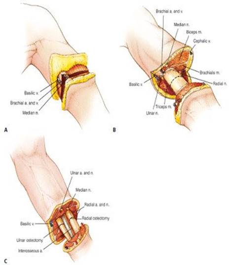
TECH FIG 1 • A. Anterior–posterior “fishmouth” flaps are used. The skin and superficial fascia are divided perpendicular to the skin surface. B,C. Osteotomies are performed at the appropriate location, as determined by the preoperative imaging studies. B. Above-elbow amputation. C. Belowelbow amputation. The radius and ulna are transected at equal lengths. (Courtesy of Martin M. Malawer.)

TECH FIG 2 • Muscle groups are positioned tightly and securely over the transected bone ends. A. Above-elbow amputation. B. Below-elbow amputation. C,D. Superficial fascia and skin are closed over closedsuction drains. C. Final closure for above-elbow amputation. D. Belowelbow amputation with closed-suction drains. E. Closed-over surgical wound. An epineural catheter, which provides continuous flow of local analgesics (specifically 0.25% bupivacaine), was inserted into the nerve sheath to control postoperative pain. (A–D: courtesy of Martin M. Malawer.)

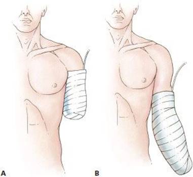
FIG 3 • A rigid dressing is used to decrease postoperative pain and edema. A. Above-elbow amputation. B. Below-elbow amputation. (Courtesy of Martin M. Malawer.)
POSTOPERATIVE CARE
A rigid dressing is applied immediately postoperatively to decrease pain and edema and facilitate maturation of the stump (FIG 3). Care must be taken to protect the skin that directly overlies the bone.
Stump edema rarely is a significant problem in the upper extremity, and prosthesis training should begin as soon as possible after surgery.
Continuous suction is required for 3 to 5 days, and perioperative intravenous antibiotics are continued until the drainage tubes are removed.
Active and passive range-of-motion exercises around the shoulder and elbow (if it exists) are practiced as tolerated.
COMPLICATIONS
Wound dehiscence
Deep infection
Loss of elbow motion (when above-elbow amputation is done)
Phantom limb pain