
Jacob Bickels and Martin M. Malawer
BACKGROUND
 Resection of the proximal tibia includes the removal of one half to two thirds of the tibia along with a portion of all muscles that insert on it, as well as the entire popliteus muscle, in combination with an extra-articular resection of the proximal tibiofibular joint. The peroneal nerve is preserved.
Of all anatomic locations in which major bone resections and prosthetic reconstructions are done, the proximal tibia is considered to be the site in which surgery is the most complicated, where rates of complications are highest, and whose functional outcome is poorest. The major reasons are the lack of muscle coverage along the anteromedial aspect of the tibia, the relatively small caliber of the blood vessels around the leg, and the need to include the insertion site of the extensor mechanism in the removed surgical specimen. In the past, these difficulties made it impossible to perform limb-sparing surgery, and above-knee amputations were the only surgical option for malignant tumors at this site.
The limb-sparing technique illustrated in this chapter offers a safe approach to the dissection of popliteal vessels and to the resection and replacement of the proximal one third to two thirds of the tibia. Preoperative evaluation of tumor extent requires a detailed understanding of the anatomy and careful evaluation by CT, MRI, bone scintigraphy, and biplane angiography.
Types of possible reconstructions include primary arthrodesis, prosthetic replacement, and allograft replacement. We prefer prosthetic replacements because of the high rates of nonunion and infections associated with allograft reconstruction and the poor function of an arthrodesed knee. The use of a gastrocnemius rotational flap is a key factor in achieving adequate soft tissue coverage of the prosthesis and in restoring function of the extensor mechanism.
ANATOMY
Knee Joint and Cruciate Ligaments
The knee joint is seldom directly invaded by tumors of the proximal tibia. When it does occur, invasion is usually the result of a pathologic fracture, contamination due to improper biopsy technique, or tumor extension along the cruciate ligaments. The presence of hemarthrosis is suggestive of intra-articular disease.
Involvement of the cruciate ligaments is often not determined until the time of surgery, although an MRI is a reliable means of determining cruciate ligament involvement preoperatively. An extra-articular resection (ie, en bloc resection of the proximal tibia, joint capsule, and femoral condyles) should be considered if nodules are identified on the cruciate ligaments.
Extensor Mechanism
The attachment site of the extensor mechanism at the tibial tuberosity is resected en bloc with the proximal tibia. Reconstruction of this mechanism is essential for a functioning extremity.
Popliteal Trifurcation
The popliteal artery divides into the anterior tibial artery, the posterior tibial artery, and the peroneal artery at the inferior border of the popliteus muscle. The popliteal trifurcation is actually composed of two bifurcations. The first is found where the anterior tibial artery arises from the popliteal artery, which then continues as the tibioperoneal trunk. The anterior tibial artery, the first branch, arises at the inferior border of the popliteus muscle.
The second bifurcation is found where the peroneal artery and the posterior tibial artery arise from the tibioperoneal trunk; thus, this bifurcation is distal to the anterior tibial takeoff. It is almost always necessary to ligate the anterior tibial artery at the time of resection, while the other vessels must be identified before ligation.
A unique and fortuitous anatomic feature is that the popliteus muscle covers the posterior surface of the tibia, which affords an excellent boundary between the posterior soft tissue extension from the tibia and the neurovascular bundle of the lower extremity. This is in contrast to what occurs at the distal femur, in which the posterior aspect is covered solely by the popliteal fat.
Tibiofibular Joint
The proximal tibiofibular joint is located close to the posterolateral aspect of the proximal tibia. Histologic studies show that tumors involving the proximal tibia have a high incidence of extension and involvement of the periscapular tissues of the tibiofibular joint.
To obtain a satisfactory surgical margin while performing a resection of the proximal tumor, it is necessary to remove this joint en bloc (ie, perform an extra-articular resection). This is a routine procedure for all high-grade sarcomas of the proximal tibia.
Subcutaneous Location of the Tibia
The entire medial aspect of the tibia lies in a subcutaneous location and remains there after resection and reconstruction approaches had been carried out. This had been a major source of primary and secondary infections, which, in turn, frequently necessitated above-knee amputation.
Today, the routine transfer of the medial gastrocnemius muscle anteriorly, to cover the prosthesis, is considered a reliable method of prosthetic coverage and one that also provides a method of extensor mechanism reconstruction. It is a simple and reliable means of decreasing the incidence of infection, flap necrosis, and secondary amputation.4
INDICATIONS
Primary bone sarcomas of the proximal tibia (FIG 1A,B)
Benign-aggressive tumors associated with extensive bone destruction (FIG 1C–G)
Metastatic tumors associated with extensive bone destruction
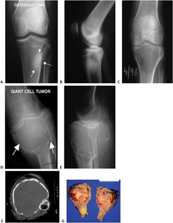
FIG 1 • AP (A) and lateral (B) plain radiographs showing osteosarcoma of the proximal tibia in a 17-year-old female patient. After treatment with neoadjuvant chemotherapy, this patient was referred for proximal tibia resection with endoprosthetic reconstruction. C. AP plain radiograph showing giant cell tumor of the proximal tibia in a 48-year-old female patient. This radiograph was done for persistent knee pain and this large lesion, which filled the metaepiphyseal regions, was missed by the physician who initially interpreted the film. AP (D) and lateral (E) plain radiographs were taken again 5 months later because of additional symptoms. This time, there was evidence of overwhelming extensive bone destruction. F. Axial computed tomography showing tumor filling the entire metaphysis of the proximal tibia with thinning and ballooning of the cortices. The patient was treated with proximal tibia endoprosthetic reconstruction. G. A sagittal cut through the surgical specimen shows the large tumoral mass and the cortical destruction.
The major contraindications to limb-sparing surgery are neurovascular involvement and compromise as well as extensive soft tissue tumor involvement that precludes adequate prosthetic coverage.
IMAGING AND OTHER STAGING STUDIES (FIG 2)
CT and MRI
CT and MRI are useful to determine the extent of cortical destruction and intramedullary and soft tissue extensions of the primary tumor. These data are essential for determining the level of tibial resection, which is 3 to 5 cm distal to the area of intramedullary tumor involvement.
MRI can also reveal skip lesions, which may affect the extent of tibial resection.
Angiography
Biplane angiography (FIG 3) is used for local arterial evaluation, especially if CT has revealed posterior soft tissue extension. The anteroposterior view is used to evaluate the popliteal bifurcation; of particular relevance is the integrity of the posterior tibial artery, which may be the sole blood supply to the leg after resection.

FIG 2 • AP (A) and lateral (B) plain radiographs, computed tomography (C), and magnetic resonance (MR) imaging (D) showing osteosarcoma of the proximal tibia. These studies show that the tumor is associated with minimal destruction of the cortices and that there is no soft tissue extension. The MR scan also accurately depicts the distal point of the intramedullary tumor extension. These findings will assist in determining the osteotomy level and the amount of surrounding soft tissue to be resected en bloc with the tumor. AP plain radiograph (E) and computed tomography (F) showing osteosarcoma of the proximal tibia with cortical destruction and soft tissue extension of stage IIB osteosarcoma.
The lateral view is essential for evaluating the interval between the tibia and the neurovascular bundle. For example, the popliteus muscle often separates a posterior tumor mass from the vessels. This is reflected as a clear interval on the lateral angiogram and serves as an indication that there is an adequate resection margin.
Ligation of the anterior tibial artery is almost always required.
The peroneal artery may be involved by tumors that have a large posterior compartment. Two of the major vessels may be ligated in a young patient without jeopardizing the possibility of a viable and functional extremity.
The posterior tibial artery is almost never involved by tumor.
SURGICAL MANAGEMENT
There are three major steps involved in successful resection and reconstruction of tumors of the proximal tibia:
Resection of the tumor
 Prosthetic reconstruction of the skeletal defect and knee joint
Reconstruction of the extensor mechanism and soft tissue coverage of the prosthesis with a gastrocnemius flap1,3

FIG 3 • An angiogram showing a lateral view of the popliteal artery. The space between the tumor in the proximal tibia and the popliteal bifurcation is best visualized by this study. The popliteal artery (P), tibioperoneal trunk (TP), and anterior tibia (AT) arteries are all identifiable. The soft tissue posterior to the tumor mass (curved arrow) must be free of cancer along the popliteal artery and tibioperoneal trunk. The popliteus muscle covers the bone in this interval and usually protects the vessels from tumor invasion. A tumor (T) blush (small arrows) is seen anteriorly. (Reprinted with permission from Clin Orthop Relat Res 1989;239:231–248.)
TECHNIQUES
INCISION
 A single anteromedial incision is made, beginning proximally at the distal third of the femur and continuing to the distal third of the tibia. The approach includes excision of biopsy sites with a margin of at least 2 cm. Medial and lateral thick flaps of skin and subcutaneous tissue including the fascia are developed to decrease the likelihood of flap ischemia (TECH FIG 1).
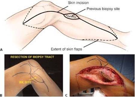
TECH FIG 1 • Illustration (A) and photograph (B) showing the anteromedial incision used for exposure of the proximal tibia and neurovascular bundle. It begins at the distal third of the femur and continues to the distal third of the tibia, and includes excision of the biopsy site, which remains attached to the underlying bone. C. Thick fasciocutaneous flaps are developed. (A: Reprinted with permission from Clin Orthop Rel Res 1989;239:231–248.)
EXPLORATION OF THE POPLITEAL FOSSA AND DETACHMENT OF THE VASCULAR BUNDLE
The popliteal trifurcation must be explored early to determine whether the tumor is operable, especially if its soft tissue component extends posteriorly. The popliteal space and trifurcation are exposed by detaching the medial gastrocnemius muscle and by splitting the soleus muscle (TECH FIG 2A–C).
The popliteal artery can be easily identified and it can be traced distally around the popliteus muscle. Care must be taken to identify and protect all major vascular branches.
Applying posterior traction proximal to the popliteal artery permits visualization of the takeoff of the anterior tibial artery and its accompanying veins. The anterior tibial vessels are individually ligated, allowing the entire neurovascular bundle to fall away from the posterior aspect of the tibia or tumor (TECH FIG 2D).
If the mass is a large one, the peroneal artery may need to be ligated as well, leaving the posterior tibial artery as the single blood supply to the leg. Further posterior mobilization of the popliteal vessels is achieved by ligation of the inferior geniculate vessels.
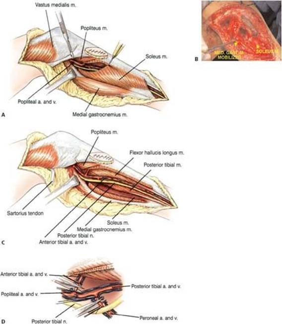
TECH FIG 2 • Illustration (A) and operative photograph (B) showing exploration of the popliteal artery trifurcation, which is required to determine the feasibility of resectability. The medial flap is continued posteriorly, and the medial hamstrings are released 2 to 3 cm proximal to their insertion to expose the popliteal fossa. The popliteal vessels are identified, and the trifurcation is initially explored through the medial approach. The medial gastrocnemius is partially mobilized, and the soleus muscle is split to expose the neurovascular structures. Care is taken to preserve the medial sural artery, which is the main pedicle to the medial gastrocnemius muscle. If the interval between the posterior aspect of the tibia and the tibioperoneal trunk (separated by the popliteus muscle) is free of tumor, resection can proceed. C.Dissection and exposure of the neurovascular bundle is often difficult because the tumor has distorted the normal anatomy. It requires splitting of the soleus muscle for most of its length. Care should be taken to identify and protect all major vascular branches before any ligation. The anterior tibial artery, which is the first takeoff of the popliteal artery, is located at the inferior border of the popliteus muscle. As it passes directly anterior through the interosseous membrane, it tethers the entire neurovascular bundle. D. Ligation of the anterior tibial vessels allows the entire neurovascular bundle to fall away from the posterior aspect of the tibia. (A,C,D: Reprinted with permission from Clin Orthop Relat Res 1989;239:231–248.)
EXPOSURE OF THE KNEE JOINT AND REMOVAL OF THE PROXIMAL TIBIA
The capsule is transected circumferentially about 1 cm away from the tibia and the patellar tendon to avoid contamination (TECH FIG 3A,B). The cruciate ligaments are carefully examined: if there is any evidence of tumor invasion into the joint space, the femoral condyles are later resected en bloc with the proximal tibia.
The patellar tendon is sectioned 1 to 2 cm proximal to the tibial tubercle, and the entire capsule of the knee is detached circumferentially by electrocautery 1 to 2 cm from the tibial insertion.
The posterior capsule is carefully dissected under direct vision after the popliteal vessels have been mobilized by ligation of the inferior geniculate vessels.
The cruciate ligaments are then sectioned close to the femoral attachments. To release the specimen, the tibia is osteomized 3 to 5 cm distal to the lesion, as determined by CT and MR imaging (TECH FIG 3C–E). The intermuscular septum is released under direct vision.
An intra-articular resection of the knee joint is then completed.
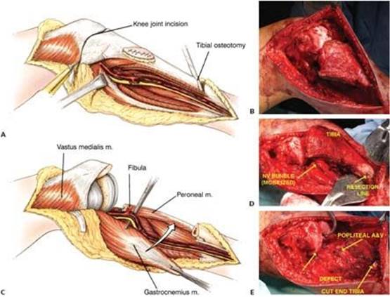
TECH FIG 3 • Illustration (A) and operative photograph (B) showing circumferential detachment of the joint capsule 1 cm away from the tibia and the patellar tendon. Illustration (C) and operative photographs (D,E) showing osteotomy of the proximal tibia 3 to 5 cm distal to the lesion. The intermuscular septum is released, after which an intra-articular resection of the knee joint is completed. (A,C: Reprinted with permission from Clin Orthop Rel Res 1989;239:231–248.)
PROSTHETIC RECONSTRUCTION
Design features of currently used proximal tibia prostheses include modular components with an anterior metal loop for the attachment of the patellar tendon, porous coating for soft tissue incorporation, side holes for securing adjacent muscles, and a rotating-hinge knee mechanism (TECH FIG 4).
Uncemented prostheses are preferentially used for reconstruction after resection of a primary bone sarcoma in an adolescent or a young adult, whereas cemented prostheses are used for reconstruction after resection of metastatic tumors.

TECH FIG 4 • Current design features of proximal tibia endoprostheses include modular components with an anterior metal loop for the attachment of the patellar tendon, porous coating for soft tissue incorporation, side holes for securing adjacent muscles, and a rotating-hinge knee mechanism (Howmedica, Mahwah, NJ).
RECONSTRUCTION OF THE EXTENSOR MECHANISM AND MEDIAL GASTROCNEMIUS FLAP
The remaining patellar tendon stump is advanced distally and secured tightly to the prosthesis with a 3-mm Dacron tape (Deknatel, Falls River, MA), which provides immediate mechanical fixation.
An autologous bone graft, taken from the cut femoral condyles, is wedged and packed tightly between the porous-coated segment of the prosthesis and the tendon, facing both surfaces (TECH FIG 5). This will create a new bone–tendon junction.
The soleus muscle is pulled anteriorly to cover the middle segment of the prosthesis and the medial gastrocnemius is used to cover its proximal segment (TECH FIG 6). The medial gastrocnemius muscle is detached at its muscle–tendon junction and interface with the lateral gastrocnemius, mobilized, and rotated anteriorly to cover the prosthesis. At its upper pole, the muscle flap is sutured to the patellar tendon to reinforce the prosthesis and bone graft reconstruction.
Suction drains are positioned along the muscle envelope, and the fasciocutaneous flaps are pulled and closed, usually leaving a gap over the medial gastrocnemius flap, which necessitates coverage with a split-thickness skin graft taken from the ipsilateral thigh (TECH FIG 7).
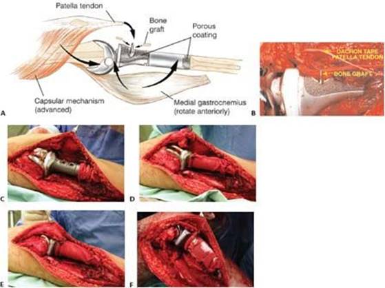
TECH FIG 5 • A. Illustration showing reconstruction of the extensor mechanism, which has three components: attachment of the patellar tendon to the prosthesis, reinforcement of the attachment site with bone graft, and overlying medial gastrocnemius flap. B. Intraoperative photograph showing the patellar tendon sutured to the prosthesis and underlying bone graft. C–F. Alternatively, a circumferential polyethylene terephthalate (Trevira) tube may be applied on the prosthesis to which the patellar tendon and surrounding muscles can be sutured.2 (A:Courtesy of Martin M. Malawer.)


TECH FIG 6 • Soft tissue coverage around the prosthesis. (A: Reprinted with permission from Clin Orthop Relat Res 1989;239:231–248; B,C: Courtesy of Martin M. Malawer.) D. The soleus muscle is pulled anteriorly to cover the middle segment of the prosthesis, and the medial gastrocnemius is transposed to cover its proximal segment. The medial sural artery to the medial gastrocnemius muscle is carefully preserved. E. The medial gastrocnemius muscle is detached at its muscle–tendon junction and interfaced with the lateral gastrocnemius, mobilized, and rotated anteriorly to cover the prosthesis. F. It is sutured to the border of the anterior muscles and the patellar tendon, forming a complete soft tissue envelope around the prosthesis.
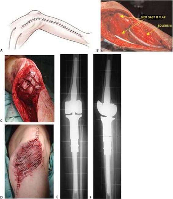
TECH FIG 7 • A–D. Suction drains are positioned along the muscle envelope, and the fasciocutaneous flaps are pulled and closed, usually leaving a gap over the medial gastrocnemius flap that necessitates coverage with a split-thickness skin graft taken from the ipsilateral thigh. E,F. AP and lateral plain radiographs showing a proximal tibia endoprosthetic reconstruction. (A: Reprinted with permission from Clin Orthop Relat Res 1989;239:231–248.)

POSTOPERATIVE CARE
The extremity is kept elevated and in full extension to avoid tension on the reconstructed patellar tendon for 5 days. Continuous suction is required for 3 to 5 days, and perioperative intravenous antibiotics are continued until the drainage tubes are removed.
If there is no evidence of significant swelling by the end of the fifth day, the patient is allowed to walk with weight bearing as tolerated for 10 to 15 minutes at a time. If the extremity remains free of swelling, a gradual increment in exercise is allowed. The knee is kept fully extended in a knee immobilizer for 6 weeks, by the end of which gradual passive and active flexion of the knee joint are allowed.
OUTCOMES
Proximal tibia resections are associated with considerably higher rates of flap ischemia, deep infection, and prosthetic loosenings than limb-sparing resections at other sites, such as the proximal humerus and the proximal and distal aspects of the femur.
The lower survival figures for prosthetic replacements at the proximal tibia (about 80% at 10 years compared to 95% for the others) are most likely attributable to the complexity of the surgical procedure and the soft tissue reconstruction, breakage of the polyethylene component, and mechanical failure. Impairment of the extensor mechanism is still the most prominent functional compromise after this resection.
The incidence of infection has been dramatically decreased by the use of a gastrocnemius muscle flap. Strict adherence to postoperative management guidelines has also decreased the incidence of limb edema, wound problems, and magnitude of extensor mechanism dysfunction.
COMPLICATIONS
Limb edema
Flap ischemia to full-thickness necrosis
Deep periprosthetic infection
Dysfunctional extensor mechanism and extension lag
Prosthetic loosening
REFERENCES
1. Bickels J, Wittig JC, Kollender Y, et al. Reconstruction of the extensor mechanism after proximal tibia endoprosthetic replacement. J Arthroplasty 2001;16:856–862.
2. Gosheger G, Hillmann A, Lindner N, et al. Soft-tissue reconstruction of megaprostheses using a Trevira tube. Clin Orthop Relat Res 2001; 393:264–271.
3. Malawer MM, McHale KA. Limb-sparing surgery for high-grade malignant tumors of the proximal tibia: surgical technique and a method of extensor mechanism reconstruction. Clin Orthop Relat Res 1989; 231:231–248.
4. Malawer MM, Price WM. Gastrocnemius transposition flap in conjunction with limb-sparing surgery for primary bone sarcomas around the knee. Plast Reconstr Surg 1984;73:741–750.