
Mohamed Khalid, Nilesh M. Chaudhari, and Thomas R. Hunt III
DEFINITION
 Malunion results when a fracture fragment heals in incorrect anatomic alignment.
ANATOMY
Metacarpals and phalanges are tubular structures with a smooth dorsal surface covered by the extensor tendon and its expansions.
Metacarpals are triangular in cross section. The medial and lateral surfaces meet at the volar ridge, providing attachment to the interossei. These attachments together with the intermetacarpal ligaments proximally and distally help splint fractured bones, making functionally significant malunions of the ring and small metacarpals less common.
Phalanges are bean-shaped in cross section. The volar aspects of the proximal and middle phalanges are in intimate relation to the flexor digitorum profundus (FDP) and superficialis (FDS) tendons, particularly in the region of the annular pulleys (FIG 1).
 As a result, the tendons are vulnerable to damage from drills and screws used in a dorsovolar direction. This problem is especially significant in the region of the annular pulleys, where the tendons are strapped against the volar cortex, rendering them vulnerable to damage.
PATHOGENESIS
Malunions most often occur secondary to lack of treatment or inadequate nonoperative care.
Malunion following internal fixation is uncommon, but when present usually results from inadequate stability or poor patient compliance.
Extra-articular malunions (EAM) often are multiplanar, but usually there is one major component to the deformity that causes the functional deficit.5
The more proximal the malunion, the greater the deformity.
Just 1 degree of rotation at the fracture site may translate to 5 degrees at the finger tip.4
5 degrees of fracture malrotation can cause 1.5 cm of digital overlap when the fingers are flexed.2
Soft tissue pathology such as neurovascular deficits, trophic changes, joint contractures, and tendon adhesions can coexist.
Results of corrective osteotomy are significantly poorer in the presence of such complicating factors.10
NATURAL HISTORY
Significant EAM can cause crossing or scissoring of fingers, pain due to distortion of joints, disturbance of muscle/tendon balance, and reduction of grip strength.10
EAMs associated with shortening can lead to an extension lag proportional to the degree of shortening. The effect is more pronounced in proximal phalanges compared to metacarpals.10
Intra-articular malunion (IAM) with a significant step (0.5 mm) or gap (1 mm) may cause joint surface incongruity, synovitis, capsular loosening or stiffness, and, ultimately, painful posttraumatic arthrosis.10,3
PATIENT HISTORY AND PHYSICAL FINDINGS
The value of a good history and physical examination cannot be overemphasized. The decision as to whether surgical treatment is to be offered depends almost entirely on a history suggestive of a significant functional impairment or pain.
Injury specifics
The original injury and method(s) of treatment
Location
 Phalanx versus metacarpal
Extra-articular versus intra-articular versus combined deformities
History of complicating factors, eg, infection and chronic mediated pain syndrome
Duration of malunion, particularly relevant in deciding surgical strategy (reducing the fracture vs osteotomy)
Associated injuries such as soft tissue defects and neurovascular injuries
Specific patient characteristics
Skeletal maturity
Hand dominance
Degree of deformity, swelling, stiffness, weakness of grip, and pain
Occupation and avocational pursuits as well as patient expectations and goals
Ability to cooperate with postoperative therapy regimen

FIG 1 • Structures on the volar aspect of the metacarpals and phalanges. The flexor digitorum profundus (FDP) and flexor digitorum superficialis (FDS) tendons are intimately associated with the volar aspect of the phalanges and, to a lesser extent, the metacarpals. This dissected specimen also depicts the vinculae (V) and the A-1 and A-2 annular pulleys. (From http://www.turntillburn.ch.)
IMAGING AND OTHER DIAGNOSTIC STUDIES
Good-quality radiographs taken in three precise planes (anteroposterior, lateral, and oblique) are sufficient for simple EAMs.
Radiographs of the opposite hand are helpful in preoperative planning for complex EAMs.
IAMs and combined malunions may require CT scans with three-dimensional reconstruction.
DIFFERENTIAL DIAGNOSIS
Fibrous nonunion
Nonunion with soft tissue contracture
Sequelae of epiphyseal injury or growth arrest
Erosive arthritis
NONOPERATIVE MANAGEMENT
Hand therapy is directed toward maximizing the range of motion (ROM) of the digits, promoting optimal tendon excursion, and improving the grip strength.
In less dramatic deformities, physical therapy is the first line treatment. Many patients will gain enough functional improvement that they decide to “live with” the deformity.
Initiation of therapy allows the opportunity to assess the patient's personality with respect to compliance and realistic expectations.
SURGICAL MANAGEMENT
Timing of Correction
Treatment of nascent malunions results in improved outcomes.
IAMs must be corrected as soon as possible if there is a significant articular step and no overwhelming technical difficulties are anticipated.10
In the case of an EAM, after 6 to 8 weeks from the injury, a “wait and watch” policy before osteotomy is advisable to see whether the malunion causes significant functional or cosmetic problems.
Location of Correction
At or near the apex of the deformity for angular and complex EAMs
In the proximal metaphysis of the malunited bone for rotational EAMs. With improved osteotomy techniques and fixation implants, a proximal metacarpal osteotomy is no longer recommended for treatment of a P-1 rotational malunion.10
Type of Osteotomy
For angular EAMs, a closing wedge osteotomy is preferable, especially in the setting of intrinsic tightness. This approach is most commonly used for dorsal apex metacarpal malunions. An opening wedge osteotomy is best in the setting of an extension lag and pseudoclaw deformity, which are more commonly seen in apex volar phalangeal malunions. An incomplete osteotomy may be used for either of these cases.
For rotational and combined rotational/angular EAM correction, a complete osteotomy is required.4 Metacarpal neck EAM from a previous Boxer's fracture without significant shortening may be corrected with a pivot osteotomy.9
Condylar advancement osteotomy8 is suitable for IAM correction in many cases.
Severity of Deformity
Malunion does not always mandate a corrective osteotomy. Patients possess a significant capacity to adapt to minor deformities. For instance, slight overlap of adjacent digits due to rotational malunion may be unsettling and unsightly, but it is consistent with good hand function.5 Similarly, a proximal diaphyseal malunion of the small-finger metacarpal can contribute to tendon imbalance and flexion contracture of the proximal interphalangeal joint, but the hand may function effectively.7
Multifragment IAMs and those with established posttraumatic arthrosis are best treated by arthrodesis or arthroplasty rather than repositioning osteotomy.
Preoperative Planning
In addition to precise evaluation of the bony deformity, careful assessment of the soft tissue envelope, gliding capacity of the flexor and extensor tendons, joint mobility, and neurovascular status is critical.
Plan for adjunct procedures (eg, tenolysis, capsulotomy) that may be required.
Determine the optimal location for placement of internal fixation.
Decide on opening or closing wedge osteotomy. In the presence of an extension lag, an opening wedge is preferred, whereas, in the presence of intrinsic tightness, a closing wedge is preferred.
Provide for soft tissue coverage as needed.
Preoperative templates are created for bony correction.
The proximal and distal fragments are each outlined then superimposed over an outline of the contralateral uninjured bone.
The type and location of the osteotomy, the size of the bone graft needed (in the case of an opening wedge osteotomy), as well as the method of fixation are determined.
In the rare cases requiring large corticocancellous interposition grafts, iliac crest bone graft harvest is planned.
Positioning
The patient is positioned supine with the shoulder abducted to 90 degrees, elbow extended, and the extremity on an arm table.
Place a proximal arm, non-sterile tourniquet.
If required, prep for ipsilateral iliac crest graft harvest.
Perform an examination under anesthesia to determine joint ROM and stability.
Approach
A dorsal approach through a dorsal skin incision in the intermetacarpal space is used for the second through fourth metacarpals (FIG 2A).
A midaxial approach through a midaxial skin incision at the junction of the wrinkled dorsal and smooth volar skin is used for the fifth metacarpal and the proximal and middle phalanges (FIG 2B).
Coronal plane correction is best accomplished with a lateral buttress plate placed over the bone graft (FIG 2C,D).
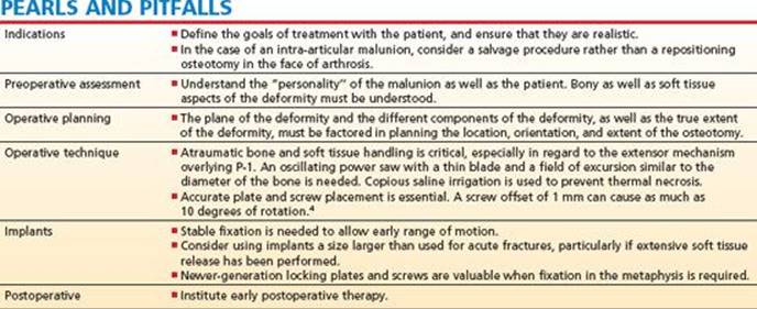
Dorsal plates should be avoided in the phalanges due to extensor tendon adhesions and resulting loss of motion.
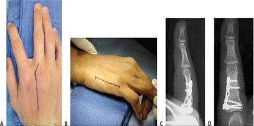
FIG 2 • A. Skin incision used for a dorsal approach to a third metacarpal malunion. The longitudinal limb of the skin incision runs between the metacarpals, and depending on whether the malunion is proximal or distal, the appropriate end is curved. B. Skin incision at the junction of the glabrous skin for an osteotomy of the fifth metacarpal. A similar mid-axial incision is employed for phalangeal malunion correction. C,D. Coronal plane correction of a proximal phalangeal malunion. The plate has been placed laterally to avoid interfering with the extensor mechanism, as well to avoid damage to the flexor tendons while drilling and inserting screws.
TECHNIQUES
INCOMPLETE OSTEOTOMY FOR ANGULAR CORRECTION
Metacarpal Closing Wedge Osteotomy
 Make a dorsal incision in the interval between either the index–long or ring–small metacarpals. depending on the bone to be treated (see Fig 2A).
 An incision at the junction of the glabrous skin often is appropriate for small metacarpal malunion correction (see Fig 2B).
Retract the extensor tendon to expose the metacarpal (TECH FIG 1A).
Make a dorsolateral incision through the metacarpal's periosteum, and carefully free this layer from the dorsum of the metacarpal with a no. 15 blade (TECH FIG 1B).
At completion of the operation, this periosteal and muscle layer will be closed, serving to protect the extensor tendons from the underlying internal fixation.
Subperiosteally expose the circumference of the bone at the planned osteotomy site.
Pass two small Hohmann retractors, one radially and one ulnarly, to protect the tendons and neurovascular structures.
Take care not to put undue tension on these structures.
Precisely identify the apex of the deformity by determining the intersection between the true anatomic axis of both the proximal and distal fragments.
Place a 0.35-mm K-wire parallel to the proximal fragment and under radiographic guidance mark the anatomic axis using diathermy or a marking pen.
Mark the distal fragment in a similar manner.
Design the osteotomy around the intersection of these two marks (TECH FIG 1C).
Plan the cuts perpendicular to the long axis of each fragment.
The size of the bone wedge to be removed is determined based on preoperative templates and intraoperative measurements.
Center and apply a sixor seven-hole 2.0to 2.7-mm compression plate to the dorsum of one fragment using two screws.
Moderately tighten the screws.
Plan for six cortices of fixation proximal and distal to the osteotomy if possible.
Juxta-articular osteotomies are best stabilized using condylar plates, T-plates, or Y-plates. Locking plates also may be of value in these cases.
Remove one screw and rotate the plate away from the osteotomy site.
Create an incomplete osteotomy, starting on the dorsal convex surface and using a water-cooled sagittal saw or sharp osteotome.
Complete the distal bone cut before making the proximal bone cut.
An elastic pillar of bone is left intact volarly on the concave side to act as a hinge.
In some cases, complete correction and osteotomy reduction can be obtained only if the volar cortex is cut and only the volar periosteum is left intact as the hinge.
Correction is adequate when the true anatomic axes of the proximal and distal fragments are parallel (TECH FIG 1D).
The dorsal plate often will serve as a guide to reduction when it sits flat on the dorsum of both fragments.

TECH FIG 1 • A. Extensor tendons have been retracted to expose the dorsal surface of the metacarpal sagittal plane malunion. B. The deep subtendinous layer of the metacarpal is demonstrated. Note that the periosteum is still intact. This layer is repaired covering the implant to prevent tendon adhesions. C,D. Method of using Kwires to determine the apex of the deformity. After removing a wedge, the size of which is determined by preoperative templating (C), deformity correction is confirmed when the K-wire markings are observed to be parallel (D). E. Dorsally applied T-plate with three screws distal and three screws proximal to the osteotomy.
Re-apply the plate, tightening the two screws. Reduce the osteotomy, and secure the other fragment by applying the other side of the plate in compression (TECH FIG 1E).
Insert the remaining screws and assess reduction clinically and radiographically.
Close the periosteal and muscle layer between the plate and the extensor tendons with absorbable suture, and close the skin in the usual manner.
Place a forearm-based splint with the wrist mildly extended and the metacarpophalangeal (MP) joints immobilized in 60 to 70 degrees of flexion. The proximal interphalangeal (PIP) joints are left free.
Phalangeal Opening Wedge Osteotomy
Make a mid-axial skin incision (TECH FIG 2A).
Protect against injury to the dorsal sensory nerve branch (TECH FIG 2B).
Incise the lateral band as required (TECH FIG 2C), and expose the circumference of the bone subperiosteally at the site of the planned osteotomy.
Use a “no touch” technique with the extensors and insert small Hohmann retractors to visualize the bone and the deformity.
Apply K-wires to precisely locate the site of the deformity and serve as a guide for correction in the manner detailed earlier (TECH FIG 2D,E).
Make an incomplete osteotomy on the concave side at the apex of the deformity perpendicular to the distal fragment.
Contouring of the bone graft is simplified if the osteotomy is made perpendicular to the distal fragment. This leaves only the proximal portion of the graft irregular.
Provisionally stabilize the fragments with a longitudinal K-wire and assess clinically and radiographically.


TECH FIG 2 • A. Lateral incision for proximal phalangeal osteotomy. B. Dorsal cutaneous nerve. C. Lateral approach to the proximal phalanx. The sagittal band has been cut and elevated to expose the proximal phalanx. D.Method of determining the apex of the deformity for an opening wedge osteotomy. E. With the deformity adequately corrected, the wire markings are parallel or overlapping. F. The corticocancellous graft has been inserted into the defect correcting the deformity.
Harvest either a corticocancellous wedge of bone or cancellous bone from the dorsal distal radius just proximal and ulnar to Lister's tubercle.
The size of the graft is determined by preoperative templating and intraoperative measurement.
Contour the graft using a water-cooled sagittal saw.
Insert the graft to correct the deformity and apply a lateral six-or seven-hole 1.5to 2.0-mm compression plate (TECH FIG 2F).
Plan for six cortices of fixation proximal and distal to the osteotomy, if possible.
Juxta-articular osteotomies are best stabilized using condylar plates, T-plates, or Y-plates. Locking plates also may be of value.
If possible, close the thin periosteal layer between the plate and the extensor tendons with absorbable suture, and close the skin in the usual manner.
Do not repair the lateral band. Check the correction clinically and compare with the preoperative pictures.
Place a forearm-based splint with the wrist mildly extended and the MP joints immobilized in 60 to 70 degrees of flexion. The IP joints are immobilized in full extension.
COMPLETE OSTEOTOMY FOR ROTATIONAL AND COMBINED ROTATIONAL/ANGULAR MALUNIONS
Perform a dorsal approach for malunions of the second through fourth metacarpals and a lateral approach for the fifth metacarpal and phalangeal malunions, as detailed earlier.
Identify and mark the true anatomic axis of the proximal and distal fragments using 0.35-mm K-wires under radiographic guidance in the manner already reviewed. Define the apex of the angular deformity (see Tech Figs 1C and 2D,E).
Insert one K-wire proximal and one distal to the malunion, perpendicular to the long axis and in a true dorsalvolar direction. This defines the rotational deformity.
In the manner detailed previously, perform the osteotomy (opening vs closing) needed to correct the angular portion of the malunion using a water-cooled sagittal saw or a sharp osteotome.
Insert a longitudinal K-wire to temporarily stabilize the fragments.
Early correction of the angular malunion aids in plate contouring and placement.
Select a suitable plate (1.5 to 2 mm for P-1 and 2.0 to 2.7 mm for metacarpals), contour it to the lateral bony surface of the proximal fragment, align it to the anatomic axis, and insert screws through the plate fixing it to that fragment.
Harvest, contour, and insert bone graft if required.
Remove the longitudinal K-wire and correct the rotational portion of the malunion by bringing the dorsalvolar K-wires into a parallel position while still maintaining angular correction (TECH FIG 3A).
Secure the distal fragment to the plate in a compression mode.
Fine-tune the rotational alignment while maintaining the angular correction by using a gliding hole rotation plate (TECH FIG 3B,C).
Check the correction and range of motion clinically (TECH FIG 3D).
Close the wound and splint as previously discussed.

TECH FIG 3 • A. K-wires previously were inserted in the dorsovolar plane, perpendicular to the dorsal surface of the proximal and distal fragments. The position of these K-wires defines the degree of rotational malunion. After correction of the sagittal plane deformity, the K-wires are manipulated into a parallel position to achieve rotational correction. B,C. Use of the rotation plates (in this case, VariAx Hand Locking Plate Module [Stryker]). The screw in the perpendicular gliding hole is positioned (but not tightened) ulnar or radial, depending on the direction of the rotational correction desired. The osteotomy is compressed, and the screws in the parallel oblong holes are tightened first. The rotational correction is then obtained, and the gliding hole screw is tightened to obtain controlled correction. D.View after correction of a combined rotational and angular malunion of the fifth metacarpal. (B,C: Courtesy of Stryker Osteosynthesis.)
CONDYLAR ADVANCEMENT OSTEOTOMY
Condylar advancement osteotomy avoids the problem of handling a small condylar malunion (TECH FIG 4A), which is difficult to fix securely and is susceptible to osteonecrosis.9
Make a sweeping dorsal, curved skin incision over the involved MP or PIP joint.
MP: Incise the sagittal band and then the capsule.
PIP: Enter the interval between the lateral band and the central slip and incise the capsule.
Protect the origin of the collateral ligament and its accompanying vascularity.
Carefully dissect the extensor tendon gently off the bone over the region of the proposed osteotomy.
Evaluate the condition of the joint. If significant arthrosis is present, consider a salvage procedure rather than a repositioning osteotomy.
Resect a wedge of bone between the condyles with a water-cooled sagittal saw (TECH FIG 4B).
Make a counter-cut in the diaphysis and advance the mal-united condyle distally to restore articular congruity (TECH FIG 4C).
Stabilize the mobilized fragment using interfragmentary screws (TECH FIG 4D).
Insert the first screw parallel with the joint to ensure precise joint reduction.

TECH FIG 4 • Condylar advancement osteotomy for unicondylar malunion.
POSTOPERATIVE CARE
If adequate stability is obtained at the time of surgery, remove the postoperative splint 3 to 5 days after surgery and initiate protected motion.
Initiate an early active and active assisted ROM program.
When not performing ROM exercises, rest the hand in a volar splint in a functional position (MP joints flexed to 60–70 degrees and IP joints fully extended), apply a compression bandage, and elevate.
Progress to passive ROM exercises, and use reverse blocking exercises to strengthen and rebalance the extensors.
If needed, and if healing is progressing appropriately, use static or dynamic splints to address pending joint contractures.
Encourage functional use of the hand long before the radiographs show complete bony consolidation.
OUTCOMES
Encouraging results have been reported. In the largest reported series of 59 osteotomies Buchler et al4 reported the following:
A 100% union rate
Satisfactory correction of the deformity in 76% of cases
A net gain in active ROM in 89% of the patients
Excellent and good functional results in 96% of patients requiring bony corrections only and 64% for those requiring bony and soft tissue correction
COMPLICATIONS
Incomplete or inadequate correction (up to 24% of patients)
Iatrogenic damage to soft tissues (up to 4% of patients)
Residual stiffness
REFERENCES
1. Büchler U, Gupta A, Ruf S. Corrective osteotomy for posttraumatic malunion of the phalanges in the hand. J Hand Surg Br 1996;21:33–42.
2. Freeland AE, Jabaley ME, Hughes JL. Fracture repair: metacarpals and carpals. In Freeland A, Jabaley M, Hughes J, eds. Stable Fixation of the Hand and Wrist. New York: Springer-Verlage, 1986:35–71.
3. Light TR. Salvage of intra-articular malunions of the hand and wrist. The role of realignment osteotomy. Clin Orthop Relat Res 1987;214: 130–135.
4. Opgrande JD, Westphal SA. Fractures of the hand. Orthop Clin North Am 1983;14:779–792.
5. Ring D. Malunion and nonunion of the metacarpals and phalanges. J Bone Joint Surg Am 2005;87A:1380–1388.
6. Rosenwasser MP, Quitkin HM. Malunion and other posttraumatic complications in the hand. In: Berger R, Weiss A, eds. Hand Surgery. Philadelphia: Lippincott Williams & Wilkins, 2003:207–230.
7. Strauch RJ, Rosenwasser MP, Lunt JG. Metacarpal shaft fractures: The effect of shortening on the extensor tendon mechanism. J Hand Surg Am 1998;23:519–523.
8. Teoh LC, Yong FC, Chong KC. Condylar advancement osteotomy for correcting condylar malunion of the finger. J Hand Surg Br 2002; 27:31–35.
9. Thurston AJ. Pivot osteotomy for the correction of malunion of metacarpal neck fractures. J Hand Surg Br 1992;17:580–582.
10. Vahey JW, Wegner DA, Hastings H. Effect of proximal phalangeal fracture deformity on extensor tendon function. J Hand Surg Am 1998;23:673–681.