
Richard Y. Kim and Melvin P. Rosenwasser
DEFINITION
 Scapholunate instability occurs as a result of injury to the scapholunate interosseous ligament (SLIL).
Instability can be categorized based on physical and radiographic findings.
 Static instability: abnormal alignment of the scaphoid and lunate evident on routine radiographs
Dynamic instability: abnormal alignment of the scaphoid and lunate present only on stress radiographs
Predynamic instability: no radiographic abnormalities present, but history and physical findings consistent with a SLIL injury
Reduction and association of the scaphoid and the lunate (the RASL procedure) is used to correct scapholunate instability.
ANATOMY
The SLIL can be divided into three components: dorsal, palmar, and proximal. Of these, the dorsal component is the thickest and contributes the most to scapholunate stability.1
Normally, the interval between the scaphoid and the lunate measures less than 3 mm, but this can vary between patients. The interval should be compared to the contralateral wrist (FIG 1A).
The normal angle between the scaphoid and the lunate measures 46 degrees with the wrist in neutral position (FIG 1B).5
With wrist flexion and extension, there is 25 degrees of obligatory rotation motion between the scaphoid and the lunate. With radial and ulnar deviation, there is 10 degrees of normal motion.7
PATHOGENESIS
SLIL injury typically occurs after a fall onto an extended wrist. The combination of axial load, wrist extension, intercarpal supination, and ulnar deviation leads to supraphysiologic loads across the SLIL.
Injury can also occur in association with other injuries, such as the constellation seen in perilunate dislocations and distal radius fractures.
NATURAL HISTORY
The motion of the scaphoid and that of the lunate are linked, such that both bones flex with wrist flexion and radial deviation and extend with wrist extension and ulnar deviation.2 After SLIL injury, the synchronous movement between the scaphoid and lunate is lost and the scaphoid flexes while the lunate extends.
Increased scaphoid flexion leads to point stress at the radiostylo–scaphoid juncture. This is the path to scapholunate advanced collapse and osteoarthritis.
Dorsal intercalated segment instability (DISI) occurs because of unlinked lunate extension, which creates a scapholunate diastasis and allows for descent and altered kinematics (FIG 2). This results in pain, weakness, and progressive osteoarthritis.
Over time, a progressive pattern of degenerative arthritis termed scapholunate advanced collapse (SLAC) occurs.10
Arthritic changes first arise between the radial styloid and the scaphoid (stage 1), followed by progression of arthritis into the proximal scaphoid fossa (stage 2). Next, the midcarpal joint becomes involved (stage 3), in particular the capitolunate joint, and eventually pancarpal arthritis is the final result (stage 4).

FIG 1 • A. The scapholunate interval normally measures less than 3 mm. B. The scapholunate angle normally measures 46 degrees with the wrist in neutral position.

FIG 2 • Dorsal intercalated segment instability (DISI) occurs as a result of lunate extension. Consequently, the capitate and distal carpal row migrate proximally and translate dorsally.
PATIENT HISTORY AND PHYSICAL FINDINGS
History should include details of prior wrist trauma, especially in regard to mechanism and timing.
Acute injuries are those that have occurred within 3 weeks, subacute between 3 weeks and 3 months, and chronic greater than 3 months before presentation.3 Dates are unreliable, but radiographic changes suggest many are acute-on-chronic injuries.
After acute trauma, there is usually a repairable scapholunate ligament, whereas in the setting of subacute or chronic injury, the ligament is resorbed or mechanically unsound. The presence of adequate ligament tissue for repair outweighs the reported time since injury.
Instability may be the result of cumulative trauma, and the patient may present with a history of multiple wrist sprains that ultimately produce chronic wrist pain.
Physical examination includes the following:
Direct palpation of the wrist: Tenderness in this region corresponds to scapholunate ligament injury. May also see fullness or thickness, corresponding to dorsal capsule synovitis.
Range of motion: Pain with range of motion may indicate instability, synovitis, and chondral wear.
Watson scaphoid shift test: Pain over the scaphoid tubercle with radial deviation indicates SLIL injury.
Assessment of both normal and aberrant motion
Provocative maneuvers
Examination of the contralateral uninjured wrist is essential to assess radiographic findings of minimal diastasis or DISI, which may be part of a hyperlaxity syndrome.
IMAGING AND OTHER DIAGNOSTIC STUDIES
Plain radiographs and stress views are critical in diagnosis and consist of:
Neutral posteroanterior (PA), lateral, and oblique views
PA views in ulnar and radial deviation
Clenched-fist PA view in pronation
Contralateral wrist films should always be taken for comparison.
Radiographic evidence of SLIL injury includes:
Scapholunate diastasis greater than 3 mm. Comparison should be made with the contralateral side, as ligamentous laxity can produce a normal scapholunate interval of greater than 3 mm (FIG 3A).4
Scaphoid cortical ring sign, which occurs when the scaphoid is in a flexed posture and the distal tubercle aligns with the proximal scaphoid (FIG 3A).
Scapholunate angle greater than 60 degrees (FIG 3B)6
DISI deformity pattern with capitate dorsal translation and decreased carpal height measurements
The accuracy of MRI is affected by the strength of the magnet and the use of dedicated wrist coils.
Sensitivity ranges from 63% to 92%.
Specificity ranges from 86% to 100%, depending on the degree of injury and the type of MRI used.6,8
DIFFERENTIAL DIAGNOSIS
Scaphoid fracture
Capitolunate arthritis
Scaphotrapeziotrapezoidal (STT) arthritis
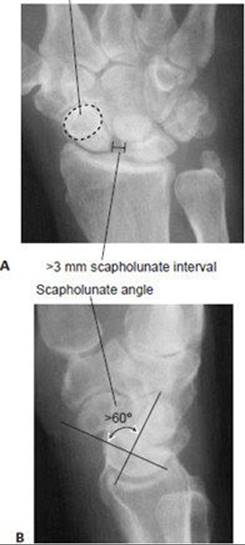
FIG 3 • A. A widened scapholunate interval (>3 mm) and a scaphoid cortical ring sign are seen on an AP view of the wrist. B. An obtuse scapholunate angle (>60 degrees) is appreciated on a lateral view of the wrist.
Midcarpal instability
Dorsal carpal ganglion
Dorsal impaction syndrome
NONOPERATIVE MANAGEMENT
Normal clinical alignment with persistent wrist pain with or without a Watson sign is managed with resting splints until symptoms resolve.
Occasionally intra-articular steroid injections are performed.
SURGICAL MANAGEMENT
Contraindications to RASL
A repairable SLIL
A primary ligament repair is preferable if a ligament of adequate tissue quality is present.
This is most likely seen in acute injuries (less than 3 weeks) but may be possible in chronic injuries. For this reason, arthroscopy is recommended before performing a RASL procedure to evaluate the quality of the SLIL.
Presence of significant capitolunate or pancarpal arthritis
If significant midcarpal, radiolunate, or radioscaphoid arthritis is present, a salvage procedure, such as a proximal row carpectomy or a limited wrist fusion, may be a better treatment option.
Focal arthritis between the scaphoid and radial styloid is not a contraindication since a radial styloidectomy is routinely performed during the RASL procedure.
Positioning
The procedure is performed with the patient supine and the arm on a standard hand table.
The operating table should be rotated 90 degrees to facilitate the use of the image intensifier during the procedure.
Approach
The RASL procedure can be performed either arthroscopically or via an open dorsal approach.
The arthroscopic RASL should be attempted only after obtaining experience with the open technique, or if already a master arthroscopist.
The open technique is performed using a dorsal intercarpal ligament–sparing approach.
TECHNIQUES
DORSAL LIGAMENT–SPARING CAPSULOTOMY
 Make a longitudinal incision on the dorsal wrist, staying just ulnar to the tubercle of Lister (TECH FIG 1A).
Bluntly dissect the soft tissue down to the level of the extensor retinaculum, taking care to preserve any dorsal veins and cutaneous nerve branches wherever possible.
Incise obliquely the extensor retinaculum parallel to the course of the extensor pollicis longus (EPL) tendon (TECH FIG 1B).
 This will open the third and fourth extensor compartments.
The EPL is retracted radially and the fourth compartment tendons are retracted ulnarly.
Make an oblique incision through the dorsal wrist capsule parallel and proximal to the dorsal intercarpal ligament (DIC) (TECH FIG 1C).
The dorsal radiocarpal ligament (DRC) should be identified and preserved.
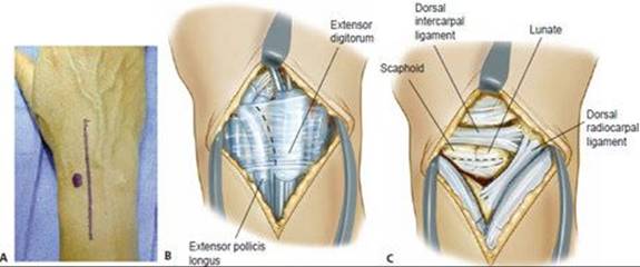
TECH FIG 1 • A. A dorsal midline incision is made just ulnar to the tubercle of Lister. B. An oblique incision is made through the extensor retinaculum parallel to the extensor pollicis longus (EPL) tendon. The EPL is retracted radially and the fourth compartment tendons are retracted ulnarly. C. An oblique incision is made through the dorsal wrist capsule parallel and proximal to the dorsal intercarpal ligament. The dorsal radiocarpal ligament should also be identified and preserved.
STYLOIDECTOMY
Once the capsulotomy is performed, identify the scapholunate interval and the SLIL and inspect the radiocarpal and intercarpal joints.
If significant arthrosis is present in areas other than the radiostyloscaphoid articulation, a salvage procedure is indicated.
Perform a second incision in the midaxial line over the first dorsal compartment (TECH FIG 2A).
The major branch of the superficial radial nerve should be seen and isolated with a vessel loupe.
Release the first compartment retinaculum and retract the tendons dorsally.
Incise the capsule longitudinally through the wrist capsule, thereby exposing the radial styloid (TECH FIG 2B).
Elevate the periosteum overlying the radial styloid and use an osteotome to perform a radial styloidectomy.
Remove enough of the radial styloid that radial deviation of the wrist does not cause impingement of the scaphoid and radius.
Too aggressive of a radial styloidectomy will compromise the volar radioscaphocapitate ligament, which originates from the base of the radial styloid.
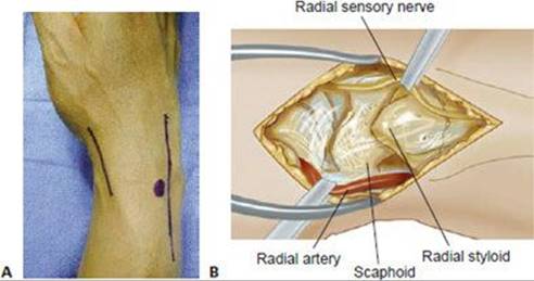
TECH FIG 2 • A. A longitudinal incision is made over the first dorsal extensor compartment. B. The first compartment is released and a longitudinal incision is made down to the radial styloid.
PREPARATION AND REDUCTION OF THE SCAPHOLUNATE JOINT
Place a 0.062-inch Kirschner wire in the lunate and another in the scaphoid to serve as joysticks (TECH FIG 3A).
To bring the lunate out of extension, place the Kirschner wire in the most proximal portion of the exposed dorsal surface, angled from proximal to distal.
Similarly, to bring the scaphoid out of flexion, place the Kirschner wire in the most distal portion of the exposed dorsal surface, angled from distal to proximal.
Keep in mind the eventual path of the Herbert screw when placing the Kirschner wires and try to avoid this area in both bones.
Remove the articular cartilage of the scapholunate joint using a side-cutting burr (TECH FIG 3B).
The joysticks can be used to separate the two bones to better visualize the articular surfaces.
Remove the cartilage until cancellous bone and punctate bleeding are visualized.
Reduce the scaphoid and lunate by flexing the lunate and extending the scaphoid (TECH FIG 3C).
A Köcher clamp is used to hold the reduction (TECH FIG 3D).
Verify the reduction with an image intensifier (TECH FIG 3E).

TECH FIG 3 • A. 0.062-inch Kirschner wires are placed in the lunate and scaphoid to serve as joysticks. B. A side-cutting burr is used to remove the cartilage within the scapholunate joint. C. The joysticks are used to extend the scaphoid and to flex the lunate. D. A Köcher clamp is used to hold the reduction. E. The reduction is verified with an image intensifier.
HERBERT SCREW PLACEMENT
The path of the Herbert screw should be through the center of rotation of both the scaphoid and the lunate.
Introduce the Herbert jig through the radial incision and place the insertion point of the jig on the scaphoid waist.
Through the dorsal incision, introduce the end of the jig and rest it on the proximal ulnar corner of the lunate.
Do not violate the lunotriquetral interosseous ligament.
The insertion angle of the screw should be roughly parallel to the radial inclination of the distal radius, 20 degrees (TECH FIG 4A).
Once the jig is in proper position and both bones are properly measured, drilled, and tapped, insert the Herbert screw.
Countersink the screw into the scaphoid so that it is not palpable.
Correct screw placement should be confirmed by fluoroscopy (TECH FIG 4B,C).
Remove the Köcher clamp and the joystick wires.
Take the wrist through a full range of motion to assess for any restrictions in motion and to confirm that the scaphoid and lunate remain reduced.

TECH FIG 4 • A. The insertion angle of the Herbert screw should be roughly parallel to the radial inclination of 20 degrees. B,C. The position of the screw is verified with an image intensifier.
WOUND CLOSURE
Release the tourniquet and achieve hemostasis using Bovie or bipolar electrocautery.
Close the dorsal and radial capsular incisions and extensor retinaculum (once the EPL and fourth compartment tendons are placed back into their respective compartments) using 3-0 absorbable monofilament suture.
Close the skin using 5-0 nylon suture and apply a sterile bulky dressing and volar thumb spica splint.

POSTOPERATIVE CARE
The wrist is kept immobilized for 4 to 6 weeks.
After 4 to 6 weeks, the thumb spica splint is removed, a removable splint is applied, and range-of-motion therapy is initiated.
Over time, therapy is advanced to strengthening exercises around 3 months postoperatively.
OUTCOMES
Rosenwasser et al3 reported on a series of 21 patients with a mean of 32 months of follow-up.
In this group, 95% of patients returned to their previous occupations. One-year postoperative scapholunate angles and intervals were corrected to within normal limits, with the scapholunate angle being corrected from 69 degrees preoperatively to 40 degrees postoperatively, and the scapholunate interval being corrected from 4.1 mm preoperatively to 1.4 mm postoperatively.
One patient was converted to a partial wrist fusion secondary to screw migration and failure of reduction. Another patient required removal of the screw 4 years postoperatively secondary to radial impingement and still demonstrated scapholunate stability after screw removal.
COMPLICATIONS
Residual instability
Screw migration
Superficial radial sensory nerve injury
REFERENCES
1. Berger RA, Imeada T, Berglund L, et al. Constraint and material properties of the subregions of the scapholunate interosseous ligament. J Hand Surg Am 1999;24A:953–962.
2. Garcia-Elias M, Geissler WB. In Green DP, ed. Green’s Operative Hand Surgery, 5th ed. Philadelphia: Elsevier/Churchill Livingstone, 2005.
3. Lipton CB, Ugwonali OF, Sarwahi V, et al. Reduction and association of the scaphoid and lunate for scapholunate ligament injuries (RASL). Atlas Hand Clin 2003;8:249–260.
4. Linscheid RL. Scapholunate ligamentous instabilities (dissociations, subdislocations, dislocations). Ann Chir Main 1984;3:323–330.
5. Linscheid RL, Dobyns JH, Beabout JW, et al. Traumatic instability of the wrist: diagnosis, classification, and pathomechanics. J Bone Joint Surg Am 1972;54A:1612–1632.
6. Rosenwasser MP, Miyasajsa KC, Strauch RJ. The RASL procedure: reduction and association of the scaphoid and lunate using the Herbert screw. Tech Hand Up Extrem Surg 1997;1:263–272.
7. Ruby LK, Cooney WP III, An KN, et al. Relative motion of selected carpal bones: a kinematic analysis of the normal wrist. J Hand Surg Am 1988;13A:1–10.
8. Schädel-Höpfner M, Iwinska-Zelder J, Braus T, et al. MRI versus arthroscopy in the diagnosis of scapholunate ligament injury. J Hand Surg Br 2001;26:17–21.
9. Schmitt R, Christopoulos G, Meier R, et al. Direct MR arthrography of the wrist in comparison with arthroscopy: a prospective study on 125 patients [in German]. Rofo 2003;175:911–919.
10. Watson HK, Weinzweig J, Zeppieri J. The natural progression of scaphoid instability. Hand Clin 1997;13:39–49.