
David J. Slutsky
DEFINITION
 Tears of the dorsal radiocarpal ligament (DRCL) are more common than previously suspected. They are best seen through a volar radial portal and are amenable to arthroscopic repair.
Tears of the DRCL have been implicated in both volar and dorsal intercalated segmental instabilities, and they also have a role in midcarpal instability.3,5
DRCL tears appear to be part of a spectrum of radialand ulnar-sided carpal instability, as evidenced by the frequent association with scapholunate and lunotriquetral ligament injuries as well as triangular fibrocartilage (TFC) tears.
Isolated DRCL tears can be solely responsible for wrist pain. The presence of an associated DRCL tear when seen in combination with a scapholunate, lunotriquetral, or TFC tear connotes a greater degree or duration of carpal instability and portends a poorer prognosis after treatment.12
Good results are obtained after arthroscopic repair of isolated DRCL tears. Results of DRCL repairs are less predictable when seen in combination with other types of carpal pathology.11
Recognition of this condition and further research into treatment methods is needed.
ANATOMY
The DRCL is an extracapsular ligament on the dorsum of the wrist. It originates on the tubercle of Lister and moves obliquely in a distal and ulnar direction to attach to the tubercle of the triquetrum. Its radial fibers attach to the lunate and lunotriquetral interosseous ligament.4
The dorsal intercarpal (DIC) ligament originates from the triquetrum and extends radially to attach onto the lunate, the dorsal groove of the scaphoid, and then the trapezium.
The lateral V configuration of the DRCL and the DIC functions as a dorsal radioscaphoid ligament.
 It can vary its length by changing the angle between the two arms while maintaining its stabilizing effect on the scapholunate joint during wrist flexion and extension.
This would require changes in length far greater than any single fixed ligament could accomplish.14
When viewed from a volar radial portal, the DRCL is seen immediately ulnar to the 3-4 portal, just underneath the lunate (FIG 1A).
The actual fibers of the DRCL may not be seen unless there is a tear present, since it is normally covered by an epiligamentous sheath (FIG 1B).
PATHOGENESIS
It is instructive to consider the wrist as having a number of primary and secondary stabilizers.
The scapholunate interosseous ligament (SLIL), the lunotriquetral interosseous ligament (LTIL), and the triangular fibrocartilaginous complex (TFCC) are the primary stabilizers.
The capsular ligaments, including the radioscaphocapitate, radiolunotriquetral, ulnolunate, ulnotriquetral, dorsal radiocarpal, and dorsal intercarpal ligaments, can be thought of as secondary stabilizers.6
A chronic tear of a primary stabilizer may culminate in the attenuation or tearing of the secondary stabilizer.
This is seen in patients with a triquetrolunate dissociation of more than 6 months’ duration, in whom arthroscopy often reveals fraying of the ulnolunate ligaments and ulnotriquetral ligaments.17
DRCL tears appear to be part of a spectrum of radialand ulnar-sided carpal instability, as evidenced by the frequent association with SLIL, LTIL, or TFC tears. They have also been associated with midcarpal instability.10
The DRCL tear may occur after or precede these injuries.
In a recent study, 35 of 64 patients who underwent arthroscopy for the diagnosis and treatment of refractory wrist pain were noted to have associated DRCL tears, for an overall incidence of 55%.9
Five patients had an isolated DRCL tear.
13 patients in this series had SLIL instability, tear, or both; 7 of 13 (54%) also had a DRCL tear. Of this subgroup four patients had Geissler stage 1 or 2 instability and three had a Geissler stage 3 or 4 tear.
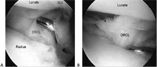
FIG 1 • A. View of an intact dorsoradiocarpal ligament (DRCL) from the VR portal. SLIL, scapholunate interosseous ligament. The hookprobe is in the 3-4 portal. B. DRCL tear (*). (From Slutsky DJ. Arthroscopic repair of dorsal radiocarpal ligament tears. Arthroscopy J Arthroscopic Relat Surg 2002;18:E49.)
Seven patients had LTIL instability, tear, or both; 2 of 7 (28%) also had a DRCL tear. Of this subgroup one patient had Geissler stage 2 instability and one had a Geissler stage 3 or 4 tear.
Two patients had a capitohamate ligament tear; one of these patients also had a DRCL tear.
Seven patients had a solitary TFCC tear; 6 of 7 (86%) were in association with a DRCL tear. One patient had a chronic ulnar styloid nonunion and a DRCL tear. There was TFCC fraying but no tear or detachment.
Two or more lesions were present in 23 patients; DRCL tears were present in 12 patients (52%). Sixty-two percent of the combined lesions that were associated with a DRCL tear also included a TFCC tear.
NATURAL HISTORY
The natural history of DRCL tears is not completely certain.9
Unrecognized DRCL tears may be a cause for treatment failures in patients with persistent dorsal wrist pain.
In nondissociative carpal instability, the pain is believed to be caused by dynamic joint incongruity.1 Chronic detachment of the ulnar sling on the triquetrum has been implicated as a cause of wrist pain in these cases.16 It is plausible that impingement of the torn edge of the DRCL against the lunate can have a similar effect.
The DRCL, DIC, and dorsal SLIL are richly innervated, and 80% of nerve endings are in the epiligamentous sheath.2
DRCL repair may allow a reinnervation of nerve endings and proprioceptive restoration (similar to anterior cruciate ligament repairs).
The frequent observation of a DRCL tear in association with the altered kinematics caused by chronic radiocarpal or ulnocarpal instability might be attributed to plastic deformation of the DRCL with cyclical wrist motion that ultimately culminates in a tear.6
When combined with other wrist pathology, the presence of a DRCL tear signifies a greater degree or longer duration of carpal instability and connotes a worse prognosis with regard to treatment.
A classification of DRCL tears was devised based on the presence or absence of associated carpal pathology (Table 1).7
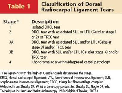
Each successive stage denotes a longer standing or more severe condition, and this has a negative impact on the prognosis.
An isolated DRCL tear does not necessarily lead to other intracarpal ligament or TFCC tears.8
PATIENT HISTORY AND PHYSICAL FINDINGS
A typical patient with an isolated DRCL tear presents with complaints of intermittent dorsal midline wrist pain that may be sporadic but last 2 or 3 days; the pain is precipitated by repetitive loading or torquing movements of the wrist.
When there is an underlying SLIL or LTIL tear or instability or TFCC tear, the pain may be more persistent and localized to the radial or ulnar side of the wrist.
There are no physical findings that are pathognomonic of a DRCL tear. When there is no other associated wrist pathology, the diagnosis can be made only at the time of arthroscopy.
Patients with isolated DRCL tears tend not to have localizing carpal tenderness and typically have a normal wrist examination, although some are mildly tender over the tubercle of Lister.11,12
Positive physical findings are usually related to any associated wrist pathology. In patients who have scapholunate instability, scaphoid tenderness and a positive scaphoid shift test are usually present.
When there is an associated TFC tear, the patient will often have tenderness over the ulnar capsule and may have crepitus and pain with ulnar loading of the pronated wrist.
If midcarpal instability is present, the patient may have a positive midcarpal shift test.
IMAGING AND OTHER DIAGNOSTIC STUDIES
Imaging studies are of mostly of value for ruling out associated carpal pathology since they are ineffective at making the diagnosis of an isolated DRCL tear.
Plain radiographs and arthrograms are normal.
The MRI is typically normal, although in one patient in the author’s series an MRI was wrongly interpreted as showing a dorsal ganglion due to a high fluid signal intensity over the dorsal capsule (FIG 2).
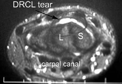
FIG 2 • MRI of an axial view of the carpus at the level of the carpal canal depicting a dorsoradiocarpal ligament tear. S, scaphoid; L, lunate.
DIFFERENTIAL DIAGNOSIS
Dynamic scapholunate instability
Scapholunate ligament tear
Dorsal wrist syndrome15
NONOPERATIVE MANAGEMENT
Patients should be treated with at least 1 month of wrist splinting, nonsteroidal anti-inflammatories, and activity modification with avoidance of repetitive gripping and lifting.
Failure to respond is an indication for a radiocarpal cortisone injection followed by 1 additional month of splinting.
Patients who continue to have wrist pain should then undergo imaging studies to rule out associated intracarpal pathology.
SURGICAL MANAGEMENT
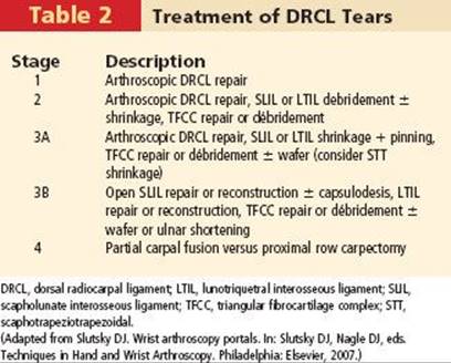
An arthroscopic repair is especially indicated for stage 1 (ie, isolated) DRCL tears, since the results are quite favorable.
Repairs may also be considered in stage 2 and 3A DRCL tears, where the associated interosseous ligament tear or TFCC tear is treated arthroscopically.
Stage 3B and stage 4 tears will likely be unresponsive to a DRCL repair since the outcomes are quite variable in the face of combined pathologies (Table 2).
Preoperative Planning
Preoperative investigations should include plain radiographs to rule out a static carpal instability pattern.
A double-row wrist arthrogram or an MR arthrogram is performed to assess the intracarpal ligaments and TFC.
Positioning
The patient is positioned supine on the operating table with the arm abducted.
Some method of overhead traction is useful. This may include traction from the overhead lights or a shoulder holder along with 5to 10-lb sand bags attached to an arm sling. A traction tower such as the Linvatec tower (Conmed, Linvatec Corp, Largo, FL) or the ARC wrist traction tower designed by Dr. William Geissler (Arc Surgical LLC, Hillsboro, OR) greatly facilitates instrumentation.
A 2.7-mm 30-degree angled arthroscope with a camera attachment is necessary.
A fiberoptic light source, video monitor, and printer are also standard equipment.
Newer digital systems provide superior video quality compared with analog cameras and allow direct writing to a CD or DVD.
A 3-mm hook-probe is needed for palpation of intracarpal structures.
A motorized shaver and suction punch forceps are useful for débridement.
Some type of diathermy unit such as the Oratec radiofrequency probe (Smith and Nephew, NY) is needed if augmentation of the repair with capsular shrinkage is desired.
A variety of curved and straight 18-gauge spinal needles are used for passage of an absorbable 2-0 suture for the outside-in repair.
A suture lasso or grasper is needed to retrieve the suture ends.
Approach
An inside-out arthroscopic repair technique of the DRCL ligament was initially performed.
An outside-in technique is technically easier and is now preferred.
The standard dorsal portals are established, including the 3-4 and 4-5 portals, a midcarpal radial portal, and a midcarpal ulnar portal.
A 6U portal is used for outflow.
A standard arthroscopic survey is performed.
TECHNIQUES
VR PORTAL 16
 A 2-cm longitudinal incision is made in the proximal wrist crease, exposing the flexor carpi radialis (FCR) tendon sheath (TECH FIG 1A).
The sheath is divided and the FCR tendon retracted ulnarly.
The radiocarpal joint space is identified with a 22-gauge needle and the joint inflated with saline (TECH FIG 1B).
A blunt trocar and a cannula are introduced through the floor of the FCR sheath, which overlies the interligamentous sulcus between the radioscaphocapitate ligament and the long radiolunate ligament.
A 2.7-mm 30-degree angled arthroscope is inserted through the cannula (TECH FIG 1C).
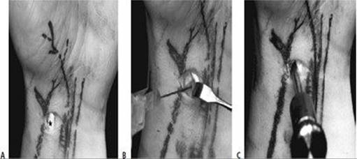
TECH FIG 1 • Establishing the volar radial portal. A. Skin incision for VR portal. FCR, flexor carpi radialis tendon. B. Saline injection of radiocarpal joint. C. Insertion of cannula through floor of the FCR sheath. (From Slutsky DJ. Volar portals in wrist arthroscopy. J Am Soc Surg Hand 2002;2:225–232.)
LIGAMENT REPAIR
The repair is performed by inserting a curved 21-gauge spinal needle through the 4-5 portal while viewing through the arthroscope, which is inserted in the VR portal (TECH FIG 2A–C).
A 2-0 absorbable suture is threaded through the spinal needle and retrieved with a grasper or suture snare inserted through the 3-4 portal (TECH FIG 2D, E).
A curved hemostat is used to pull either end of the suture underneath the extensor tendons, and the knot is tied either at the 3-4 or 4-5 portal (TECH FIG 2F).
With dorsal traction, the encircling suture pulls the torn DRCL up against the dorsal capsule, preventing it from impinging into the joint.
The repair may be augmented with thermal shrinkage if the torn edge of the DRCL is voluminous (TECH FIG 2G).
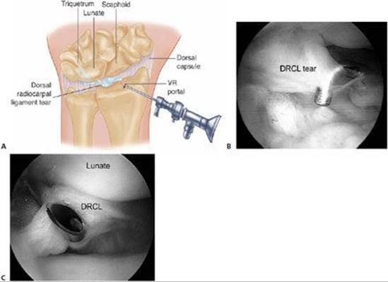
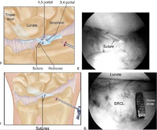
TECH FIG 2 • Outside-in dorsoradiocarpal ligament (DRCL) repair. A. Drawing of DRCL tear. B. Arthroscopic view of DRCL tear from the VR portal. C. Insertion of curved spinal needle through edge of DRCL tear. D.Outside-in technique using two spinal needles and a suture retriever. E. Arthroscopic view of suture loop before tying. F. Drawing of completed repair. G. Repair augmented with thermal shrinkage after suture has been tied. (B,C,E,G: Courtesy of Slutsky DJ. Clinical applications of volar portals in wrist arthroscopy. Tech Hand Up Extrem Surg 2004;18:229–238.)
POSTOPERATIVE CARE
After an isolated DRCL repair the patient is placed in a below-elbow splint with the wrist in neutral rotation.
Finger motion and edema control are instituted immediately. At the first postoperative visit the sutures are removed and the patient is placed in a below-elbow cast for a total immobilization time of 6 weeks.
Wrist motion with use of a removable splint for comfort is instituted after cast removal.
Gradual strengthening exercises are added after 8 weeks.
Dynamic wrist splinting is instituted at 10 weeks if needed.
OUTCOMES
The five patients who underwent an isolated DRCL repair graded their pain as none or mild.9
No patient required pain medication and all had returned to their previous occupation without restriction.
The preand postoperative wrist motion was unchanged in four of these patients, with less than 15% loss of motion in the fourth patient.
Grip strengths were 90% to 130% of the opposite side.
A dorsal capsulodesis was performed in the seven patients with scapholunate instability.
Three of these patients graded their pain level as none or mild, with both returning to full duty.
Four graded their pain as moderate or severe, with all four changing their occupation.
Four patients underwent DRCL repair or shrinkage and LTIL pinning.
Two patients had no pain, and two had chronic, moderate pain.
Seven patients underwent DRCL repair and TFCC repair or débridement, wafer resection, or both.
Two had no pain (with wafer resection), two had occasional mild pain, and three had chronic moderate pain.
Of the patients with combined injuries who underwent a DRCL repair and treatment for associated tears of the SLIL, LTIL, or TFCC, seven of nine had chronic moderate pain.
COMPLICATIONS
There were no complications related to the DRCL repair as described.
Potential complications from use of a volar radial portal would include injury to the radial artery or the palmar cutaneous branch of the median nerve.
Use of capsular shrinkage is still unproven and cannot as yet be considered a standard of care of the treatment of intercarpal ligament injuries.
REFERENCES
1. Bednar JM, Osterman AL. Carpal instability: evaluation and treatment. J Am Acad Orthop Surg 1993;1:10–17.
2. Hagert E, Garcia-Elia M, Forsgren S, et al. Immunohistochemical analysis of wrist ligament innervation in relation to their structural composition. J Hand Surg Am 2007;32A:30–36.
3. Horii E, Garcia-Elias M, An KN, et al. A kinematic study of lunotriquetral dissociations. J Hand Surg Am 1991;16A:355–362.
4. Mitsuyasu H, Patterson RM, Shah MA, et al. The role of the dorsal intercarpal ligament in dynamic and static scapholunate instability. J Hand Surg Am 2004;29A:279–288.
5. Moritomo H, Viegas SF, Elder KW, et al. Scaphoid nonunions: A 3dimensional analysis of patterns of deformity. J Hand Surg Am 2000;25A:520–528.
6. Short WH, Werner FW, Green JK, et al. Biomechanical evaluation of the ligamentous stabilizers of the scaphoid and lunate, part III. J Hand Surg Am 2007;32A:297–309.
7. Slutsky DJ. Arthroscopic dorsal radiocarpal ligament repair. In: Slutsky DJ, Nagle DJ, eds. Techniques in Hand and Wrist Arthroscopy. Philadelphia: Elsevier, 2007.
8. Slutsky DJ. The incidence of dorsal radiocarpal ligament tears in patients having diagnostic wrist arthroscopy for wrist pain. J Hand Surg Am 2008;33A:332–334.
9. Slutsky DJ. The incidence of dorsal radiocarpal ligament tears in the presence of other intercarpal derangements. Arthroscopy 2008;24: 526–533.
10. Slutsky D. Arthroscopic repair of dorsoradiocarpal ligament tears. J Arthroscopic Relat Surg 2005;21:86e1–86e8.
11. Slutsky DJ. Management of dorsoradiocarpal ligament repairs. J Am Soc Surg Hand 2005;5:167–174.
12. Slutsky DJ. Arthroscopic repair of dorsal radiocarpal ligament tears. Arthroscopy 2002;18:E49.
13. 13. Slutsky DJ. Wrist arthroscopy through a volar radial portal. Arthroscopy 2002;18:624–630.
14. Viegas SF, Yamaguchi S, Boyd NL, et al. The dorsal ligaments of the wrist: anatomy, mechanical properties, and function. J Hand Surg Am 1999;24A:456–468.
15. Watson HK, Weinzweig J. Physical examination of the wrist. Hand Clin 1997;13:17–34.
16. Watson HK, Weinzweig J. Triquetral impingement ligament tear (tilt). J Hand Surg Br 1999;24B:321–324.
17. Zachee B, De Smet L, Fabry G. Frayed ulno-triquetral and ulno-lunate ligaments as an arthroscopic sign of longstanding triquetro-lunate ligament rupture. J Hand Surg Br 1994;19B:570–571.