
Ross J. Richer, Craig S. Phillips, and Leon S. Benson
DEFINITION
 Instability of the extensor digitorum tendons at the metacarpophalangeal (MCP) joint has been subdivided into two categories: subluxation and dislocation.
 Subluxation of the extensor digitorum tendons at the MCP joint is defined as lateral displacement of the tendon with its border reaching beyond the midline, but remaining in contact with the condyle during full MCP joint flexion.
Dislocation describes the condition in which the extensor tendon is located in the groove between the metacarpal heads.12
Instability of the extensor digitorum tendons at the MCP joint usually occurs in patients with underlying inflammatory conditions (ie, rheumatoid arthritis).
Traumatic injury to the sagittal bands, particularly the radial sagittal band, can cause instability of the extensor tendon. Although ulnar-sided injuries have been reported, the overwhelming majority of injuries occur to the radial sagittal band.
Instability of the extensor tendon is relatively rare in nonrheumatoid patients.
The sagittal bands are sometimes referred to as the “shroud” ligament because of the way they cover, or wrap, the MCP joint.
Sagittal band injuries are classified as type I, II, or III depending on the degree of extensor tendon instability.12
Traumatic extensor tendon subluxation at the MCP joint level is classified as type II injury; dislocation is type III. These injuries have been given the eponym “boxer's knuckle.5
Not all injuries to the sagittal bands result in extensor tendon subluxation. Clinical examination will identify those patients in which extensor tendon instability has occurred.
Factors influencing treatment include symptoms and time elapsed since injury.
ANATOMY
The digital extensor mechanism at the level of the MCP joint consists of the extensor tendon, sagittal bands, and volar plate. The sagittal bands are part of a complex extensor retinacular system that includes the triangular ligament between the lateral bands, the transverse retinacular ligament, and the oblique retinacular ligament at the proximal interphalangeal (PIP) joint level (FIG 1A).
The sagittal bands are dynamic structures that envelop the extensor tendons, centering them over the MCP joint during flexion, preventing bowstringing during hyperextension, and controlling tendon excursion. The sagittal bands insert onto the volar plate overlying the MCP joint (FIG 1B).13
The sagittal bands are the primary stabilizers of the extensor digitorum tendons at the MCP joints, and their integrity is essential for normal extensor tendon function.10,13,15,18

FIG 1 • A. Anatomic representation of the extensor mechanism including the sagittal bands within the digit. DIP, distal interphalangeal joint; PIP, proximal interphalangeal joint; MP, metacarpophalangeal joint. B. Functional depiction of the sagittal band. The sagittal bands connect the extensor tendon to the base of the proximal phalanx and volar plate, thereby extending the MP joint.
When the MCP joint is maintained in neutral extension, the sagittal bands are oriented perpendicular to the tendon.
The sagittal bands are anatomically and physiologically distinct from the deeper collateral ligaments.
The radial sagittal band is often thinner and longer than its ulnar counterpart.
The greatest tension on the sagittal bands occurs with wrist and MCP flexion and radioulnar deviation.
The lumbrical muscles function to flex the MCP joint and extend the interphalangeal (IP) joint through the lateral bands. They originate on the flexor digitorum profundus (FDP) tendon and traverse on the radial aspect of the digit inserting into the extensor expansion.
The intermetacarpal ligaments are stout ligaments that originate and insert on adjacent metacarpal necks. These ligaments pass dorsal to the lumbrical tendons and volar to the interosseous tendons.
PATHOGENESIS
The mechanism of sagittal band injury commonly involves a direct blow to a flexed MCP joint.
Injury may result indirectly from forced flexion or directly from shear forces across the sagittal band.
Other described mechanisms include forceful deviation of the digit against resistance, usually with the MCP joint extended.
In open injuries, the sagittal band is usually lacerated.
Sometimes laceration of the junctura tendinum can also lead to extensor tendon subluxation.
Extensor tendon subluxation typically occurs with at least 50% disruption of the proximal sagittal band.18 The extensor tendon no longer remains centralized over the MCP joint through flexion, but rather subluxates ulnarly.
It has been suggested that frequency of injury among the digits is related to the cross-sectional diameter of the sagittal band, the extent of distal attachment, and the length of the sagittal band.6,12,13 The long finger is most commonly injured.
It has been suggested that traumatic subluxation occurs when there is tearing of both the superficial and deep layers of the sagittal band enveloping the extensor tendon.7
When underlying inflammatory conditions are present, the sagittal bands become attenuated and atrophic, allowing for atraumatic subluxation of the extensor tendons into the troughs (usually ulnar) between metacarpal heads.
NATURAL HISTORY
Symptoms from acute injuries typically resolve within 3 weeks with appropriate treatment. However, pain can persist for up to 9 months before fully dissipating.5
When sagittal band injuries associated with discomfort, swelling, and subluxation are neglected, patients will experience ongoing symptoms that may worsen over time. The extensor tendon may become fixed in the valley between the metacarpal heads, leading to loss of extension and deviation of the digit. These patients will require surgical treatment for resolution of their symptoms.6,12,14
PATIENT HISTORY AND PHYSICAL FINDINGS
This chapter deals with traumatic subluxation. Treatment protocols for inflammatory subluxation differ and are beyond the scope of this chapter.
A critical aspect of treatment involves understanding the circumstances surrounding the injury. This information will help identify those at risk for infection in open injuries (eg, clean laceration, fight bite), or the possibility of underlying systemic disease contributing to closed injuries caused by low-energy trauma.
Shortly after injury soft tissue swelling may obscure the alignment of the tendon over the MCP joint.
Initially after traumatic injury to the sagittal bands with subsequent extensor tendon instability, symptoms and signs include the following:
Localized pain
Swelling over the involved MCP joint
Limited motion (FIG 2A)
Limited or deviated MCP joint extension, or both (FIG 2B)
Weak MCP extension
A potentially painful snapping of the tendon over the MCP joint with active flexion (FIG 2C)
Ulnar deviation deformity and difficulty adducting (or abducting in the case of the index) the affected finger early or late
Chronic cases of tendon instability often exhibit pain during MCP joint flexion, such as during grip, along with localized tenderness and swelling over the injured sagittal band.14
MCP extension can be actively maintained when the joint is passively placed into extension; however, difficulty is usually encountered when attempts are made to extend the MCP joint from flexion or when flexing the MCP joint from full extension.

FIG 2 • A. Lack of complete active digital extension at the metacarpophalangeal joint associated with a sagittal band disruption. B. Ulnar deviation of the long finger associated with a radial sagittal band disruption. C. Dislocation of the long finger extensor tendon into the ulnar trough of the fourth web space (arrow). (A,B: Courtesy of Brian Hartigan.)
Methods for examining extensor tendon instability over the MCP joint include the following:
Assess sagittal bands throughout MP range of motion.
Assess swelling, open injuries, and so forth. Determine location of pathology.
Palpate over the MCP joints and in the groove between the metacarpal heads.
 Sagittal band injuries will exhibit pain with superficial palpation. In contrast, pain associated with collateral ligament injury is usually deeper, within the groove between the metacarpal heads.
Perform tendon instability examination.
Ask the patient to flex the MCP joint and wrist. This position places the maximum amount of ulnar force on the extensor tendon at the MCP joint. This will help to determine the amount of instability.
Pain provocation test: With the distal and proximal IP joints extended and the MCP joint flexed, ask the patient to try to extend the MCP joint against resistance.
IMAGING AND OTHER DIAGNOSTIC STUDIES
A standard radiographic series, including posteroanterior, lateral, and oblique views of the MCP joints, is obtained.
These views will exclude any mechanical or bony pathology limiting extension of or predisposing the sagittal band to dislocate.
A Brewerton view (AP view with dorsal surface of the fingers touching the cassette and the MCP joints flexed 45 degrees) or stress views may be needed to rule out collateral ligament avulsion injury.
Magnetic resonance imaging (MRI) has been used with success to identify patients with sagittal band injuries, especially when the physical examination is obscured by swelling and patient discomfort. MRI with the injured MCP joint flexed facilitates the diagnosis.
Acute injuries demonstrate morphologic and signal intensity abnormalities within and around the sagittal bands on axial T1and T2-weighted images, together with poor definition, focal discontinuity, and focal thickening.4
Dynamic ultrasound has been reported as a useful modality for diagnosis of extensor tendon subluxation when swelling obscures the physical examination.9
DIFFERENTIAL DIAGNOSIS
MCP joint collateral ligament injury
Trigger finger
Ulnar nerve palsy
Congenital sagittal band deficiency
Extensor digitorum communis tendon rupture
Radial nerve injury
Junctura tendinum disruption
MCP joint arthritis
NONOPERATIVE MANAGEMENT
In our experience, most symptomatic patients presenting within 3 weeks of injury with acute sagittal band disruptions and extensor tendon instability can be treated successfully nonoperatively with a splint.12
Success in the literature varies, however. Studies have shown that 44% to 100% of patients treated conservatively will be asymptomatic at an average of 13.5 months.2,3,6,12
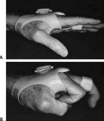
FIG 3 • Typical splint used for the conservative treatment of sagittal band disruption. The interphalangeal joints are free and no more than 30 degrees of metacarpophalangeal joint flexion is allowed.
Certainly, except in special circumstances such as the professional athlete, conservative therapy should initially be attempted.
Although several different protocols and splints have been described, most share one common objective: maintaining the MCP joint in neutral (full extension) for a period of weeks. In all situations, motion of the IP joint should be accommodated and encouraged.
A hand-based, custom orthoplast splint holding the involved digit in 0 to 20 degrees of MCP joint flexion (FIG 3) is worn for 4 to 6 weeks, depending on the patient's progress at the 2- and 4-week follow-up visits.
After 6 weeks of MCP immobilization in extension, the splint is weaned except for sporting endeavors and other heavy activities, in which case the splint is used for another 2 weeks. Buddy taping can provide long-term support as may be indicated.
Active range-of-motion activities are initiated and slowly progressed to gentle passive flexion of the involved MCP joint.
Thereafter, unrestricted use of the hand is promoted. It is unusual to need formal hand therapy; however, when excessive joint stiffness is present and radiographs fail to document any bony pathology, a short course of therapy along with modality use can be helpful.
If the injury clearly is not responding to immobilization, surgery is recommended.
SURGICAL MANAGEMENT
Operative indications
Patients with painful extensor tendon instability more than 3 weeks after the injury
Patients whose injury has failed to respond to nonsurgical management and have persistent, painful tendon instability beyond 6 weeks of conservative care
Professional athletes5 and other high-demand individuals
When possible, direct repair of the sagittal band should be performed.
Although we believe that this is usually not possible more than 8 weeks after injury, Hame and Melone5 reported on 11 direct repairs at an average of 3.3 months out from time of injury. No patient had prior splinting. All patients were asymptomatic with full recovery of range of motion and return to professional sports at an average of 5 months.
Carroll et al2 reported on five patients who underwent reconstruction after failed conservative management. All patients regained full, asymptomatic range of motion.
If tissue deficiency or scarring exists, reconstruction as opposed to primary repair will be required.
Preoperative Planning
With open injuries, the surgeon should determine if the cause was related to a bite. In this situation, MCP joint contamination is likely and surgical irrigation and débridement as well as antibiotic treatment is warranted. When severe contamination is present, delayed sagittal band repair is indicated.
Concomitant MCP joint capsular injury is possible. Once surgically exposed, methylene blue injection into the joint, out of the zone of injury, can help to reveal any rents in the MCP joint capsule. These defects should be débrided with subsequent irrigation of the joint. Afterward, no capsular repair is necessary.5
The surgeon should be prepared to perform either a repair or a reconstruction.
Local anesthesia with sedation is preferred, but regional or general anesthesia is acceptable.
Positioning
The patient is placed supine on the operating table with the affected hand outstretched onto a hand table.
A tourniquet is applied to the arm and inflated to the appropriate pressure before starting the procedure.
TECHNIQUES
EXPOSURE
 A curvilinear incision is placed dorsally over the ulnar aspect of the affected MCP joint.
 This is used for primary sagittal band repair.
A longitudinal incision is centered dorsally over the affected MCP joint.
This is used for reconstructive cases requiring greater exposure.
Sensory branches of the radial or ulnar nerves, or both, are identified and protected.
The extensor tendon is exposed, the tear identified, and scar tissue débrided.
The MCP capsule, which is deep to the extensor tendon, is usually left undisturbed; however, when MCP joint pathology needs to be addressed, the capsule may be incised.
PRIMARY REPAIR
The sagittal band disruption is identified and the extensor tendon centralized (TECH FIG 1A,B).
Excess tissue is excised from the area between the torn sagittal band and the common extensor tendon.
The sagittal fibers are then repaired using 4-0 or 5-0 nonabsorbable suture (Ethibond). The knots are buried where possible.
The repair is performed with the joint in 60 to 70 degrees of flexion to avoid tension on the repair and stiffness of the joint.
The joint is flexed and extended to ensure midline stability (TECH FIG 1C–E).
The wound is closed with interrupted 4-0 nylon sutures.

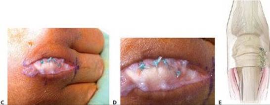
TECH FIG 1 • A. Traumatic extensor tendon dislocation (ulnar) over the metacarpophalangeal (MCP) joint (arrowhead) with the MCP joint extended. B. Extensor tendon subluxation over the MCP joint with the joint flexed.C. Primary repair of the sagittal band with the MCP joint extended. D. Primary sagittal band repair with the MCP joint flexed. The extensor tendon remains centralized dorsally with flexion. E.Primary repair of the deficient sagittal band. (A–D: Courtesy of Brian Hartigan.)
RECONSTRUCTION WITH EXTENSOR TENDON SLIP (CARROLL/KILGORE) 2,8 (AUTHORS' PREFERRED TECHNIQUE)
Release of the ulnar sagittal band may be necessary to mobilize the scarred tendon dorsally and radially (TECH FIG 2A,B).
A distally based radial or ulnar slip of extensor tendon (about one third) is fashioned and routed deep to the intact extensor tendon (TECH FIG 2C).
In a distal-to-proximal direction, the tendon graft is then looped around the radial collateral ligament (if subluxed ulnarly) (TECH FIG 2D).
Once proper tension is determined, the slip is then sutured back to the main tendon with interrupted, nonabsorbable suture or woven through the tendon in a Pulvertaft fashion (TECH FIG 2E,F).
As with all reconstruction techniques, tension is determined by taking the joint through a full range of motion and documenting stability dorsally.
The wound is closed with interrupted 4-0 nylon sutures.


TECH FIG 2 • A. Long finger extended at the metacarpophalangeal (MCP) joint, still with evidence of extensor dislocating into the ulnar trough. B. Extensor tendon dislocating when the MCP joint is flexed (arrow). C. A distally based slip of the extensor tendon is fashioned (white arrow). The ulnarly subluxed extensor tendon is indicated with a blue arrow. The red asterisk indicates the distal aspect of the digit. D. The distally based slip of extensor tendon (black arrowhead) has been rerouted volar to the radial collateral ligament (yellow arrowhead) from a distal to proximal direction. E. The distally based slip of extensor tendon has been secured to the extensor tendon proximal to the MCP joint (black arrowhead). The remaining ulnar sagittal band was repaired to prevent radial subluxation of the extensor tendon (red arrowhead). F. Reconstruction of the sagittal band using a distally based radial slip of extensor tendon wrapped around the radial collateral ligament (RCL) and reattached to the extensor tendon (with a weave).
DYNAMIC LUMBRICAL MUSCLE TRANSFER (SEGALMAN 16)
The lumbrical muscle is identified on the radial side of the joint and mobilized (TECH FIG 3A,B).
Begin proximally by separating the lumbrical muscle from the more dorsal interossei.
Once the lumbrical muscle is separated, continue distally to identify its tendinous insertion.
The lumbrical tendon is harvested just proximal to its insertion into the lateral band (TECH FIG 3C).
With the extensor tendon reduced, an isometric point in the extensor tendon must be identified. This is achieved by gently ranging the finger or asking the patient to flex. Once it is identified, a small longitudinal slit is made and the lumbrical tendon is passed through from volar to dorsal (TECH FIG 3D).
Tension is set appropriately while gently ranging the finger to confirm the absence of subluxation. The tendon is sutured back to itself using interrupted, nonabsorbable suture.
The wound is closed with interrupted 4-0 nylon sutures.

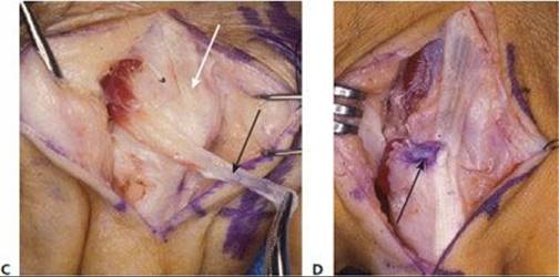
TECH FIG 3 • Technique using the lumbrical muscle for dynamic extensor tendon stabilization. A. Ulnar dislocation of the extensor tendon over the long finger metacarpophalangeal (MCP) joint (arrow). B.Surgical exposure identifying the extensor dislocation (black arrow) with a large chronic defect in the radial sagittal band (white arrow). C. The lumbrical muscle–tendon unit is isolated and mobilized for transfer (black arrow). The extensor tendon is indicated by the white arrow. D. The lumbrical tendon is woven into the extensor tendon (arrow), now stabilizing the extensor tendon during MCP motion. (Courtesy of Keith Segalman, MD.)
SAGITTAL BAND RECONSTRUCTION TO THE DEEP TRANSVERSE INTERMETACARPAL LIGAMENT (WATSON)
A 4-cm, distally based slip of extensor tendon consisting of no more than one-third the tendon width is harvested starting proximal to the MCP joint on the affected side (TECH FIG 4A).
This segment of tendon is then passed through a small slit in the remaining tendon at the level of the deep transverse metacarpal ligament to prevent further propagation of the tendon split.
The segment is then passed around or through the deep transverse intermetacarpal ligament using a curved clamp (TECH FIG 4B).
The free end of the tendon graft is then woven through and sutured to the remaining extensor tendon once it has been centralized and properly tensioned using nonabsorbable suture (TECH FIG 4C).
Wounds are closed with interrupted 4-0 nylon sutures.
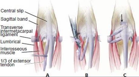
TECH FIG 4 • A. A distally based slip of extensor tendon constituting no more than one-third the width of the tendon is harvested. B. The slip of extensor tendon is rerouted from proximal to distal, around the deep intermetacarpal ligament. C. The tendon slip is then attached to the extensor tendon (usually radially) through a weave distal to the metacarpophalangeal joint.
CENTRALIZATION USING JUNCTURA TENDINUM
A longitudinal incision is centered dorsally over the affected MCP joint.
The extensor tendon is identified and held in a centralized position.
The MCP joint is flexed to reveal the more proximal, ulnar-sided junctura tendinum to the adjacent tendon.
The junctura tendinum is released from its ulnar-sided insertion into the adjacent tendon.
It is then brought over to the radial side of the affected finger, still in continuity with the tendon, and sutured to the palmar portion of the remaining sagittal band after correct tension has been set to centralize the tendon.
Wounds are closed with interrupted 4-0 nylon sutures.

POSTOPERATIVE CARE
Wounds are sterilely dressed immediately after the procedure and a splint is applied.
A volar and dorsal splint is used with the wrist slightly extended, the MCP joints at 0 to 30 degrees flexion, and the IP joints extended.
On postoperative day 5, the patient is seen in the office. Sutures are removed and a short-arm cast is applied with the wrist slightly extended, the MCP joints at 0 to 30 degrees of extension, and the IP joints free.
Sometimes, in the compliant elderly patient, we favor a hand-based splint fabricated to include the MCP joints in 30 degrees of flexion and the IP joints free.
Several postoperative protocols have been described for nondynamic reconstructions.
Inoue and Yukihisa6 placed the involved finger in a plaster cast for 3 weeks with the MCP joint in neutral or slightly flexed, allowing active IP joint motion.
Carrolletal2 splinted the MCP joint neutral for 6 weeks. At 2 weeks after surgery they began PIP joint range of motion, and at 6 weeks active range of motion at the MCP was initiated.
Watsonetal17 used a splint and Kirschner wire to immobilize the MCP joint at 15 to 20 degrees of flexion for 3 weeks.
Hame and Melone5 used cast immobilization of the MCP joint in 60 to 70 degrees of flexion for 6 weeks, with active flexion, but not extension, allowed.
For dynamic transfers, the patient is immobilized for 4 weeks in a short-arm cast with the wrist in neutral, MCP joints in extension, and the PIP joints free. Active motion is begun 4 weeks after surgery and strengthening at 6 weeks. Therapy is then continued for 6 to 8 weeks.
OUTCOMES
Rayanetal13 treated three type II injuries nonoperatively with 3 weeks of splinting the MCP joint at 0 degrees of extension, followed by 2 to 3 weeks of protected range of motion out of the splint three times a day, with a final 4 weeks of buddy splinting. They reported full range of MCP joint motion and no tenderness or pain with resisted digital abduction in all three patients. However, one patient did experience residual painless subluxation.
Carrolletal2 treated nine subluxed extensor tendons. Four were treated nonoperatively with 6 weeks of splinting the MCP joint in 0 degrees of extension, followed by range-of-motion therapy. Five were treated operatively using a slip of extensor tendon looped around the collateral ligament. After splinting and therapy, all patients were pain-free with full extension and active flexion to 90 degrees or more. There were no recurrences of symptoms in either group and no complications in the surgical group.
Watsonetal17 described 16 patients treated operatively with a slip of extensor tendon looped through the deep transverse metacarpal ligament. They reported an average MCP joint flexion of 90 degrees postoperatively, with no subluxation of the tendon. All patients were pain-free. There were no complications and no need for further surgery.
Hame and Melone5 reported on eight professional athletes who underwent immediate repair of sagittal band injuries with subluxation of the extensor tendon. There were 11 injured fingers in total. Seven of the 11 had capsular injuries; they were all débrided but not repaired. Each athlete demonstrated full range of motion postoperatively and all returned to professional sport at 5 months on average. No additional intervention was necessary and there were no complications.
COMPLICATIONS
Complications are rare. Most series in the literature do not report any complications.
With nonoperative therapy, possible complications include joint stiffness, skin irritation from splinting, and failure of treatment.
With operative therapy, possible complications include infection, joint stiffness, injury to neurovascular structures, and failure of treatment with recurrent subluxation or dislocation either in a radial or ulnar direction.
REFERENCES
1. Araki S, Ohtani T, Tanaka T. Acute dislocation of the extensor digitorum communis tendon at the metacarpophalangeal joint. J Bone Joint Surg Am 1987;69A:616–619.
2. Carroll C, Moore JR, Weiland AJ. Posttraumatic ulnar subluxation of the extensor tendons: a reconstructive technique. J Hand Surg Am 1987;12A:227–231.
3. Catalano LW, Gupta S, Ragland R, et al. Closed treatment of nonrheumatoid extensor tendon dislocations at the metacarpophalangeal joint. J Hand Surg Am 2006;31A:242–245.
4. Drape JL, Dubert T, Silbermann P, et al. Acute trauma of the extensor hood of the metacarpophalangeal joint: MR imaging evaluation. Radiology 1994;192:469–476.
5. Hame S, Melone C. Boxer's knuckle in the professional athlete. Am J Sports Med 2000;28:879–882.
6. Inoue G, Yukihisa T. Dislocation of the extensor tendons over the metacarpophalangeal joints. J Hand Surg Am 1996;21A:464–469.
7. Ishizuki M. Traumatic and spontaneous dislocation of extensor tendon of the long finger. J Hand Surg Am 1990;15A:967–972.
8. Kilgore ES, Graham WP, Newmeyer WL, et al. Correction of ulnar subluxation of the extensor communis. Hand 1975;7:272–274.
9. Lopez-Ben R, Lee DH, Nicolodi DJ. Boxer knuckle (injury of the extensor hood with extensor tendon subluxation): diagnosis with dynamic US: report of three cases. Radiology 2003;228:642–646.
10. Milford LW Jr. Retaining Ligaments of the Digit of the Hand. Philadelphia: WB Saunders, 1968:26–27.
11. Pfirrmann CWA, Theumann NH, Botte MJ, et al. MRI Imaging of the metacarpophalangeal joints of the fingers. Radiology 2002;222:447–452.
12. Rayan GM, Murray D. Classification and treatment of closed sagittal band injuries. J Hand Surg Am 1994;19A:590–594.
13. Rayan GM, Murray D, Chung KW, et al. The extensor retinacular system at the metacarpophalangeal joint: an anatomical and histological study. J Hand Surg Br 1997;22B:585–590.
14. Saldana MJ, McGuire RA. Chronic painful subluxation of the metacarpophalangeal joint extensor tendons. J Hand Surg Am 1986; 11A:420–423.
15. Scheweitzer T, Rayan G. The terminal tendon of the digital extensor mechanism, part I: anatomic study. J Hand Surg Am 2004;29A:898–902.
16. Segalman KA. Dynamic lumbrical muscle transfer for correction of posttraumatic extensor tendon subluxation. Tech Hand Up Ext Surg 2006;10:107–113.
17. Watson HK, Weinzweig J, Guidera PM. Sagittal band reconstruction. J Hand Surg Am 1997;22A:452–456.
18. Young CM, Rayan GM. The sagittal band: anatomic and biomechanical study. J Hand Surg Am 2000;25A:1107–1113.