
Catherine M. Curtin and Amy L. Ladd
DEFINITION
 Cubital tunnel syndrome is a compression neuropathy of the ulnar nerve that occurs at or around the level of the elbow (cubis is Latin for “elbow”).
Cubital tunnel syndrome is the second most common compression neuropathy of the upper limb requiring treatment, after carpal tunnel syndrome.
ANATOMY
The ulnar nerve is the terminal branch of the medial cord of the brachial plexus, with contributions from C8 and T1 nerve roots.
The ulnar nerve traverses the cubital tunnel, a fibro-osseous tunnel at the elbow. The medial epicondyle, the olecranon, the medial collateral ligament of the elbow (which forms the floor), and the fibrous retinaculam extending from the medial epicondyle to the olecranon make up the anatomic landmarks (FIG 1).11
Any of several possible sites of compression of the ulnar nerve around the elbow can result in cubital tunnel syndrome. All of these sites should be considered when selecting the type of surgical decompression.
 The arcade of Struthers is a controversial site of compression, because it is found in only a minority of patients. If present, it is found approximately 8 cm proximal to the medial epicondyle and consists of a fascial band running from the medial head of the triceps to the intermuscular septum.15
The medial intermuscular septum is a fascial band from the coracobrachialis to the medial humeral epicondyle, especially thick at its attachment to the epicondyle. The ulnar nerve may rest or scissor over the septum as it crosses from the anterior to the posterior compartment, as it approaches the medial epicondyle, or after an anterior transposition if it is not adequately excised.
The arcuate ligament of Osborne at the cubital tunnel, which is the fibrous band extending from the medial epicondyle to the olecranon, can cause stenosis of the cubital tunnel and, thus, ulnar nerve compression.
Distally, the nerve can be compressed as it passes between the two heads of the flexor carpi ulnaris, especially if each muscle head from the medial epicondyle and the olecranon converge close to the elbow joint.
The presence of an anconeus epitrochlearis (FIG 2), an anomalous thin muscle extending from the triceps or olecranon to the medial epicondyle, also can cause ulnar nerve compression.
The medial antebrachial cutaneous nerve and the medial brachial cutaneous nerve both emanate directly from the medial cord and are thus not ulnar nerve branches, but they importantly may lie in the surgical field. They are usually found deeper than expected, along the fascia of the triceps, brachialis, and flexor carpi ulnaris.
PATHOGENESIS
Cubital tunnel syndrome is a compressive neuropathy. Several anatomic factors make the ulnar nerve susceptible to compression at the elbow.
The nerve is superficial at the level of the elbow, making it susceptible to minor and major trauma, ranging from mild repetitive contusion to high-energy injury.
The bony tunnel and its soft tissue support between the olecranon and medial epicondyle may be shallow, either inherently or traumatically, promoting subluxation, “perching” on the epicondyle, and microtrauma.
Elbow flexion increases pressure on the nerve and decreases the volume of the cubital tunnel, resulting in compression of the nerve.5
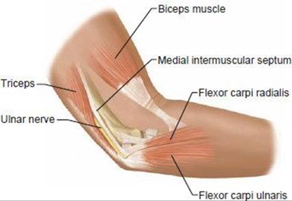
FIG 1 • Anatomy of the cubital tunnel.
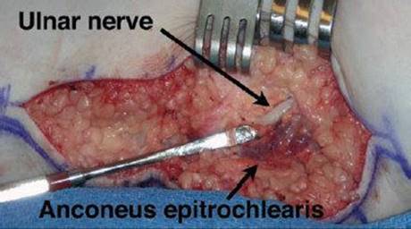
FIG 2 • An anomalous anconeus epitrochlearis encountered overlying the cubital tunnel. Anterior is at top and posterior at bottom; the forearm is to the left.

FIG 3 • A. “Perched” ulnar nerve. The nerve subluxates anteriorly, sitting on top of the medial epicondyle with the elbow in flexion. B. Wasting of first dorsal interosseous nerve. (A: Copyright Amy Ladd, MD.)
NATURAL HISTORY
Without operative intervention, about half of mild cases can resolve with activity modification.10
No long-term studies have been done of the natural history for severe disease.
PATIENT HISTORY AND PHYSICAL FINDINGS
Subjective complaints include numbness in the small and ring fingers, often with accompanying burning pain around the medial epicondyle. Symptoms may be worse at night.
As the disease progresses, patients may complain of weakness or clumsiness of their hands. More advanced disease will demonstrate wasting of the intrinsics and clawing of the ring and small fingers.
Systemic diseases such as diabetes, amyloidosis, or alcoholism may cause peripheral neuropathy, which can mimic the symptoms of a compressive neuropathy.
A smoking history is important, not only for impaired vascularity, but because it may point to the rare Pancoast tumor, an apical lung tumor, which causes plexus compression, mimicking the symptoms of cubital tunnel syndrome.
Elbow trauma can create deformity, causing ulnar nerve compression. Deformities include a cubitus valgus, cubitus varus, or malunion. The elbow trauma can be remote and result in tardy ulnar nerve palsy.
Look for atrophy of the intrinsic muscles of the hand or a clawed posture of the ring and small fingers. Check for masses around the elbow.
Palpate the elbow and hand to evaluate for tender masses or other anomalous elbow anatomy.
Put the elbow through its range of motion and assess whether the ulnar nerve subluxates or perches at the medial epicondyle with elbow flexion (FIG 3A).
Visible atrophy of the first dorsal interosseous nerve correlates with significant ulnar nerve compression and can indicate significant motor impairment (FIG 3B).
Perform a sensory examination of the hand, using SemmesWeinstein monofilaments to obtain threshold measurements. Evaluate sensation on the ulnar dorsum of the hand. If sensation is normal, it suggests the problem may be distal, at the level of Guyon's canal.
Clinical tests that can help with diagnosis include the following:
Tinel's test. This test may not be specific, because many normal individuals will have a positive Tinel's response to percussion.
Elbow flexion test. This test is sensitive for cubital tunnel syndrome.
Crossed finger test. This test demonstrates weakness of dorsal and palmar interossei.
Froment's sign. A positive Froment's sign indicates weakness of the adductor pollicis.
Wartenberg's sign (in which the small finger assumes an abducted posture with finger extension). This sign is the result of weakness in the palmar interossei, resulting in unopposed ulnar pull of the extensor digiti quinti.
IMAGING AND OTHER DIAGNOSTIC STUDIES
Radiographs of the elbow define the bony architecture and its alterations: masses, erosions, arthritis, and previous trauma. An axial view is helpful to evaluate the cubital canal (FIG 4).
Normal results on electrodiagnostic studies (eg, nerve conduction and electromyography) do not exclude the diagnosis of cubital tunnel syndrome; the syndrome may be present but not severe.
These tests localize the area of compression if the nerve conduction is measured at short segment intervals.
Several positive electrodiagnostic findings suggest ulnar compression:
 Motor conduction across the elbow less than 50 m/sec.13
Focal slowing of nerve velocity across the elbow

FIG 4 • Axial view of the elbow demonstrates a hooked osteophyte within the cubital tunnel, as well as calcification in the bursa and osteophyte. (Copyright Amy Ladd, MD.)
Fibrillation potentials or positive waves suggest axonal degeneration, representing a poorer prognosis for complete recovery.
MRI and CT may occasionally be helpful as ancillary imaging studies to define soft tissue aberrancies and localize bone abnormalities such as osteophytes in the cubital tunnel.
DIFFERENTIAL DIAGNOSIS
Cervical spine disease affecting C8 and T1
Compression of the inferior aspect of the brachial plexus from shoulder trauma
Apical lung tumor (Pancoast tumor)
Thoracic outlet syndrome
Entrapment of the ulnar nerve at the wrist (Guyon's canal)
NONOPERATIVE MANAGEMENT
Activity modification
Ulnar nerve protection limiting microtrauma to the nerve through elbow padding and limiting direct pressure on the nerve
Minimize prolonged elbow flexion, especially at night, through sleep modifications or splints.
Splinting
Splints to prevent elbow flexion; rigid splints are more effective but are less tolerated by patients. If persistent paresthesias exist, a trial of temporary full-time use is recommended. For milder cases, the splint is worn only at night.2
Nonoperative treatment requires a trial of several months before determining its success.
SURGICAL MANAGEMENT
Surgical intervention should be considered for patients presenting with motor involvement or permanent sensory changes, or for those who have failed nonoperative treatment.
Preoperative Planning
Review the history and physical examination.
Review plain radiographs for evidence of old trauma, valgus or varus deformity, or loose bodies.
Electrodiagnostic testing and examination may correlate with postoperative results.
Body habitus, especially the presence of abundant adipose tissue around the elbow, may help the surgeon select a subcutaneous transposition—a procedure with less dissection—rather than a more extensive but protective procedure such as an intraor submuscular transposition.
A patient with a visible and symptomatic subluxating nerve may be considered for a medial epicondylectomy.
Patients with severe disease with muscle wasting are less likely to have complete recovery.5
Positioning
The patient usually is placed in the supine position.
If a sterile tourniquet is preferred, drape out the forequarter. A standard tourniquet may be used, but position it high in the axilla, with good padding. A proximally placed tourniquet can be challenging to position in the obese arm in either circumstance, because the tourniquet tends to gap distally. It is worth the extra time to position it properly, because adequate hemostasis and visualized proximal dissection are important aspects of ulnar nerve surgery.
The patient's shoulder is externally rotated and abducted on an arm table.
The tourniquet is inflated after exsanguination of the arm.
Folded towels stabilize and elevate the elbow (FIG 5).
An obese patient with sleep apnea under peripheral nerve block (most commonly supraor infraclavicular block) may require slight truncal elevation, which may be vexing for the surgeon.
Approach
The choice of technique depends on the severity of symptoms, the patient's body habitus, the presence of elbow anatomic pathology, and the surgeon's preference.9
The three general types of release are in situ release, in situ release with medial epicondylectomy, and anterior transposition (subcutaneous, intramuscular, and submuscular).
Table 1 summarizes the surgical options for treating cubital tunnel syndrome.

FIG 5 • The arm is draped, the sterile tourniquet is placed proximally, and a bump under the elbow assists visualization. Alternatively, a proximal tourniquet may be placed before the arm is draped.

TECHNIQUES
IN SITU RELEASE
 Center the longitudinal incision just anterior to the medial epicondyle, making an incision about 8 cm long (TECH FIG 1A).
Dissect through the fat, down to the level of the medial epicondyle.
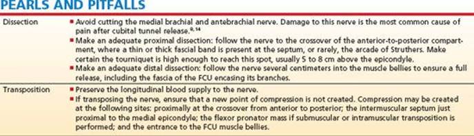
Preserve the branches of the medial brachial and antebrachial cutaneous nerves. Although the course is variable, branches can be found from 6 cm proximal to 6 cm distal to the medial epicondyle and often are at the level of the fascia8 (TECH FIG 1B,C).
Identify the ulnar nerve and dissect it free proximally until it pierces the medial intermuscular septum. Release any areas of constriction.
Take the dissection distal to the level of the medial epicondyle and release the band spanning from the medial epicondyle to the olecranon.
Preserve the branches of the ulnar nerve: the first is the articular sensory branch, followed by the motor branches to the flexor carpi ulnaris (FCU) and flexor digitorum profundus (FDP). The FCU branches are found proximally with appearance of the muscle.
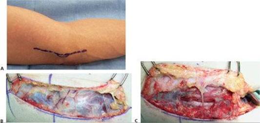
TECH FIG 1 • A. The standard incision, centered just anterior or posterior to the medial epicondyle. B,C. Preservation of crossing medial brachial and antebrachial nerves. The cutaneous nerves lie deep in the fat, typically on the fascia. Here two branches are encountered before and after fasciotomies to expose the nerve. (Copyright Amy Ladd, MD.)
The distal dissection proceeds through the thick arcade of fascia of the flexor pronator aponeurosis. Two layers exist: a superficial layer that covers both heads of the FCU, and a deeper one that overlies the nerve as it traverses between the two heads. Continue fascial release into the muscle for several centimeters to ensure that there are no areas of entrapment within the muscle belly, taking care to preserve nerve branches to the muscle.
Gently palpate to ensure that the entire ulnar nerve is free from compressive bands.
Range the elbow and check for smooth ulnar nerve excursion. If perching (snapping) over the medial epicondyle occurs, consider medial epicondylectomy. This is often a preclinical determination.
Close the soft tissues using the surgeon's preferred technique.
Typically, no drain is placed.
Place the arm in a bulky supportive dressing or a posterior plaster elbow splint with flexion of about 60 degrees. Remove the splint according to wound care and the surgeon's mobilization preference.
IN SITU RELEASE WITH MEDIAL EPICONDYLECTOMY
The incision and dissection are the same as the in situ release.
Excise a strip of the tough fascial intermuscular septum as it attaches to the medial epicondyle to minimize the nerve “scissoring” over the firm edge.
Once the nerve is free of all areas of entrapment, a longitudinal incision is made slightly anterior to the medial epicondyle with a knife or electrocautery, reflecting the periosteum to reveal the bony prominence of the epicondyle. Carefully protect the ulnar nerve; gentle retraction with a saline-lubricated ¼-inch Penrose drain on a short hemostat is sufficient.
Expose the medial epicondyle subperiosteally.
Remove the prominence of the epicondyle, which is most acute in its posterior position, removing 2 to 3 mm of prominence and 6 to 8 mm in length. Use a small, sharp osteotome and smooth with a file while protecting the nerve (TECH FIG 2A).
Place bone wax over the raw bone. This minimizes postoperative hematoma.
The periosteum is closed with buried sutures, either braided absorbable or nonabsorbable, minimizing contact with the nerve.
Check that the nerve glides, rather than perches, when the elbow is flexed and extended before closure of the skin (TECH FIG 2B).
Because of potential bony bleeding, a drain is recommended.
Apply a posterior plaster splint for 10 to 14 days, with protected mobilization thereafter.
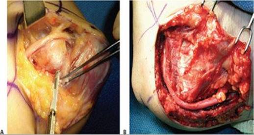
TECH FIG 2 • Medial epicondylectomy. A. The medial epicondyle is exposed, and the most prominent aspect is removed. We recommend removal of the most prominent and inferior portion, 2 to 3 mm in depth, to avoid disruption of the medial collateral ligament. B. Once the epicondylectomy is performed and the fascia closed, the elbow is flexed to visualize smooth movement of the nerve. The nerve no longer perches on the medial epicondyle. (Copyright Amy Ladd, MD.)
ANTERIOR SUBCUTANEOUS TRANSFER
The incision and dissection are the same as for the in situ release, except that the incision may have to be slightly longer.
Release the nerve at every potential level of entrapment.
Circumferentially dissect the nerve to allow it to be moved anterior to the medial epicondyle. Free all posterior attachments to allow for maximal anterior excursion.
Excise the intermuscular septum from the crossover of the ulnar nerve, anterior to posterior in the proximal dissection, all the way to its tough attachment at the medial epicondyle.
Preserve the longitudinal vasculature accompanying the nerve to prevent devascularization of the nerve. Use caution around the medial epicondyle and the most fibrous part of the intermuscular septum, where lies an external but vulnerable large venous leash.
Develop the interval between the skin and the fascia overlying the flexor pronator muscle mass anterior to the medial epicondyle, about 4 cm.
Transpose the nerve to lie anterior to the medial epicondyle (TECH FIG 3A).
The nerve should lie in its new position without any tension or areas of compression. An intraneural dissection to release the motor branches to the FCU may be required proximally.
To prevent the nerve from subluxating, a 1-cm fasciodermal sling is constructed from the fascia overlying the flexor pronator mass (ie, the FCU, FCR, and the pronator teres)3 (TECH FIG 3B). This flap is sutured to the skin. This flap prevents the nerve from sliding back to its old position.
Care must be taken to ensure that this flap does not become a new area of compression.
No drain is required.
Apply a posterior plaster splint for 10 to 14 days, with protected mobilization thereafter.
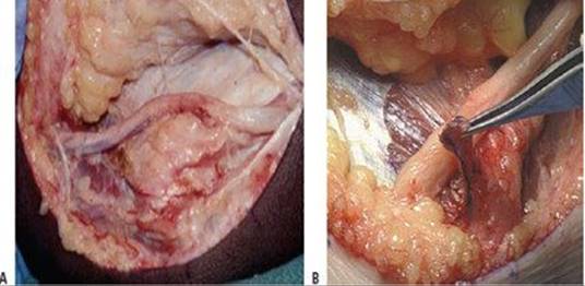
TECH FIG 3 • Anterior subcutaneous transposition. A. The subcutaneous flap at the level of the flexor pronator fascia has been developed and the nerve transposed anteriorly. B. A 1-cm fascial sling is developed from the flexor pronator mass to provide an inferior restraint for the transposed nerve. (A: Courtesy of Thomas R. Hunt, III, MD. B: From Glickel SZ, Barron OA, Eaton RG, et al. Stabilized subcutaneous ulnar nerve transposition with immediate range of motion. Video J Orthop 2000; www.vjortho.com/cgi/content/abstract/2511.)
ANTERIOR INTRAMUSCULAR TRANSPOSITION
The nerve is fully released, as described for the subcutaneous transposition.
The interval between the skin and the fascia is developed anterior to the medial epicondyle, to about 4 cm.
Transpose the nerve so that its rests along the flexor pronator mass (ie, FCU, FCR, and the pronator teres).
A trough slightly bigger than the nerve is carved out of the muscles along this anterior course (TECH FIG 4). Release any fascial bands found within the muscle substance.
 Flex the elbow and place the nerve in the trough.
Suture fascia over the nerve, creating a tunnel.
Range the elbow to ensure that there is no kinking or tethering of the transposed nerve.
The arm is immobilized with a pronated forearm in an elbow splint for 2 to 3 weeks at 45 to 60 degrees of flexion with progressive protected mobilization.
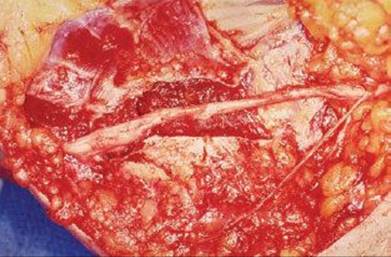
TECH FIG 4 • Intramuscular transposition. The nerve is placed in a tunnel in the muscle, and the fascia is closed. (Courtesy of William Kleinman, Indiana Hand Center.)
ANTERIOR SUBMUSCULAR TRANSPOSITION
The nerve is fully released as described with the preceding procedures, and the skin flap is developed similarly to the intramuscular procedure.
Divide the flexor pronator mass about 1 cm distal to its insertion on the medial epicondyle, either as a straight incision or in a V-Y fashion (TECH FIG 5A).
Lift the flexor pronator mass distally at the level of the FDS muscle. There is a loose areolar plane between these FDS muscle. There is a loose areolar plane between these muscle bellies.
The median nerve and brachial artery lie in this plane. Transpose the ulnar nerve in the medial position (TECH FIG 5B).
Take care to avoid injury to the medial collateral ligament complex
Flex the elbow and repair the flexor pronator mass with 3-0 Ethibond suture.
Place a drain.
The arm is immobilized with a pronated forearm in an elbow splint for 2 to 3 weeks at 45 to 60 degrees of flexion with progressive protected mobilization.

TECH FIG 5 • Submuscular transposition. The flexor pronator mass is incised (A), and the nerve is passed deep to the flexor pronator muscle mass (B). Sutures are in place to repair the muscle origin following use of a simple straight incision. (A: Copyright Amy Ladd, MD. B: Courtesy of Thomas R. Hunt, III, MD.)
POSTOPERATIVE CARE
Postoperative care instructions are given individually with the discussion of each technique. In general, the more extensive the dissection, the more protected postoperative splinting and mobilization is required. Strengthening may begin a few weeks after an in situ decompression, for example, and 6 to 8 weeks following a submuscular transposition.
OUTCOMES
Overall, all procedures have a success rate of about 90% for mild cases. The rate of total relief decreases as severity of disease increases.9
Postoperative outcomes are proportional to disease severity: ie, severe disease is less likely to achieve full recovery.5
Recent studies suggest that outcomes are similar for the different procedure types.1,4,10
COMPLICATIONS
Pain at the elbow
Decreased sensation around the scar
Incomplete symptom relief
Painful neuroma of cutaneous nerves
Symptomatic subluxating nerve
Injury to motor branches to the FCU
REFERENCES
1. Bartels R, Verhagen W, Gert J, et al. Prospective randomized controlled study comparing simple decompression versus anterior subcutaneous transposition for idiopathic neuropathy of the ulnar nerve at the elbow. Part 1. Neurosurgery 2005;56:522–530.
2. Dellon AL, Hament W, Gittelshon A. nonoperative management of cubital tunnel syndrome: An 8-year prospective study. Neurology 1993;43:1673–1677.
3. Eaton RG, Crowe JF, Parkes JC. Anterior transposition of the u1nar nerve using a noncompressing fasciodermal sling. J Bone Joint Surg Am 1980;62A:820–825.
4. Gervasio O, Gambardella G, Zaccone C, et al. Simple decompression versus anterior submuscular transposition of the ulnar nerve in severe cubital tunnel syndrome: A prospective randomized study. Neurosurgery 2005;56:108–117.
5. Hironori M, Yoshizu T, Maki Y, et al. Long-term clinical and neurologic recovery in the hand after surgery for severe cubital tunnel syndrome. J Hand Surg Am 2004;29;373–378.
6. Iba K, Wada T, Aoki M, et al. Intraoperative measurement of pressure adjacent to the ulnar nerve in patients with cubital tunnel syndrome. J Hand Surg Am 2006;31;553–558.
7. Kleinman WB, Bishop AT. Anterior intramuscular transposition of the ulnar nerve. J Hand Surg Am 1989;14:972–979.
8. Lowe JB, Maggi SP, Mackinnon SE. The position of crossing branches of the medial antebrachial cutaneous nerve during cubital tunnel surgery in humans. Plast Reconstr Surg 2004; 114:692–696.
9. Mowlavi A, Andrews K, Lille S, et al. The management of cubital tunnel syndrome: a meta analysis of clinical studies. Plast Reconstr Surg 2000;106:327–334.
10. Nabhan A, Ahlhelm F, Kelm J, et al. Simple decompression or subcutaneous anterior transposition of the ulnar nerve for cubital tunnel syndrome. J Hand Surg Am 2005;30:521–524.
11. O'Driscoll SW, Horii E, Carmichael SW, et al. The cubital tunnel and ulnar neuropathy. J Bone Joint Surg Br 1991;73B:613–617.
12. Padua L, Aprile I, Caliandro P, et al. Natural history of ulnar entrapment at elbow. Clin Neurophysiol 2002;113:1980–1984.
13. Practice parameter for electrodiagnostic studies in ulnar neuropathy at the elbow: summary statement. American Association of Electrodiagnostic Medicine, American Academy of Physical Medicine and Rehabilitation, American Academy of Neurology. Arch Phys Med Rehabil 1999;80:357–360.
14. Sarris I, Göbel F, Gainer M, et al. Medial brachial and antebrachial cutaneous nerve injuries: effect on outcome in revision cubital tunnel surgery. J Reconstr Microsurg 2002;18:665–670.
15. Siqueira MG, Martins RS. The controversial arcade of Struthers. Surg Neurol 2005;64S:S17–S20.