
David Ring
DEFINITION
 Fracture of the olecranon process is common, usually displaced, and nearly always treated operatively.
Important injury characteristics include displacement, comminution, and subluxation or dislocation of the elbow, and all are accounted for in the Mayo classification (FIG 1).6
Fracture-dislocations of the olecranon can be anterior (trans-olecranon) or posterior (the most proximal type of posterior Monteggia according to Jupiter and colleagues3) in direction.2,3,9,10
Open injuries are unusual.
ANATOMY
The greater sigmoid notch of the ulna is formed by the coronoid and olecranon processes and forms a nearly 180-degree arc capturing the trochlea.
The region between the coronoid and olecranon articular facets is the nonarticular transverse groove of the olecranon, a common location of fracture and a place where precise articular reduction is not critical.
The triceps has a broad and thick insertion from just superior to the point of the olecranon and the tip of the olecranon process that can be used to enhance fixation of small, osteoporotic, or fragmented fractures and can be split longitudinally, if needed, when applying a plate.
PATHOGENESIS
Fractures of the olecranon are most often the result of a direct blow to the point of the elbow, but occasionally they result from indirect forces during a fall on the outstretched hand.
NATURAL HISTORY
Stable nondisplaced or minimally displaced fractures are uncommon.
The majority of olecranon fractures are displaced and benefit from operative treatment.
The occasional untreated displaced simple olecranon fracture demonstrates a slight flexion contracture, some weakness of extension, no arthrosis, and little if any pain.
In contrast, undertreated or poorly treated fracture-dislocations lead to severe arthrosis with or without instability.
Even well-treated complex injuries are at risk for stiffness, heterotopic ossification, arthrosis, and occasionally nonunion.
PATIENT HISTORY AND PHYSICAL FINDINGS
Knowledge of the characteristics of the patient (age, gender, medical health) and the injury (mechanism, energy) will help the surgeon understand the injury and determine optimal treatment.
First the patient is assessed for life-threatening injuries (ATLS protocol) and any medical problems that may have contributed to the injury.
A secondary survey is performed to identify any other fractures, ipsilateral arm injuries in particular.
The skin is carefully inspected for any wounds associated with the fracture.
The pulses are palpated, capillary refill inspected, and an Allen test performed if necessary.
Peripheral nerve function is assessed.
Patients with high-energy injuries, particularly those with ipsilateral wrist or forearm injuries, are at risk for compartment syndrome. If the clinical examination is suggestive or unreliable (owing to problems with mental status), compartment pressure monitoring should be performed.
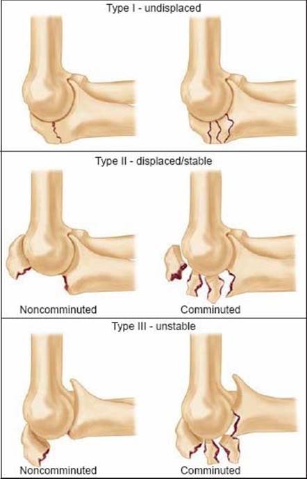
FIG 1 • The Mayo classification of olecranon fractures accounts for the factors that will influence treatment decisions: displacement, comminution, and dislocation or subluxation of the articulations.
IMAGING AND OTHER DIAGNOSTIC STUDIES
Anteroposterior (AP) and lateral radiographs are used for initial characterization of the injury.
Radiographs after reduction or splinting, or oblique views can be useful.
Computed tomography (CT) is useful for characterization of fracture-dislocations. In particular, 3D CT reconstructions can be useful for assessment of the coronoid and radial head.
DIFFERENTIAL DIAGNOSIS
Elbow dislocation
Monteggia and Essex-Lopresti fracture-dislocations of the forearm
Distal humerus fracture
NONOPERATIVE MANAGEMENT
Nonoperative management is appropriate for the rare fracture of the olecranon that is less than 2 millimeters displaced with the elbow flexed 90 degrees.
Four weeks of splint immobilization followed by active assisted mobilization of the elbow will usually result in a healed fracture and good elbow function.
SURGICAL MANAGEMENT
The vast majority of olecranon fractures are displaced and merit operative treatment.
Transverse, noncomminuted fractures not associated with fracture-dislocation are treated with tension band wiring.4,8
Comminuted fractures and fracture-dislocations are treated with dorsal contoured plate and screw fixation.1–3
The treatment of fracture-dislocations requires attention to the coronoid, radial head, and lateral collateral ligament.2,9–11
Preoperative Planning
The fracture characteristics that determine treatment are defined on radiographs and CT.
Templating the surgery with tracings of the radiographs is a useful way of running through the surgery in detail before performing it, familiarizing oneself with the anatomy, anticipating problems, and ensuring that all of the implants and equipment that might be necessary are available.
Positioning
In most patients a lateral decubitus position with the arm over a bolster or support is best.
Some patients with fracture-dislocations that require both medial and lateral access may be positioned supine with the arm supported on a hand table.
A sterile pneumatic tourniquet is used.
Approach
A dorsal longitudinal skin incision is used.
TECHNIQUES
TENSION BAND WIRING
Reduction and Kirschner Wire Fixation
 Blood clot and periosteum are cleared from the fracture site to facilitate reduction.
Limited periosteal elevation is performed at the fracture site to monitor reduction.
A large tenaculum clamp is used to secure the fracture in a reduced position (TECH FIG 1A,B). A drill hole can be made in the dorsal cortex of the distal fragment to facilitate clamp application.
Two 1.0-mm smooth Kirschner wires are drilled across the fracture site (TECH FIG 1C).
 If these are drilled obliquely from dorsal proximal to volar distal, they will exit the anterior ulnar cortex distal to the coronoid process, providing an anchoring point of cortical bone to limit the potential for pin migration.
In anticipation of later impaction of the proximal ends of the wires, the Kirschner wires should be retracted 5 to 10 mm after drilling through the anterior ulnar cortex.
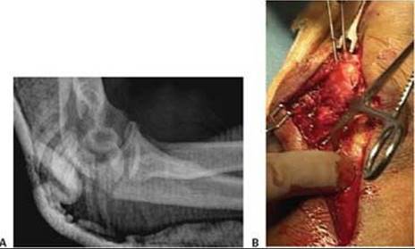

TECH FIG 1 • A. A lateral radiograph with the arm in plaster shows a transverse, noncomminuted fracture of the olecranon. B. An open reduction is held with a fracture reduction forceps. C. Two 1-mm Kirschner wires are drilled obliquely across the fracture site so that they exit the anterior ulnar cortex distal to the coronoid process. (A,B: Copyright David Ring, MD.)
Wiring
The apex of the ulnar diaphysis just distal to the flat portion of the proximal ulna is drilled with a 2.0-mm drill, with or without prior subperiosteal dissection.
 When two wires are used, a second drill hole is made a centimeter more distal.
If one wire is used, it should be 18 gauge. My preference is to use two 22-gauge stainless steel wires to limit the size of the knots, which may diminish implant prominence. The wires are passed through the drill holes. A large-bore needle can be used to facilitate passage of the wire through the drill hole (TECH FIG 2A).
The two tension wires are each passed over the dorsal ulna in a figure 8 fashion, then around the Kirschner wires, and underneath the insertion of the triceps tendon using a large-bore needle (TECH FIG 2B).
Each wire is tensioned both medially and laterally by twisting the wire with a needle holder (TECH FIG 2C,D).
 This should be done to take up slack only. These small wires will break if they are firmly tightened, which is not necessary.
The tightening should be done in a place that will make the wire knots less prominent.
After tightening the knots are trimmed and bent into the soft tissues to either side.
The Kirschner wires are then bent 180 degrees and trimmed.
These bent ends are then impacted into the proximal olecranon, beneath the triceps insertion, using an osteotome (TECH FIG 2E–H).


TECH FIG 2 • A. Two 22-gauge stainless steel tension wires are passed in a figure 8 fashion through drill holes in the ulnar shaft. B. They engage the triceps insertion proximally. C,D. The wires are tensioned on both sides. These do not need to be tight, but simply snug, with all slack taken up. Attempts to tighten these smaller 22gauge wires will break them. E. The proximal ends of the Kirschner wires are bent 180 degrees and impacted into the olecranon process, beneath the triceps insertion. F. The resulting fixation has a relatively low profile and is unlikely to migrate. G,H. Even these small wires are strong enough for active exercises to regain elbow motion. (A,B,D,F–H: Copyright David Ring, MD.)
PLATE AND SCREW FIXATION OF OLECRANON FRACTURES
Contour the plate to wrap around the proximal aspect of the olecranon or use a precontoured plate (TECH FIG 3A–C).
A straight plate will have only two or three screws in metaphyseal bone proximal to the fracture.
Bending the plate around the proximal aspect of the olecranon provides additional screws in the proximal fragment. The most proximal screws can be very long, crossing the fracture line into the distal fragment. In some cases, these screws can be directed to engage one of the cortices of the distal fragment, such as the anterior ulnar cortex.
A plate contoured to wrap around the proximal ulna can be placed on top of the triceps insertion. Alternatively, the triceps insertion can be incised longitudinally and partially elevated medially and laterally sufficiently to allow direct plate contact with bone.
If the proximal (olecranon) fragment is small, fragmented, or osteoporotic, it can be useful to add a figure 8 tension wire that engages the triceps insertion and passes over the top of the plate and around one of the screws at the metaphyseal level.

TECH FIG 3 • A. A lateral radiograph illustrates a comminuted olecranon fracture with a small proximal olecranon fragment. B. An oblique view shows the fragmentation. C. A 3.5-mm limited-contact dynamic compression plate and screws contoured to wrap around the dorsal surface of the olecranon is used for fixation. D. A 22-gauge stainless steel wire engages the triceps insertion—this is useful when the olecranon fragment is small, fragmented, or osteopenic. (Copyright David Ring, MD.)
Distally, a dorsal plate will lie directly on the apex of the ulnar diaphysis. The muscle need only be split sufficiently to gain access to this apex—there is no need to elevate the muscle or periosteum off either the medial or lateral flat aspect of the ulna.
No attempt is made to precisely realign intervening fragmentation—once the relationship of the coronoid and olecranon facets is restored and the overall alignment is restored, the remaining fragments are bridged, leaving their soft tissue attachments intact.
Bone grafts are rarely necessary if the soft tissue attachments are preserved.
If the olecranon fragment is small, osteoporotic, or fragmented, a wire engaging the triceps insertion should be used to reinforce the fixation (TECH FIG 3D).
The plate and screws will serve to hold the coronoid and olecranon facets in proper alignment and bridge fragmentation, and the wire will help ensure fixation even if screw purchase is lost.
PLATE AND SCREW FIXATION OF FRACTURE-DISLOCATIONS OF THE OLECRANON
Exposure
In the setting of a fracture-dislocation of the olecranon (TECH FIG 4A), fractures of the radial head and coronoid process can be evaluated and often definitively treated through the exposure provided by the fracture of the olecranon process.
With little additional dissection, the olecranon fragment can be mobilized proximally as one would do with an olecranon osteotomy, providing exposure of the coronoid through the ulnohumeral joint.
If the exposure of the radial head through the posterior injury is inadequate, a separate muscle interval (eg, Köcher or Kaplan intervals) accessed by the elevation of a broad lateral skin flap can be used.
If the exposure of the coronoid is inadequate through posterior injury and olecranon fracture, a separate medial or lateral exposure can be developed.

TECH FIG 4 • A. A complex anterior fracture-dislocation of the elbow. A lateral radiograph shows extensive comminution of the trochlear notch of the ulna, including the coronoid, and anterior displacement of the forearm. B,C. The coronoid fragments are connected to the dorsal metaphyseal fragments in this patient, which facilitates reduction and fixation. (A,C: Copyright David Ring, MD.)
A medial exposure, between the two heads of the flexor carpi ulnaris, or by splitting the flexor-pronator mass more anteriorly, or by elevating the entire flexor–pronator mass from dorsal to volar, may be needed to address a complex fracture of the coronoid, particularly one that involves the anteromedial facet of the coronoid process.
When the lateral collateral ligament is injured, it is usually avulsed from the lateral epicondyle. This facilitates repair that can be performed using suture anchors or suture placed through drill holes in the bone.
The fracture of the coronoid can often be reduced directly through the elbow joint using the limited access provided by the olecranon fracture (TECH FIG 4B,C).
Fixation
Provisional fixation can be obtained using Kirschner wires to attach the fragments either to the metaphyseal or diaphyseal fragments of the ulna, or to the trochlea of the distal humerus when there is extensive fragmentation of the proximal ulna.
An alternative to keep in mind when there is extensive fragmentation of the proximal ulna is the use of a skeletal distractor (a temporary external fixator; TECH FIG 5A).
External fixation applied between a wire driven through the olecranon fragment and up into the trochlea and a second wire in the distal ulnar diaphysis can often obtain reduction indirectly when distraction is applied between the pins.
Definitive fixation can usually be obtained with screws applied under image intensifier guidance.
The screws are placed through the plate when there is extensive fragmentation of the proximal ulna.
A second, medial plate may be useful when the coronoid is fragmented.
If the coronoid fracture is very comminuted and cannot be securely repaired, the ulnohumeral joint should be protected with temporary hinged or static external fixation, or temporary pin fixation of the ulnohumeral joint, depending on the equipment and expertise available.
A long plate is contoured to wrap around the proximal olecranon (TECH FIG 5B).
A very long plate should be considered (between 12 and 16 holes), particularly when there is extensive fragmentation or the bone quality is poor.
When the olecranon is fragmented or osteoporotic, a plate and screws alone may not provide reliable fixation.
In this situation, it can be useful to use ancillary tension wire fixation to control the olecranon fragments through the triceps insertion (TECH FIG 5C).

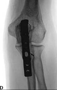
TECH FIG 5 • A. When there is diaphyseal comminution, a temporary external fixator may be useful. B. A long, 3.5-mm limited-contact dynamic compression plate is used for fixation. A 22-gauge stainless steel wire is used to enhance fixation of the comminuted olecranon fragments. C. The comminution extending into the diaphysis heals with the bridging plate. The trochlear notch is restored with good elbow function. (B,C: Copyright David Ring, MD.)
PEARLS AND PITFALLS

POSTOPERATIVE CARE
When good fixation is obtained (which occurs in most patients), active assisted and gravity-assisted elbow and forearm exercises can be initiated immediately after surgery. A delay of several days for comfort is reasonable.
If the lateral collateral ligament was repaired, the patient must be instructed not to abduct the shoulder for the first month.
If the fixation is tenuous, it is reasonable to immobilize the arm in a splint for a month or so before beginning exercises.
OUTCOMES
Nonunion is nearly unheard of after simple olecranon fractures, and early implant failure is usually due to noncompliance.6
The appeal of tension band wiring has been limited by prominence of the implants; however, if the techniques described herein are followed, few patients will request a second surgery specifically for implant removal.8
Macko and Szabo pointed out that it was initial implant prominence and not migration that led to implant-related problems after tension band wiring of olecranon fractures.5
In any case, a second surgery for implant removal is not unreasonable, and it may not be appropriate to consider this a complication.
Some surgeons have considered plate-and-screw fixation of simple, noncomminuted olecranon fractures.1 However, plates can also cause symptoms, and if only a few screws can be placed in the olecranon fragment, particularly in the setting of fragmentation or osteoporosis, it may be preferable to use the soft tissue attachments to enhance fixation rather than relying on implant–bone purchase alone.
Medial and lateral plates have been associated with early failure, malunion, and nonunion in the treatment of complex proximal ulna fractures.10,11
Dorsal plates perform better, but the elbow is often compromised in the setting of such complex injuries.
COMPLICATIONS
Implant loosening
Implant breakage
Nonunion
Malunion
Instability
Arthrosis
REFERENCES
1. Bailey CS, MacDermid J, Patterson SD, et al. Outcome of plate fixation of olecranon fractures. J Orthop Trauma 2001;15:542–548.
2. Doornberg J, Ring D, Jupiter JB. Effective treatment of fracturedislocations of the olecranon requires a stable trochlear notch. Clin Orthop Relat Res 2004;429:292–300.
3. Jupiter JB, Leibovic SJ, Ribbans W, et al. The posterior Monteggia lesion. J Orthop Trauma 1991;5:395–402.
4. Karlsson M, Hasserius R, Besjakov J, et al. Comparison of tensionband and figure-of-eight wiring techniques for treatment of olecranon fractures. J Shoulder Elbow Surg 2002;11:377–382.
5. Macko D, Szabo RM. Complications of tension-band wiring of olecranon fractures. J Bone Joint Surg Am 1985;67A:1396–1401.
6. Morrey BF. Current concepts in the treatment of fractures of the radial head, the olecranon, and the coronoid. J Bone Joint Surg Am 1995;77A:316–327.
7. O'Driscoll SW, Jupiter JB, Cohen M, et al. Difficult elbow fractures: pearls and pitfalls. AAOS Instruct Course Lect 2003;52:113–134.
8. Ring D, Gulotta L, Chin K, et al. Olecranon osteotomy for exposure of fractures and nonunions of the distal humerus. J Orthop Trauma 2004;18:446–449.
9. Ring D, Jupiter JB, Sanders RW, et al. Trans-olecranon fracturedislocation of the elbow. J Orthop Trauma 1997;11:545–550.
10. Ring D, Jupiter JB, Simpson NS. Monteggia fractures in adults. J Bone Joint Surg Am 1998;80A:1733–1744.
11. Ring D, Tavakolian J, Kloen P, et al. Loss of alignment after surgical treatment of posterior Monteggia fractures: salvage with dorsal contoured plating. J Hand Surg Am 2004;29A:694–702.