
Fotios P. Tjoumakaris and James P. Bradley
DEFINITION
 Posterior shoulder instability results in pathologic glenohumeral translation ranging from mild subluxation to traumatic dislocation. Most patients with this pathologic entity report pain in provocative positions of the glenohumeral joint, a condition referred to as recurrent posterior subluxation.
Posterior shoulder instability is much less common than anterior instability, representing about 5% to 10% of all patients with pathologic shoulder instability.2,5,10
A decision must be made regarding surgical treatment of this condition when an extended trial of conservative measures, such as physical therapy, has failed.
ANATOMY
The important stabilizing structures of the glenohumeral joint are the articular surfaces and congruity of the humerus and glenoid of the scapula, the capsular structures, the glenoid labrum, the intra-articular portion of the biceps tendon, and the rotator cuff muscles.
Pathologies of the posterior capsule and labral complex are believed to be the main contributors to posterior instability.
With the arm forward-flexed to 90 degrees, the subscapularis provides significant stability against posterior translation, and as the arm is placed in neutral, the coracohumeral ligament resists this force. With internal rotation of the shoulder (followthrough phase of throwing), the inferior glenohumeral ligament complex is the main restraint to posterior translation.1
Histologic evaluation of the posterior capsule shows it to be relatively thin and composed of only radial and circular fibers, with minimal cross-linking.
PATHOGENESIS
Posterior instability can be the result of trauma in the form of a direct blow to the anterior shoulder or may occur as the result of indirect forces acting on the shoulder, causing the combined movements of shoulder flexion, adduction, and internal rotation.13
Electrocution and seizures are the most common causes of an indirect mechanism resulting in posterior dislocation.
Patients with recurrent posterior subluxation may present with more vague symptoms, with pain being the chief complaint. Athletes may report that velocity with throwing is diminished, and a sharp pain may accompany the follow-through phase of throwing.
Other associated injuries such as superior labrum anterior posterior (SLAP) lesions, rotator cuff tears, reverse Hill-Sachs defects, and chondral injuries may be present and contribute to the pathology.4
NATURAL HISTORY
Patients with a history of a chronically locked posterior dislocation are at increased risk for the development of chondral injury and subsequent degenerative arthritis.6
Static posterior subluxations of the humeral head have been correlated with the presence of arthritis in young adults whose instability was left untreated.14
No long-term studies on the arthroscopic treatment of shoulder instability have documented a reduction in the development of osteoarthritis.
PATIENT HISTORY AND PHYSICAL FINDINGS
A thorough history is obtained, documenting whether a dislocation has occurred (as well as the need for closed reduction) or if the primary symptoms are pain.
The circumstances regarding pain are documented, namely onset (provocations), severity, ability to participate in sports, and whether symptoms are present at rest.
Any response to conservative treatment (ie, physical therapy, rest, anti-inflammatory medication) should be noted.
As with the examination of any joint, the shoulder is palpated to elicit tenderness and range of motion is documented. Any restriction in motion should be compared to the contralateral extremity, and differences between active and passive motion may indicate pain or capsular contracture.
Impingement signs are tested to determine whether any associated rotator cuff tendinitis is present.
Other examinations for posterior instability are:
 Strength testing. Weakness may be the result of deconditioning or may indicate underlying rotator cuff or deltoid pathology.
Load and shift test. The degree of pathologic subluxation is assessed, as well as any apprehension or pain experienced by the patient during provocative testing.
Jerk test. A positive jerk test indicates pathologic posterior subluxation.
Kim test. A positive Kim test suggests a posteroinferior labral tear or subluxation.
Circumduction test. A positive test result is highly suspicious of posterior subluxation or dislocation.
Sulcus sign evaluation. A positive sulcus sign suggests multidirectional instability.
IMAGING AND OTHER DIAGNOSTIC STUDIES
Plain radiographs, including a glenohumeral anteroposterior view, scapular Y view, axillary lateral view, and supraspinatus outlet view, should be obtained to rule out associated injuries, bone defects (either humeral or glenoid), or degenerative changes (FIG 1A).
Magnetic resonance (MR) arthrography is currently the best method for imaging the posterior capsulolabral structures.
Findings on MR suggestive of posterior instability are posterior humeral head translation, posterior labral injury, posterior labrocapsular avulsion, humeral avulsion of the posterior band of the inferior glenohumeral ligament, posterior glenoid bone defects, and anterior humeral head bone defects (FIG 1B).
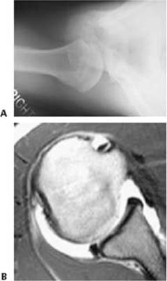
FIG 1 • A. Axillary lateral radiograph that demonstrates glenoid hypoplasia, which predisposes to posterior instability of the shoulder. B. Axial image from an MR arthrogram that demonstrates a posterior labral lesion. Contrast can be seen between the posterior labrum and the articular margin of the glenoid, indicating a labral tear or avulsion.
DIFFERENTIAL DIAGNOSIS
Posterior shoulder dislocation (may be locked)
Recurrent posterior subluxation
Multidirectional instability
Internal impingement
SLAP tear
Rotator cuff tear
Acromioclavicular joint injury
Fracture (eg, glenoid, greater tuberosity)
NONOPERATIVE MANAGEMENT
An extended period of nonoperative management is warranted in most cases of posterior shoulder instability.
Nonoperative therapy constitutes physical therapy to regain a full and symmetric shoulder range of motion, with later emphasis placed on strengthening the rotator cuff and scapular stabilizing muscles.
The premise of a conditioning program is to enable the dynamic stabilizers of the shoulder to compensate for the deficient static stabilizers (eg, capsule, labrum).
Once full motion and strength are achieved, return to sport is gradually introduced.
SURGICAL MANAGEMENT
Surgical management of posterior instability is considered when an exhaustive rehabilitation program has failed to alleviate disabling posterior subluxation, or when instability is the result of a macrotraumatic event.
Preoperative Planning
All imaging studies are again reviewed and the pathology is determined.
Any bone deficiencies, loose bodies, and concomitant rotator cuff and SLAP tears should be evaluated and treatment determined before arrival in the operating room.
An examination under anesthesia is performed before positioning to confirm the diagnosis. This examination should consist of sulcus test, load and shift test, and manual circumduction test or jerk test.
Positioning
We prefer lateral decubitus positioning because this offers greater exposure than the beach-chair position for evaluating the posterior labrum and capsule.
An inflatable beanbag and kidney rests hold the patient in the lateral position.
Foam cushions are used to pad the axilla and all bony prominences, including the fibular head (protection of the peroneal nerve).
The operative extremity is placed in 10 pounds of traction in 45 degrees of abduction and 20 degrees of forward flexion (FIG 2).
Approach
We use an all-arthroscopic technique for this procedure, with a posterior portal that is used as the main working portal (through the posterior deltoid) and an anterior portal (placed through the rotator interval) that is used for arthroscopic visualization.

FIG 2 • A. Lateral decubitus is the preferred position for arthroscopic surgery of the posterior capsule and labrum. B. The arm is placed in 10 to 15 pounds of traction and slightly abducted and forward flexed.
TECHNIQUES
PORTAL PLACEMENT
 The glenohumeral joint is first injected (posteriorly) with 50 mL of sterile saline through an 18–gauge spinal needle.
A posterior portal is established 1 cm distal and 1 cm lateral to the standard posterior portal that is used for routine shoulder arthroscopy. This portal is often in line with the lateral border of the acromion (TECH FIG 1A).
Placement of this portal more laterally than typical allows adequate access to the posterior glenoid rim for later anchor placement.
An anterior portal is established high in the rotator interval via an inside-out technique with a switching stick. As an alternative, this portal can be established with a spinal needle via an outside-in technique (TECH FIG 1B).
The anterior switching stick is then replaced with an 8.25-mm distally threaded clear cannula.
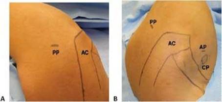
TECH FIG 1 • A. The posterior portal (PP) is marked in line with the lateral border of the acromion (AC). B. Surface landmarks identify the posterior portal (PP), acromion (AC), anterior portal (AP), and coracoid process (CP).
DIAGNOSTIC ARTHROSCOPY
With the arthroscope in the posterior portal, a diagnostic arthroscopy is performed.
The articular surfaces of the glenohumeral joint are inspected for chondral damage. The posterolateral aspect of the humeral head is inspected for any Hill-Sachs lesions (which may indicate combined anterior instability).
The anterior and inferior labrum is inspected and the glenohumeral ligaments are visualized.
The biceps tendon and superior labrum are probed to detect any pathology. Concomitant SLAP tears are common with posterior instability.
The rotator cuff is inspected (including the subscapularis tendon).
A switching stick is then placed in the posterior portal and replaced with an additional 8.25-mm distally threaded clear cannula. The arthroscope is then replaced into the anterior cannula for viewing; it remains there for the rest of the operation.
The posterior capsule and labrum are inspected and probed (TECH FIG 2).
The anterior humeral head surface is inspected for any reverse Hill-Sachs lesions, which may indicate macroinstability.

TECH FIG 2 • A. Arthroscopic view from the posterior portal showing an avulsed posterior labrum. B. A complete avulsion of the labrum off the posterior glenoid is visualized from the posterior portal.
PREPARATION OF THE GLENOID AND PLACEMENT OF SUTURE ANCHORS
Typically the posterior labrum is detached and the capsule attenuated, requiring the placement of suture anchors.
An arthroscopic rasp or chisel is used to mobilize the labrum from the glenoid rim.
The rasp is then used to débride the capsule to create an optimal environment for healing.
A motorized shaver or burr can be used on the glenoid rim to achieve a bleeding surface for healing.
Suture anchors are placed along the articular margin, not the glenoid neck, for the repair and capsular plication (TECH FIG 3A).
Typically we use three, 3-mm Bio-Suture Tak suture anchors with no. 2 FiberWire (Arthrex Inc., Naples, FL). A number of other commercially available anchors can be used in a similar fashion.
The anchor pilot holes are predrilled and the anchor is inserted with a mallet.
The anchor is placed so that the sutures are perpendicular to the glenoid rim. This facilitates passage of the most posterior suture through the torn labrum.
The anchors are evenly spaced on the posterior glenoid rim for a symmetric repair (TECH FIG 3B).
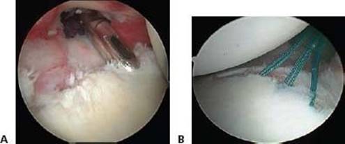
TECH FIG 3 • A. The anchor is placed on the glenoid margin. A drill is used to place a pilot hole before insertion of the anchor. B. The anchors are evenly spaced on the posterior glenoid margin to provide a symmetric and balanced repair.
LABRAL AND CAPSULAR REPAIR
A 45-degree Spectrum Hook (Linvatec Corp., Largo, FL) loaded with number 0 PDS suture (Ethicon, Somerville, NJ) is used to shuttle the suture through the capsule and labrum (TECH FIG 4A).
The suture hook is delivered through the capsule (if a plication is warranted) and under the torn labrum at the articular margin of the glenoid.
 An inferior-to-superior direction is used for this maneuver to achieve a small capsular plication.
This direction of suture passage is aimed at restoring tension to the posterior band of the inferior glenohumeral ligament.
Patients with significant instability clinically may require a more aggressive plication than those with isolated pathology to the glenoid labrum.
The PDS is fed into the glenohumeral joint and the passer is withdrawn.

TECH FIG 4 • A. A suture hook is used to shuttle the anchor limb through the capsulolabral complex. B. The PDS suture has been passed through the capsule and posterior labrum. C. The anchor limb suture is then shuttled via the PDS suture. D. The sutures are tied using arthroscopic knot-tying techniques through the posterior portal, and the capsulolabral plication is finished.
A suture grasper is then used to withdraw the most posterior suture in the anchor and the PDS that has been delivered through the capsulolabral complex.
Grabbing the more posterior suture helps to ensure that the suture limbs do not become entangled.
The PDS is then fashioned into a single loop and tied over the braided FiberWire suture.
The opposite limb of the PDS is then pulled and the FiberWire is delivered through the labrum and capsule (TECH FIG 4B,C).
Additional sutures are then shuttled in similar fashion to complete the repair.
After each suture has been shuttled through the capsule and labral complex, it is tied using arthroscopic knottying techniques (TECH FIG 4D).
We prefer to begin our repair inferiorly and advance superiorly up the posterior glenoid rim. In this way, the tension achieved with each advancing stitch can be assessed.
REPAIR COMPLETION
An arthroscopic awl is used to penetrate the posterior bare area of the humerus in an effort to achieve punctate bleeding to augment the healing response.
The posterior cannula is then withdrawn to just posterior to the level of the capsule and the posterior capsular incision is closed with a PDS suture.
A crescent Spectrum suture passer is used to penetrate one side of the capsule by the posterior capsular incision, and the suture is threaded into the joint.
The suture is retrieved through the opposite side of the incision with a penetrator and an arthroscopic knot is tied down to close the portal (TECH FIG 5).
Varying the distance of the suture from the portal incision allows additional tension to be applied to the posterior capsule.
If additional plication is warranted (such as in multidirectional instability), additional sutures can be placed in the rotator interval or anterior capsule as described elsewhere in this text.
The skin portals are closed with interrupted nylon suture and the patient is placed in a sling that allows slight abduction.

TECH FIG 5 • The repair is completed after closure of the posterior portal (suture shown in black).

POSTOPERATIVE CARE
The patient leaves the operating room in an abduction sling that can be removed for passive range-of-motion exercises at home.
We allow 90 degrees of forward elevation and external rotation to 0 degrees by 4 weeks after surgery.
The sling is discontinued 6 weeks after surgery and activeassisted range-of-motion exercises and gentle passive rangeof-motion exercises are progressed.
Pain-free, gentle internal rotation exercises are instituted at 6 weeks.
At 2 to 3 months after surgery, range of motion is progressed to achieve full passive and active range of motion.
Stretching exercises can be instituted for any deficiency in motion at this point.
After 4 months, the shoulder is often pain-free and eccentric rotator cuff strengthening is begun.
At 5 months, isotonic and isokinetic exercises are advanced.
At 6 months, throwing athletes undergo isokinetic strength testing.
If 80% of the strength and endurance of the contralateral extremity is attained, a throwing program is begun.
Full, competitive throwing is typically not attained until 12 months after surgery.
Nonthrowing athletes are often released to a sport-specific program by 6 months, when 80% of their strength has returned.
OUTCOMES
Arthroscopic posterior stabilization has achieved good results with respect to recurrence of instability and return to sport in athletes.
Studies have shown rates of recurrence of 0% to 8% and rates of return to sport of 89% to 100%.3,7,15
COMPLICATIONS
Recurrent instability
Stiffness
Infection
Neurovascular injury
REFERENCES
1. Blasier RB, Soslowsky LJ, Malicky DM, et al. Posterior glenohumeral subluxation: active and passive stabilization in a biomechanical model. J Bone Joint Surg Am 1997;79A:433–440.
2. Boyd HB, Sisk TD. Recurrent posterior dislocation of the shoulder. J Bone Joint Surg Am 1972;54A:779.
3. Bradley JP, Baker CL 3rd, Kline AJ, et al. Arthroscopic capsulolabral reconstruction for posterior instability of the shoulder: a prospective study of 100 shoulders. Am J Sports Med 2006;34: 1061–1071.
4. Gartsman GM, Hammerman SM. Superior labrum anterior and posterior lesions: when and how to treat them. Clin Sports Med 2000; 19:115–124.
5. Hawkins RJ, Koppert G, Johnston G. Recurrent posterior instability (subluxation) of the shoulder. J Bone Joint Surg Am 1984;66A: 169.
6. Keppler P, Holz U, Thielemann FW, et al. Locked posterior dislocation of the shoulder: treatment using rotational osteotomy of the humerus. J Orthop Trauma 1994;8:286–292.
7. Kim SH, Ha KI, Park JH, et al. Arthroscopic posterior labral repair and capsular shift for traumatic unidirectional recurrent posterior subluxation of the shoulder. J Bone Joint Surg Am 2003;85A: 1479–1487.
8. Kim SH, Park JC, Jeong WK, et al. The Kim test: a novel test for posteroinferior labral lesion of the shoulder: a comparison to the jerk test. Am J Sports Med 2005;33:1188–1192.
9. Kim SH, Park JC, Park JS, et al. Painful jerk test: a predictor of success in nonoperative treatment of posteroinferior instability of the shoulder. Am J Sports Med 2004;32:1849–1855.
10. McLaughlin HL. Posterior dislocation of the shoulder. J Bone Joint Surg Am 1952;34A:584.
11. Pollock RG, Bigliani LU. Recurrent posterior shoulder instability. Diagnosis and treatment. Clin Orthop Relat Res 1993;291:85–96.
12. Silliman JF, Hawkins RJ. Classification and physical diagnosis of instability of the shoulder. Clin Orthop Relat Res 1993;291:7–19.
13. Tibone JE, Bradley JP. The treatment of posterior subluxation in athletes. Clin Orthop 1993;291:124–137.
14. Walch G, Ascani C, Boulahia A, et al. Static posterior subluxation of the humeral head: an unrecognized entity responsible for glenohumeral osteoarthritis in the young adult. J Shoulder Elbow Surg 2002;11:309–314.
15. Williams RJ III, Strickland S, Cohen M, et al. Arthroscopic repair for traumatic posterior shoulder instability. Am J Sports Med 2003; 31:203–209.