
Matthew L. Ramsey
DEFINITION AND PATHOGENESIS
 Posttraumatic conditions of the elbow represent a variety of disorders involving the elbow as a result of previous injury. Included among the posttraumatic conditions are:
 Posttraumatic arthriti.
 Primary pathology involves posttraumatic degeneration of the articular surface.
Secondary pathologies can include contracture, loose bodies, and heterotopic bone.
Nonunion of the distal humeru.
Total elbow arthroplasty (TEA) is considered when reconstruction of the nonunion is deemed impossible or undesirable.
Dysfunctional instability of the elbo.
This is a special clinical situation where the fulcrum for stable elbow function is lost. The forearm may be dissociated from the brachium (FIG 1).
Chronic instability (dislocation.
Chronic ligamentous instability of the elbow can lead to articular degeneration, particularly in the elderly, osteopenic patient.
Treatment for posttraumatic conditions is individualized depending on the underlying pathology as well as the functional demands and age of the patient.
PATIENT HISTORY AND PHYSICAL FINDINGS
The patient history is directed at gaining information about the initial injury, treatments undertaken, complications of treatment, presenting complaints, and patient expectations.
Detailed investigation of the patient’s symptoms should include questions regarding the degree of pain, presence of instability or stiffness, and mechanical symptoms of catching, or locking.
The physical examination of the elbow should follow a systematic approach:
Inspection of the elbo.
Presence and location of previous skin incisions or persistent wounds
Alignment of the extremity at rest
Prominent hardware
Range of motion (ROM.
Active ROM is assessed and compared to the opposite side. The degree of motion, smoothness of motion, and feel of the endpoint are established.
Normal active ROM varies, but it should be symmetrical with the opposite unaffected side. Range of motion should be from near full extension (may have hyperextension) to 130 to 140 degrees of flexion. Normal forearm rotation is an arc of 170 degrees, with slightly more supination than pronation.
Functional ROM has been defined as a flexion– extension arc from 30 degrees to 130 degrees and a pronation–supination arc from 50 degrees of pronation and 50 degrees of supination.10
Passive range of motion (PROM) is then assessed and compared to the active motion arc.
Palpation of the elbow should systematically review all of the bony and soft tissue structures of the elbow.
The ulnar nerve needs to be carefully assessed. If previously surgically manipulated, its location should be identified if possible.

FIG 1 • Radiograph demonstrating dissociation of the forearm from the brachium in a patient with an inadequately treated fracture of the distal humerus with resultant nonunion.
Motor function of the elbow should be assessed, in particular the flexor (biceps and brachialis) and extensor (triceps) function.
IMAGING AND OTHER DIAGNOSTIC STUDIES
Orthogonal radiographic views of the elbow are mandatory (FIG 2).
A good lateral radiograph can typically be obtained.
A useful anteroposterior (AP) radiograph can be difficult, particularly if the patient has significant flexion contracture. A poor AP radiograph can make assessment of the joint space difficult, typically resulting in overestimating the amount of joint destruction.
Oblique radiographs can be helpful in obtaining more detail.
CT scans are particularly helpful in assessing the integrity of the bone and establishing whether the joint space is reasonably preserved.
Three-dimensional reconstructions provide a better understanding of any deformity.
Magnetic resonance imaging is rarely needed in the assessment of a posttraumatic joint and is therefore used sparingly.

FIG 2 • AP and lateral radiographs of the elbow in a patient with posttraumatic arthritis of the elbow.
DIFFERENTIAL DIAGNOSIS
Nonunion or malunion of the distal humerus
Posttraumatic stiffness of the elbow
Chronic dislocation of the elbow
NONOPERATIVE MANAGEMENT
The success of nonoperative management depends on specific features of the pathology and the motivation and goals of the patient.
Activity modification is used to reduce the forces across the elbow.
Range of motion of the elbow should be maintained. Aggressive efforts to regain lost motion can aggravate the joint.
External bracing is occasionally used to support an unstable extremity. However, in general, bracing is poorly tolerated and functionally limiting.
SURGICAL MANAGEMENT
Surgical management of traumatic conditions of the elbow is directed at addressing the underlying cause of disability and should take into consideration the patient’s age, physical requirements, and expectations.
Surgical Options
Interposition Arthroplasty2,7
Indication.
Patients with pain or loss of range of motion who have failed to respond to nonoperative management
Posttraumatic arthritis in patients who are either too young for TEA or who are unwilling to accept the functional restrictions with TEA
The patients who do best following interposition are those with painful loss of motion when there is no requirement for aggressive, heavy use of the extremity.
Contraindication.
Active infection (septic arthritis with persistent infection)
Grossly unstable elbow
Marked angular deformity
Pain without associated functional loss
Inadequate bone stock
Patients unable or unwilling to follow postoperative instructions
Total Elbow Replacement3–5,8,11,14,15
Patients with posttraumatic conditions of the elbow tend to be younger than other patients undergoing TEA.
In this group of patients, TEA should be considered in patients who:
Have failed to respond to appropriate nonoperative management
Are not appropriate candidates for other surgical options
Are willing to adopt a more sedentary lifestyle
Have no absolute contraindications to the procedure
Preoperative Planning
Interposition Arthroplasty
Graft option.
Achilles tendon allograft has the advantage of no donor site morbidity. It can also be used to reconstruct the collateral ligaments if necessary.
Dermis or fascia lata autogenous graft
Dermal tissue allograft
Revisio.
The salvage for a failed interposition arthroplasty is a TEA.1
Interposition arthroplasty should not be undertaken unless the surgeon is comfortable performing a total elbow replacement in the face of failure.
Total Elbow Replacement
Implants are described in terms of their physical linkage (linked, unlinked, or linkable) and based on their constraint (constrained, semiconstrained, minimally constrained).
Linkage is determined by whether the components are physically linked.
Constraint is a more poorly defined quality of the implant. It depends on the geometry of the implant and its interaction with stabilizing soft tissues about the elbow.6
Implant selection in posttraumatic arthriti.
Linked (semiconstrained) designs: Linked implants have the advantage of being universally applicable to all posttraumatic conditions of the elbow.
Unlinked designs: The requirement for the use of unlinked designs in posttraumatic conditions of the elbow is integrity of the collateral ligaments and limited deformity such that normal anatomic relationships can be re-established.
Linkable designs: Linkable designs have been developed to take advantage of the features of an unlinked implant while capturing the universal applicability of the linked implants. They can be converted from unlinked to linked either at the time of an initial surgery if stability cannot be conferred or remotely if instability becomes an issue postoperatively.
Positioning
Interposition arthroplast.
Supine with the arm across the chest and a bump under the ipsilateral shoulder
Alternatively, the lateral decubitus position with the arm over an arm holder
Total elbow replacement
Patients are placed supine on the operating table with a bump under the ipsilateral shoulder. The arm should be freely mobile through the shoulder to allow manipulation of the joint throughout surgery. The arm can then be placed across the body on a bump or externally rotated through the shoulder and flexed at the elbow (FIG 3).

FIG 3 • Patient positioning for total elbow arthroplasty with the arm across the body supported on a bolster.
TECHNIQUES
INTERPOSITION ARTHROPLASTY
 Posterior skin incision: Develop medial and lateral subcutaneous flaps.
Isolate and transpose the ulnar nerve.
Perform deep exposure to the elbow through an extensile Köcher approach.9 The triceps can be partially released from the ulna to allow the triceps–anconeus composite to be mobilized (TECH FIG 1A).
Mobilize the common extensor group from the anterior capsule and release it proximally with the extensor carpi radialis longus.
Isolate the lateral ulnar collateral ligament and release it from its humeral origin (TECH FIG 1B). Perform an anterior and posterior capsular release. Supination of the forearm allows the ulna to be rotated away from the humerus. Attempt to leave the medial collateral ligament intact as it will improve postoperative stability.
Inspect the cartilage surfaces. If more than 50% of the articular surface is involved, surgery proceeds to interposition.
If extensile exposure is required, the extensile Köcher approach can be expanded to a triceps-reflecting anconeus pedicle (TRAP) approach (TECH FIG 1C,D).13
Reshape the distal humerus to conform to the olecranon. Remove the cartilage from the distal humerus and smooth the bone, but avoid aggressive resection of bone (TECH FIG 1E).
Prepare the interposition tissue. The graft of choice is up to the surgeon, but there is a growing experience with allograft Achilles tendon. In addition to being a robust graft source, it allows for reconstruction of one or both collateral ligaments (TECH FIG 2A).
Place drill holes across the supracondylar region from anterior to posterior (TECH FIG 2B). These drill holes are placed at the medial aspect of the trochlea, above the trochlear sulcus, at the lateral margin of the trochlea, and at the lateral aspect of the capitellum.
Drape the interposition tissue over the distal humerus and secure it with sutures placed through the graft from front to back. If there is collateral ligament insufficiency, the tails of the graft (especially when using Achilles tendon) can be fashioned to reconstruct the collateral ligaments (TECH FIG 2C).
Leave the radial head intact, especially if medial collateral ligament reconstruction is performed, to contribute to the valgus stability of the elbow.
Repair the lateral collateral ligament through drill holes at the center of rotation laterally. Do not tie the ligament until the external fixator is securely applied.
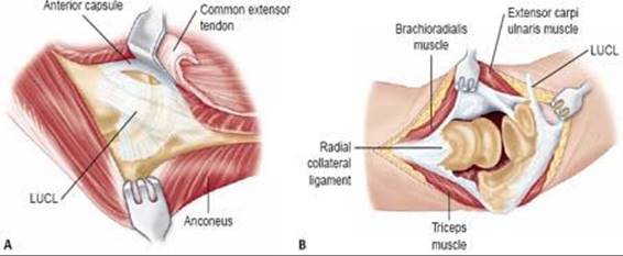

TECH FIG 1 • A. Extensile Köcher approach to the lateral elbow. The anconeus and triceps are elevated off the posterolateral capsule while the common extensor group is elevated off the anterior capsule. Exposure can be extended posteriorly with partial release of the triceps from the lateral aspect of the olecranon. B. Deep extensile exposure requires release of the lateral collateral ligament and anterior and posterior capsule. C,D. The triceps-reflecting anconeus pedicle (TRAP) approach is an alternative approach that allows extensile exposure. C. The medial interval is along the medial triceps proximally and between the anconeus and flexor carpi ulnaris. The triceps is reflected from medial to lateral (Bryan-Morrey approach) off the olecranon in continuity with the anconeus. D. The lateral interval is an extensile Köcher approach between the anconeus and the extensor carpi ulnaris, which is extended proximally along the lateral supracondylar column. The triceps–anconeus composite maintains the neurovascular pedicle to the anconeus from above while allowing extensile exposure to the joint. E. The remaining cartilage on the distal humerus is removed and the subchondral bone is reshaped. Care should be taken to retain as much subchondral bone as possible for structural support of the interposition membrane.
Hinged Elbow External Fixator
Apply a hinged external fixator to protect the interposed graft and to stabilize the joint while soft tissue healing occurs.
The axis of rotation of the elbow is defined by bony landmarks about the lateral and medial joint (TECH FIG 3A).
 The center of rotation at the lateral elbow is the center point of an arc defined by the articular surface of the capitellum.
 The center of rotation at the medial elbow is defined by tightly distributed instantaneous centers of rotation approximated by a point at the anterior inferior aspect of the medial epicondyle.
Establish an axis pin coincident with the lateral and medial centers of rotation. This is the foundation for construction of the fixator.
The type of fixator used dictates the method of pin insertion relative to the axis pin. Fixator systems that allow the humeral and ulnar pins to be placed independently and than assembled to the axis pin are easiest for the surgeon with limited experience (TECH FIG 3B).
When placing the humeral pin, take care to avoid injury to the neurovascular structures.
Pins in the proximal humerus are placed through the anterolateral aspect of the deltoid distal to the axillary nerve.
 Pins in the midshaft of the humerus are placed in the anterolateral humerus to avoid the radial nerve, which lies posteriorly.
Ulnar pins are placed along the posterolateral aspect of the ulna.

TECH FIG 2 • A. The interposition membrane is prepared with mattress sutures placed distally. The Achilles tendon allograft also permits reconstruction of the collateral ligaments if necessary. B. Drill holes are placed from posterior to anterior across the supracondylar region to secure the interposition graft. C. The interposition membrane is secured to the distal humerus. If necessary, the graft can be fashioned to reconstruct the collateral ligaments. (From Morrey BF, Larson AN. Interposition arthroplasty of the elbow. In: Morrey BF, Sanchez-Sotelo J, eds. The Elbow and Its Disorders, 4th ed. Philadelphia: Elsevier; 2009: Figure 69–6.)

TECH FIG 3 • A. Drawing demonstrating the center of rotation on the lateral and medial side of the elbow. B. Photograph demonstrating a hinged external fixator. The humeral and ulnar pins are independently attached to the hinge.
A bar is fixed to the humeral and another bar is fixed to the ulnar pins.
The hinge is loosely attached to the humeral and ulnar bars.
The joint is reduced and ligament reconstruction, if necessary, is completed.
With the joint reduced, the fixator is tightened. If desired, distraction can be applied.
TOTAL ELBOW REPLACEMENT
Surgical Approach
A straight posterior skin incision placed off the medial aspect of the olecranon is preferred. Previous incisions may modify the location of the incision. Regardless of the incision used, deep access to the medial and lateral aspect of the joint is essential.
Identify the ulnar nerve. If not previously handled surgically, the nerve is transposed anteriorly. If the nerve was previously transposed, it only needs to be identified, but not formally dissected unless the position of the nerve places it at risk during surgery.
Triceps Management
Triceps-reflecting approaches are preferred over tricepssparing approaches for posttraumatic conditions. Posttraumatic scarring and deformity can make a tricepssparing approach difficult unless a nonunited distal humeral segment is to be resected.
A Bryan-Morrey approach is typically performed (TECH FIG 4A,B).9 The medial aspect of the triceps is developed proximally while the interval between the anconeus and flexor carpi ulnaris (FCU) is developed distal to the olecranon. The triceps is reflected from medially to laterally in continuity with the anconeus. Release of the lateral and medial collateral ligaments completes the exposure and allows separation of the ulna from the humerus.
A modification of the Bryan-Morrey approach involves release of the triceps insertion onto the ulna through an extra-articular osteotomy of the dorsal tip of the ulna (TECH FIG 4C,D).16 The rationale for this modification relates to the recognized complication of triceps insufficiency that occurs with soft tissue release of the triceps. The osteotomy affords several advantages:
Bone-to-bone healing of the osteotomy is more reliable than soft tissue healing of the triceps to the ulna.
Failure of the osteotomy to heal can be identified radiographically and addressed early.
Deep Dissection
Release the collateral ligaments and capsule (TECH FIG 5). This permits the ulna to be separated from the humerus. If ligamentous integrity is necessary (ie, unlinked arthroplasty) then the lateral ulnar collateral ligament and medial collateral should be tagged with plans at reattachment via bone tunnels in the humerus during closure.
Release contracted muscles (flexor–pronator and common extensor) to correct deformity, which can result in maltracking of the TEA. Release the scarring about the elbow sufficiently to gain unencumbered access to the humerus and ulna for component implantation.
The tip of the olecranon can be removed to better visualize the trochlea.
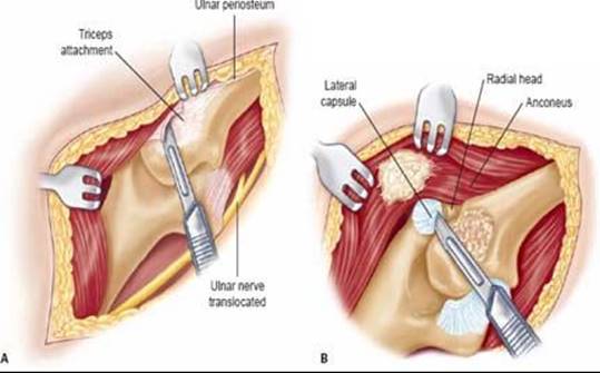

TECH FIG 4 • A,B. Bryan-Morrey triceps-reflecting approach. A. The triceps insertion is released in continuity with the anconeus from medial to lateral. B. Further dissection allows the collateral ligaments to be released. C,D. The osteo-anconeus flap approach. The triceps is reflected from medial to lateral in the distal interval between the flexor carpi ulnaris and anconeus.
Component Insertion and Completion
Insertion of total elbow implants is performed in standard fashion and is described in Chapter SE-45.
After component insertion, the triceps mechanism is repaired through bone tunnels in the ulna. When a sliver of bone is taken with the triceps insertion, transverse tunnels are made. Each limb of nonabsorbable suture is tied over the top of the triceps and bone fragment. An additional cerclage suture is brought through one of the two transverse tunnels and is brought around the tip of the olecranon, incorporating the triceps insertion. This suture counters the pull of the triceps.
The anconeus is repaired to the flexor carpi ulnaris fascia. Similarly, the medial triceps is repaired to the flexor– pronator group.
Subcutaneous ulnar nerve transposition is routinely performed.
A subcutaneous drain is placed and wound closure is performed.

TECH FIG 5 • An extra-articular osteotomy of the tip of the olecranon is performed, leaving the triceps attached to the fragment. The shoulder is externally rotated and the elbow is hyperflexed to allow separation of the ulna from the humerus.
PEARLS AND PITFALLS

POSTOPERATIVE MANAGEMENT
Interposition Arthroplasty
ROM is started as quickly as allowed by the condition of the soft tissues. In general, immediate motion is preferred. However, the prerequisite is a quiet soft tissue envelope. ROM may be assisted with a continuous passive motion machine if desired.
Patients are taught pin care, which is performed daily at home.
Patients are seen at 10 to 14 days postoperatively for staple removal and wound check and every 2 weeks thereafter until pin removal.
The external fixator is left in place for about 4 to 6 weeks and then removed in the operating room with assessment of elbow stability and motion under anesthesia.
I prefer to wait 6 weeks to allow collateral ligament healing since instability is the most common complication after fixator removal.
Rehabilitation is continued, focusing on obtaining a functional ROM.
Total Elbow Replacement
The elbow is immobilized in full extension in a well-padded anterior splint.
The arm is elevated on pillows or suspended from an IV pole to reduce swelling.
The splint is removed 24 to 48 hours after surgery.
Gentle active ROM is begun in flexion, pronation, and supination. Active extension is avoided for 6 weeks to protect the triceps repair. However, gravity-assisted extension or passive extension is permitted.
In general, formal physical therapy is rarely required to regain ROM. However, it may be beneficial in patients who struggle to regain their ROM. The general timeline of therapy is:
Phase I (0 to 6 weeks): Protect the soft tissue and begin protected active-assistive ROM.
Phase II (6 to 12 weeks): Continue to improve ROM. Begin strengthening exercises and encourage functional use of the arm.
Phase III (12 to 16 weeks): Return to normal functional activities within the restrictions for TEA.
Postoperative stiffness may be helped with splinting. Static splinting is preferred over dynamic splinting.
Restrictions: Lifetime limitations of the operated extremity include 2to 5-pound repetitive lifting and 10-lb single-event restriction.
OUTCOMES
Interposition Arthroplasty
The most predictable results for interposition occur in patients presenting with:
Stiffness and pain preoperatively
Stable elbow
One or no ligament reconstruction required at surgery
Poor results are noted when.
Pain is the only presenting complaint
Elbow is unstable
Reconstruction of both the medial and lateral collateral ligaments is needed at the time of interposition
Most studies report a 70% satisfaction rate among patients with respect to pain relief; 80% of patients regain a functional ROM.
Cheng and Morrey2 found that 67% of patients treated for rheumatoid arthritis had satisfactory relief of pain, and 75% of patients treated for osteoarthritis were satisfied at 5-year follow-up.
Total Elbow Arthroplasty
Patients undergoing TEA for posttraumatic conditions of the elbow tend to be younger and have higher demand.
TEA for posttraumatic conditions of the elbow is associated with improved clinical outcomes.
A higher complication rate is noted for posttraumatic conditions compared to other indications for TEA.
Mechanical complications such as component fracture and increased polyethylene bushing wear are more common. Causes of increased complications include.
Multiple previous surgeries
Deformity of the elbow requiring realignment of the extremity through the implant
COMPLICATIONS
Interposition Arthroplasty
Complications of interposition arthroplasty include.
Instability
Infection
Ulnar neuropathy
Resorptive bone loss
Heterotopic bone formation
Complications related to the external fixator include.
Superficial pin tract infections
Deep infection (osteomyelitis)
Pin breakage
In the literature, complications have been reported to occur in up to 25% of patients.
Total Elbow Replacement
TEA for traumatic conditions is associated with a high complication rate. Major complications include:
Infectio.
Current reports indicate an infection rate of 2% to 5% for primary TEA.
Higher infection rates are noted with posttraumatic arthritis and a history of prior surgery.
Loosening
Triceps insufficiency (an underrecognized problem)
Neurologic injury (incidence of transient ulnar neuropathy as high as 26% and permanent nerve injury up to 10%)
Wound complication.
Associated with prior surgery
Manage wound by immobilizing in extension postoperatively; use a subcutaneous drain to avoid hematoma formation. A significant postoperative hematoma should be evacuated.
Periprosthetic fracture (can occur intraoperatively or postoperatively; incidence ranges from 1% to 23%)
REFERENCES
· Blaine TA, Adams R, Morrey BF. Total elbow arthroplasty after interposition arthroplasty for elbow arthritis. J Bone Joint Surg Am 2005;87A:286–292.
· Cheng SL, Morrey BF. Treatment of the mobile, painful arthritic elbow by distraction interposition arthroplasty. J Bone Joint Surg Am 2000;82A:233–238.
· Figgie MP, Inglis AE, Mow CS, et al. Salvage of non-union of supracondylar fracture of the humerus by total elbow arthroplasty. J Bone Joint Surg Am 1989;71A:1058–1065.
· Figgie HE III, Inglis AE, Ranawat CS, et al. Results of total elbow arthroplasty as a salvage procedure for failed elbow reconstructive operations. Clin Orthop Relat Res 1987;219:185–193.
· Inglis AE, Inglis AE Jr, Figgie MM, et al. Total elbow arthroplasty for flail and unstable elbows. J Shoulder Elbow Surg 1997;6:29–36.
· Kamineni S, O’Driscoll SW, Urban M, et al. Intrinsic constraint of unlinked total elbow replacements: the ulnotrochlear joint. J Bone Joint Surg Am 2005;87A:2019–2027.
· Larson AN, Morrey BF. Interposition arthroplasty with an Achilles tendon allograft as a salvage procedure for the elbow. J Bone Joint Surg Am 2008;90A:2714–2723.
· Moro JK, King GJ. Total elbow arthroplasty in the treatment of posttraumatic conditions of the elbow. Clin Orthop Relat Res 2000;370:102–114.
· Morrey BF. Surgical exposures of the elbow. In: Morrey BF, SanchezSotelo J, eds. The Elbow and its Disorders, 4th ed. Philadelphia: Saunders Elsevier, 2009:115–142.
· Morrey BF, Askew LJ, Chao EY. A biomechanical study of normal functional elbow motion. J Bone Joint Surg Am 1981;63A:872–877.
· Morrey BF, Schneeberger AG. Total elbow arthroplast1y for posttraumatic arthrosis. AAOS Instr Course Lect 2009;58:495–504.
· Nolla J, Ring D, Lozano-Calderon S, et al. Interposition arthroplast1y of the elbow with hinged external fixation for post-traumatic arthritis. J Shoulder Elbow Surg 2008;17:459–464.
· O’Driscoll SW. The triceps-reflecting anconeus pedicle (TRAP) approach for distal humeral fractures and nonunions. Orthop Clin North Am 2000;31:91–101.
· Ramsey ML, Adams RA, Morrey BF. Instability of the elbow treated with semiconstrained total elbow arthroplast1y. J Bone Joint Surg Am 1999;81A:38–47.
· Schneeberger AG, Adams R, Morrey BF. Semiconstrained total elbow replacement for the treatment of post-traumatic osteoarthrosis. J Bone Joint Surg Am 1997;79A:1211–1222.
· Wolfe SW, Ranawat CS. The osteo-anconeus flap: an approach for total elbow arthroplast1y. J Bone Joint Surg Am 1990;72A: 684–688.