
Paul S. Shurnas, Mark E. Easley, Troy Watson, and Amy Sanders
DEFINITION
 Symptomatic hallux valgus, or bunion deformity, is a common problem seen in foot and ankle and general practice clinics.
 Historically, it is seen almost exclusively in persons who wear shoes.
It is characterized by a painful prominence at the medial aspect of the great toe.
The deformity is exemplified by lateral deviation (valgus) of the great toe proximal phalanx and medial (varus) deviation of the first metatarsal.
Juvenile hallux valgus deformity usually is a combination of valgus inclination of the metatarsal articular surface (i.e., increased distal metatarsal articular angle [DMAA]) and varus deformity of the first metatarsal.
Deformity may be classified as mild, moderate, or severe, evaluated on weight-bearing radiographs of the foot and based on the following criteria:
The degree of valgus at the metatarsophalangeal (MTP) joint or hallux valgus angle (HVA)
The degree of varus deformity of the first metatarsal or 1–2 intermetatarsal angle (IMA)
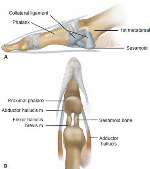
FIG 1 • Collateral ligaments and sesamoid tendon relations. A. Collateral S mesh with sesamoid complex. B. Sesamoid and tendon relations about the metatarsophalangeal (MTP) joint.
Advanced deformity is more complex, and the hallux exhibits the following:
Toe pronation noted clinically by medial rotation of the toenail
Sesamoid subluxation noted on the anteroposterior (AP) radiograph and sesamoid view
Medial capsular laxity and lateral capsular contracture
ANATOMY
The great toe MTP joint is unique when compared to the lesser MTP joints because of the sesamoid complex, unique tendon insertions, and ligamentous support about the joint (FIG 1).
The sesamoid ligaments mesh with the collateral ligaments both medially and laterally.
The tendons of the flexor hallucis brevis, abductor and adductor hallucis, plantar aponeurosis, and joint capsule coalesce to form the plantar plate, surrounding and stabilizing the first metatarsal head (FIG 2).
Because there are no true tendon insertions on the first metatarsal head, it is vulnerable to varus deviation.
An intermetatarsal facet occasionally is present between the first and second metatarsal bases, sometimes creating a rigid metatarsus primus varus.
The first metatarsal blood supply is derived from arterial supply primarily through the lateral midshaft, and its flow is distal.
Intraosseous flow is variable with respect to proximal and distal branches.
The primary arterial sources are the first dorsal and plantar metatarsal arteries and the superficial branch of the medial plantar artery.8
The DMAA is defined by the relationship between the metatarsal long axis and the distal metatarsal articular surface lateral inclination (FIG 3).
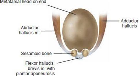
FIG 2 • Plantar plate contributions.
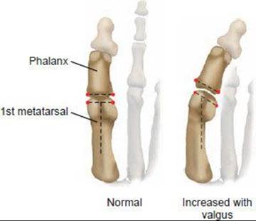
FIG 3 • Distal metatarsal angle (DMAA).
PATHOGENESIS
The concept of a hallux valgus deformity with a congruent or subluxated MTP joint is important (FIG 4).
Whereas hallux valgus with a subluxated joint usually is progressive, congruent joints tend to be static deformities.
Congruent joint hallux valgus deformity is associated with an increased DMAA and juvenile hallux valgus.
A flat metatarsal head, with little convexity, is associated with hallux rigidus.3
A rounded metatarsal head is associated with greater MTP instability and hallux valgus. As the proximal phalanx deforms laterally, the metatarsal head shifts medially, increasing both the HVA and 1–2 IMA.6
The inciting event leading to hallux valgus is poorly understood. Typically, with longstanding subluxated hallux valgus deformity, the medial capsule attenuates and the lateral capsule contracts.
The sesamoid complex remains in its physiologic position as the metatarsal head shifts medially. The weak link is thought to be the medial capsule immediately superior to the insertion of the abductor hallucis.6
Ultimately, the abductor hallucis slides plantar to the metatarsal head, leading to a lack of intrinsic muscle stability to the first MTP joint, with resultant pronation of the phalanx (FIG 5).

FIG 4 • Congruent versus subluxed joint. A. A congruent joint may be associated with hallux valgus when the DMAA is increased, as seen in juvenile hallux valgus. B. Joint in subluxed position.

FIG 5 • The abductor hallucis slides under the metatarsal head, contributing to pronation of the toe. A. End-on view of toe and nail (normal). B. End-on view with pronation.
With deformity progression, callus may develop along the plantar, medial interphalangeal (IP) joint. A lateral weight shift away from the hallux to the lesser MTP joints may occur, creating lesser MTP joint instability and further callus formation.
NATURAL HISTORY
Hallux valgus with a subluxated joint usually is progressive, and the pathogenesis described in previous sections commonly is observed over time.
Hallux valgus associated with a congruent joint tends to be more static in terms of deformity.
Subluxated and congruent deformities may become symptomatic over time due to shoe pressure, cutaneous nerve irritation, bursitis, callus formation and a painful medial eminence.
With progressive or painful deformities, other lesions may develop, such as associated lesser toe deformities, Morton's neuroma, lesser MTP joint capsular instability, stress fractures, skin ulceration, and hallux or lesser MTP joint dislocation.
PHYSICAL FINDINGS
A reddened prominence over the medial aspect of the great toe MTP joint, or “bunion,” often develops with pressure from shoe wear (Table 1).
Many patients exhibit callus formation under the second or third metatarsal heads because the displaced first metatarsal is not bearing weight in a balanced manner with the lesser metatarsal heads.
On palpation of the foot, most patients are tender over the medial eminence or show irritability in the cutaneous nerve.
A large dorsal metatarsal prominence is more commonly associated with hallux rigidus and is not typical of symptomatic hallux valgus.
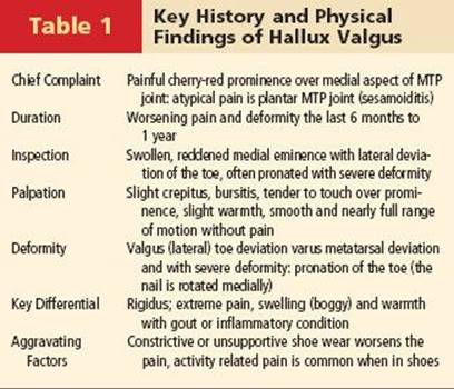
Joint range of motion in hallux valgus, even with severe deformity, usually is well preserved, without crepitance, and with minimal pain.
Range of motion also is checked while gently reducing the deformity out of valgus.
Chronic deformities or congruent joints with increased DMAA may exhibit less dorsiflexion at the MTP joint.
Palpation of the first metatarsal cuneiform (MTC) joint is performed.
Prominence, swelling, and pain with cantilever stress of the first MTC joint, if present, should be noted.
Mobility of the first MTC joint and gastrocnemius tightness are assessed.
Other painful areas are sought out, including those with callus formation, the lesser MTP joints, intermetatarsal spaces, bunionette deformities, and hammer toes.
We routinely also analyze the patient's gait, with particular attention focused on the stance phase and evaluation of hindfoot position and status of the longitudinal arch.
Hindfoot joints are examined, tendon strength is checked, and general alignment about the foot and ankle are noted. In select patients, correction of concomitant pes planovalgus deformity, either simultaneously or in a staged fashion, may be warranted, because a valgus hindfoot may predispose to progression or recurrence of hallux valgus.
Pulses are palpated, sensation is assessed, and the skin is inspected. Poor circulation should prompt a vascular evaluation, and loss of protective sensation may indicate neuropathy and may be a contraindication to corrective hallux valgus surgery. Previous forefoot incisions should be noted and considered in preoperative planning.
IMAGING AND DIAGNOSTIC STUDIES
Weight-bearing AP and lateral radiographs are routinely obtained.
MRI, CT, and bone scans are rarely indicated. The reported normal radiographic values of the hallux MTP joint are a hallux valgus angle (HVA) no greater than 15 degrees, a 1–2 IMA of no more than 9 degrees, and an IP angle of less than 10 degrees.5,6
Standardized mid-diaphyseal reference points are used to measure the HVA and IMA.
The IP angle is measured by a line bisecting the base of the proximal phalanx and the long axis of the phalanx.
Although precise measurement of the DMAA has been controversial, it is critical that it be considered, because subtotal correction and persistent deformity will result if it is not addressed at the time of surgery.
A DMAA of more than 15 degrees is considered increased.1,2 It is measured based on the AP weight-bearing radiograph (Fig. 3).
Hallux valgus interphalangeus (HVI) is measured by the IP angle. HVI has been associated primarily with hallux rigidus,3 but occasionally it occurs with hallux valgus.
Proximal first metatarsal osteotomies and distal soft tissue procedures do not correct an increased HVI; phalangeal osteotomies are required to correct HVI.
MTC instability may be indicated by a first MTC angle of more than 10 degrees or excess joint obliquity on the weight-bearing AP radiograph, plantar gapping of more than 2 mm on the lateral weight-bearing view, or an intermetatarsal os.
Although first ray hypermobility is controversial, in select patients, an increased 1–2 angle may be best corrected with a first MTC arthrodesis in lieu of a proximal first metatarsal osteotomy.
An intermetatarsal facet at the base of the first and second metatarsals may directly impede correction of the 1–2 IMA.
Hallux valgus associated with advanced arthrosis of the first MTP joint may preclude joint-preserving operations, and typically is best managed with a first MTP joint arthrodesis.
DIFFERENTIAL DIAGNOSIS
Inflammatory arthritis of many varieties can result in hallux valgus.
Traumatic hallux valgus or hallux varus can occur.
Adult or congenital flatfoot, posterior tibial tendon deficiency, generalized hyperlaxity, and first MTC instability can exacerbate hallux valgus.
Tarsal coalition or symptomatic accessory navicular with associated hallux valgus may occur.
Neuromuscular disorders are associated with hallux valgus.
NONOPERATIVE MANAGEMENT
Shoe wear modification, the mainstay of nonoperative management, includes shoe stretching, wider toe box shoes, and, occasionally, accommodative orthotics.
Toe spacers, night splints, bunion pads or posts, and other inventive devices may help reduce symptoms.
SURGICAL MANAGEMENT
More than 120 procedures have been described to surgically correct hallux valgus, including a variety of proximal first metatarsal base, shaft, and distal osteotomies.
The goals of an ideal proximal base osteotomy are reliable, powerful, predictable correction with stable fixation to allow for early weight bearing.
Proximal metatarsal opening wedge osteotomy (PMOW) with a newer low-profile plate fixation system (Arthrex, Inc., Naples, FL) is, in our opinion, nearly ideal for addressing those goals.
TECHNIQUES
PROXIMAL METATARSAL OPENING WEDGE OSTEOTOMY
 PMOW combined with a distal soft tissue procedure (ie, lateral capsular release and medial capsular placation) is considered for moderate to severe hallux valgus, hallux valgus associated with a short first ray, a 1–2 IMA of more than 12 degrees, and recurrent hallux valgus, either after a distal procedure alone or as an adjunct to a distal procedure if subtotal correction is achieved.
Two or three 3-cm incisions are used, depending on which distal procedure is being performed.
 The first longitudinal incision is centered over the medial eminence, and a simple bunionectomy is performed in a routine fashion.
 We prefer to use an inverted L-shaped capsulotomy and save bone resected from the medial eminence to use as autograft in the PMOW.
Alternatively, cancellous graft may be harvested from the lateral calcaneus through a 1to 2-cm lateral heel incision.
We make the longitudinal incision for the PMOW dorsomedially, beginning just distal to the first MTC joint.
The superficial peroneal nerve branch to the hallux and the extensor hallucis longus tendon must be identified and protected.
The osteotomy is initiated medially about 1.5 cm distal to the joint, slightly oblique (about 20–30 degrees) toward the lateral aspect of the first metatarsal base, without violating the lateral cortex (TECH FIG 1A). Minimal periosteal stripping is required.
The osteotomy is gently opened with three successive osteotomes (largest blade first) from the PMOW set, and care is take to preserve the lateral hinge of bone and soft tissue, if possible. “Stacking” osteotomes diminishes the risk of breaking the lateral cortex, which is more likely to occur when a single osteotome is used to lever the osteotomy open.
Once the osteotomy is opened, manual pressure over the medial eminence and a mini lamina spreader (supplied in the set) are used to obtain the desired correction and verified fluoroscopically.
 Alternatively, a measuring wedge (also provided in the set) may be utilized.
If the lateral cortical hinge should fail, the mini lamina spreader is quite useful.
The osteotomy site is held open and the plate applied as described, which typically reduces the lateral cortex.
To gain further support to the lateral cortex, one of the proximal screws may be placed not only through the plate but also across the osteotomy to capture the distal lateral cortex. Alternatively, an additional oblique screw may be added outside the plate.
About 5% of our cases have resulted in lateral cortex fracture with no delay in healing or modification in the postoperative protocol.
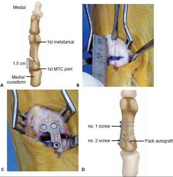
TECH FIG 1 • A. Site and position of first metatarsal opening wedge osteotomy. The proximal metatarsal opening wedge osteotomy is initiated about 1.5 cm distal medial to the first metatarsal cuneiform (MTC) joint. B. Site of osteotomy 1.5 cm distal to the first MTC joint. C. The plate has been set into position by insertion of the first screw. D. Screw placement with opening wedge plate.
Based on the authors' clinical data utilizing the oblique osteotomy, a general rule for preoperative planning is approximately 3 degrees of correction per millimeter of opening wedge.
The desired wedge is selected and the first screw is placed in the distal hole closest to the osteotomy to set the plate (TECH FIG 1B,C).
The next screw placed is in one of the proximal holes. We prefer to place both of these screws obliquely across the apex of the osteotomy (TECH FIG 1D).
If there is any concern about stability, an additional screw can be placed obliquely outside the plate to enhance the construct.
The final screw is placed distally.
With the plate securely fixed, fluoroscopy is used with the foot flat on the table to verify a congruent joint and increased DMAA or to determine whether any further correction is required.
With subluxated deformities, the PMWO is combined with a modified McBride bunionectomy using two or three incisions.
The third incision, if used, is for the first web space. However, with this technique an aggressive lateral release typically is not required.
If the joint is congruent with an increased DMAA or if still more correction is desired, a biplanar chevron incision with a long dorsal limb is used (TECH FIG 2A–C).
Any IP deformity or residual pronation can be treated with an Akin osteotomy.
The capsule is repaired through a drill hole at the metadiaphyseal junction or with mattress suture technique proximally if the tissue quality is satisfactory. The soft tissues over the osteotomy site are closed in a layered fashion, after autologous graft is impacted.
Nylon sutures are used for the skin.

TECH FIG 2 • A. Preoperative weight-bearing AP radiograph shows a severe hallux valgus deformity with a bipartite tibial sesamoid and mild degenerative changes. B. Intraoperative radiograph shows good correction of the 1–2 IMA to less than 9 degrees but an increased DMAA of 25 degrees. C. Six week postoperative weightbearing AP radiograph shows good alignment post-PMOW first metatarsal and distal biplanar chevron bunionectomy.
PEARLS AND PITFALLS
 Start slightly oblique (10–15 degrees) osteotomy at least 1.5 cm distal to the first tarsometatarsal joint.
A small lamina spreader is useful to obtain desired correction.
The opposite side plate may fit better on the base of the first metatarsal in some people.
Avoid an aggressive lateral release; pie-crusting release through the joint is usually enough.
Distal biplanar chevron osteotomy or similar correction for the DMAA is often needed.
Careful not to enter the joint with the oblique osteotomy; verify cut with radiology.
 Lateral cortex disruption can occur; temporary Kirschner wire fixation will stabilize.
Plate removal is lower in clinical studies.7
Varus overcorrection is reduced.10
Although the DMAA is not increased because of PMOW, it becomes more easily recognized and should be treated to optimize the results.
POSTOPERATIVE CARE
We prefer to use a carefully wrapped Coban dressing with a figure 8 toe cradle in the operating room and for the first 3 weeks postoperatively.
A soft Velcro bunion splint is used thereafter for 6 weeks.
The last 2 weeks are nighttime use only.
Patients are seen 5 days after surgery and every 10 to 14 days, depending on the amount of swelling they experience.
Sutures are removed 2 to 3 weeks postoperatively.
We routinely place the patient's operated foot in a short controlled ankle motion (CAM) walker immediately after surgery and allow heel weight bearing in the boot as tolerated.
Patients are allowed full weight bearing on the foot at 6 weeks, first in the boot and then with a relatively rapid transition out of the boot into a comfortable shoe.
Range of motion is initiated to the MTP joint 10 to 14 days postoperatively.
Our routine is to assess healing with weight-bearing radiographs at 3, 6, and 14 weeks postoperatively.
OUTCOMES
Wukich et al11 reported on 14 patients using PMOW with modified McBride bunionectomy for moderate and severe deformities during 1 year of follow-up. They found no instances of malunion or nonunion, and experienced excellent and reliable correction with complete patient satisfaction.
Cooper et al4 reported on 25 patients using the same technique during their first year of experience and noted excellent correction and healing and no adverse outcomes with complete patient satisfaction.
The authors reported about 2 degrees correction of 1–2 IMA per mm of opening wedge using a flat cut at the base of the metatarsal.
Sargas7 reported greater than 90% good and excellent results in a retrospective review of patients treated by proximal opening wedge osteotomy of the first metatarsal and distal procedure with a low incidence of complications and plate and screw removal.
The opposite side plate was used with excellent correction and minimal or no need for removal.
Shurnas9 reported cadaveric biomechanical results comparing proximal chevron osteotomy and PMOW, finding no difference in load to failure, ultimate strength, or stiffness.
Shurnas9 also reported the initial experience on 50 patients: 25 with at least 1 year of follow-up and 25 with 6 months to 1 year of follow-up.
The author reported about 3 degrees correction of 1–2 IMA per mm of opening wedge using an oblique osteotomy.
The mean postoperative IMA and HVA were 3 degrees and 11 degrees, respectively, with a mean change in IMA and HVA of 12 degrees and 20 degrees, respectively.
Mean time to radiographic and clinical healing was 5.8 weeks, with no instances of nonunion, malunion, or delayed union.
All patients were satisfied with their outcome, and mean range of motion was not significantly different comparing preoperative and postoperative values.
There was an insignificant increase in the mean first metatarsal protrusion distance of 1.9 mm but no instances of shortening, elevatus, or hardware failure.
A prospective study of patients who have undergone PMOW and various distal procedures for subluxed, congruent, and juvenile deformities is ongoing.
Shurnas9 reported on a retrospective review of more than 90 patients with moderate and severe hallux valgus treated by proximal opening wedge osteotomy and distal procedure with a minimum of 2 years follow-up.
The authors reported better than 90% good and excellent results.
Plate and screw removal was required in about 15%.
There were two varus deformities that required arthrodesis.
There was one nonunion in a patient with true metal allergy.
COMPLICATIONS
Five screws broken during insertion that were stabilized with an additional screw outside the plate without requiring healing delay or regimen change
Five hardware removals for symptomatic hardware
The primary author had five varus overcorrections:
Four of less than 8 degrees; the patients are completely satisfied and asymptomatic.
One of 15 degrees varus, which has been revised with follow-up pending.
The primary author had two cases of recurrence
One due to capsule repair laxity, but the patient is satisfied with a 15-degree HVA.
The other recurrence was due to technical error. The first MTC joint was penetrated, leading to instability that required a Lapidus procedure.
REFERENCES
· Coughlin MJ. Juvenile hallux valgus. In: Coughlin MJ, Mann RA, eds. Surgery of the Foot and Ankle, ed 7. St. Louis, MO: CV Mosby, 1999:270–319.
· Coughlin MJ, Carlson RE. Treatment of hallux valgus with an increased distal metatarsal articular angle: Evaluation of double and triple first ray osteotomies. Foot Ankle Int 1999;20:762–770.
· Coughlin MJ, Shurnas PS. Hallux rigidus: Demographics, etiology, and radiographic assessment. Foot Ankle Int 2003;24:731–743.
· Cooper MT, Berlet GC, Shurnas PS, et al. Proximal opening-wedge osteotomy of the first metatarsal for correction of hallux valgus. Surg Technol Int 2007;16:215–219.
· Hardy RH, Clapham JCR. Observations on hallux valgus. J Bone Joint Surg 1951;33:376.
· Mann RA, Coughlin MJ. Adult hallux valgus. In: Coughlin MJ, Mann RA, eds. Surgery of the Foot and Ankle, ed 7. St. Louis, MO: CV Mosby, 1999:150–269.
· Sargas NP. Proximal opening wedge osteotomy of the first metatarsal for hallux valgus using a low profile plate. Foot Ankle Int 2009; 30:976–980.
· Shereff MJ, Yang QM, Kummer FJ. Extraosseous and intraosseous arterial supply to the first metatarsal and metatarsophalangeal joint. Foot Ankle Int 1987;8:81–93.
· Shurnas PS. Proximal opening wedge osteotomy of the 1st metatarsal: biomechanical and clinical evaluation. Proceedings of the AAOS Annual Meeting, Chicago, March 22–26, 2006.
· Shurnas PS, Watson TS, Crislip TW. Proximal first metatarsal opening wedge osteotomy with a low profile plate. Foot Ankle Int 2009; 30:865–872.
· Wukich DK, Roussel AJ, Dial D. Opening wedge osteotomy of the first metatarsal base: A technique for correction of metatarsus primus varus using a new titanium opening wedge plate. Oper Tech Orthop 2006;16:76–81.