
John T. Campbell and Kevin L. Kirk
DEFINITION
 Disorders of the first ray are a common cause of foot and ankle problems. Arthrodesis of the hallux metatarsophalangeal (MTP) joint is a utilitarian technique in contemporary foot and ankle surgery.
Arthrodesis can effectively address a variety of conditions affecting the hallux, including deformity, inflammatory and degenerative arthritides, spasticity and neuromuscular disorders, and salvage of failed surgeries.
The most important aspect of this procedure is optimal positioning of the toe during first MTP joint arthrodesis.
ANATOMY
The bony anatomy of the first MTP joint includes the rounded first metatarsal head, which articulates with the concave, elliptically shaped base of the proximal phalanx.
Two longitudinal grooves separated by the crista, a central prominence, are located on the plantar surface of the metatarsal head. The two sesamoid bones contained in the medial and lateral tendon slips of the flexor hallucis brevis articulate with their corresponding longitudinal grooves on the inferior surface of the first metatarsal head. The flexor hallucis longus tendon runs between the two sesamoids, bypassing the MTP joint to insert distally onto the distal phalangeal base.
The extensor hallucis brevis tendon inserts into the dorsal MTP capsule and the extensor hallucis longus runs distally to insert onto the distal phalanx.
The strong, fan-shaped collateral ligaments of the MTP joint originate medially and laterally from the metatarsal head and run distally and plantarward to the base of the proximal phalanx. The metatarsosesamoid ligaments fan out in a plantar direction to the margin of the sesamoid and the plantar pad.
Distally, the two sesamoids are attached by the fibrous plantar plate to the base of the proximal phalanx, stabilizing the joint plantarly (FIG 1).
PATHOGENESIS
Common forms of degenerative arthritis that affect the hallux MTP joint include hallux rigidus and posttraumatic arthritis. Hallux rigidus may be the result of isolated trauma, with forced hyperextension and resultant chondral injury, or the result of repetitive microtrauma of the articular cartilage. Pathologic alteration in the kinematics of the first MTP joint also may lead to degenerative changes.
Chondral erosion or loss is seen dorsally on the metatarsal head and phalangeal base.
Inflammatory arthropathies can affect the hallux MTP joint, necessitating fusion. Common causes include rheumatoid arthritis, gouty arthropathy, lupus, and seronegative spondyloarthropathies. Repetitive episodes of synovitis lead to chondral loss and joint narrowing.

FIG 1 • A. Anterior and (B) lateral views of the first metatarsophalangeal joint.

FIG 2 • AP weight-bearing radiograph of the first metatarsophalangeal joint. Note the joint narrowing, extensive osteophytes, and the medial subchondral cyst.
Progressive hallux valgus or hallux varus with severe deformity, spasticity (secondary to neurologic conditions), soft tissue contracture, or arthritis, or that occurring in elderly patients, also may benefit from MTP arthrodesis.
NATURAL HISTORY
Hallux rigidus and degenerative arthritis present with progressive pain, stiffness, and osteophyte formation of the MTP joint.
Initial symptoms of inflammatory arthritides include pain and swelling from MTP synovitis; progressive disease is marked by worsening stiffness, pain, and deformity.
Hallux valgus or hallux varus deformities typically are flexible in the early stage, but over time these deformities tend to become progressively more rigid secondary to joint contracture.
All of these conditions can produce pain, difficulty with ambulation, and transfer metatarsalgia to the lesser toes.
PATIENT HISTORY AND PHYSICAL FINDINGS
Patient history
 Pain and mechanical symptoms on ambulation in hallux rigidus and degenerative arthritis
Pain at rest or in the morning with inflammatory arthritis
Pain with shoe wear over the hallux or medial eminence (bunion)
Some patients complain of the prominence over the first MTP joint.
Physical findings
Careful interview of the patient to identify contributing medical conditions, shoe wear history, previous treatment methods, and previous surgical procedures
Standing examination of the foot to assess for malalignment of the toe, including varus, valgus, or claw deformity
Gait examination to identify dynamic deformity of the foot, including forefoot supination or generalized pes planovalgus
Visible shortening of the hallux, failure of the toe to engage the ground, and lesser toe metatarsalgia or keratosis (callus) indicate mechanical unloading of the first ray.
Examination of the seated patient allows observation for callus, skin irritation, or presence of dorsal or medial bunion.
Palpation elicits tenderness about the joint. Hallux rigidus typically is tender dorsally, whereas the pain with hallux valgus is located medially over the bunion. Generalized degenerative or inflammatory arthritides exhibit diffuse tenderness about the MTP joint, and axial grinding of the phalanx against the metatarsal elicits pain.
Manipulation of the joint is performed to assess stability of the collateral ligaments and the relative flexibility or rigidity of varus or valgus toe deformity.
Range-of-motion examination often shows limited passive MTP dorsiflexion, with normal or reduced plantarflexion.
Skin irritation may be present over the dorsal exostosis or medial bunion.
Tingling, hypesthesias, or a positive Tinel (percussion) sign over the dorsal hallucal nerve may indicate nerve compression from synovitis or dorsal osteophytes.
IMAGING AND OTHER DIAGNOSTIC STUDIES
Standing AP, lateral, and oblique radiographs are the standard views for evaluation. Additional views, such as an oblique or sesamoid view, sometimes are indicated.
The weight-bearing AP view is obtained to determine the overall alignment of the MTP joint. It also can be assessed for the extent of arthritic involvement, including joint narrowing, flattening of the metatarsal head, and the presence of subchondral sclerosis, erosions or cystic changes within the metatarsal head, osteopenia, or bone loss (FIG 2). This view can also facilitate evaluation of shortening of the first ray relative to the lesser metatarsals. The oblique view also can illuminate these findings.
The lateral weight-bearing view can show dorsal metatarsal or phalangeal osteophytes and can be used to evaluate the degree of joint narrowing (particularly plantarly) and the presence of an elevated first metatarsal (FIG 3). However, the plantar two thirds of the joint can be obscured by overlapping shadows of the lesser metatarsals.
An axial sesamoid view can be an adjunctive radiograph for evaluating the metatarsal–sesamoidal articulation for narrowing or cystic changes, although involvement of the metatarsosesamoid joint occurs infrequently, except with severe arthrosis.

FIG 3 • Lateral weight-bearing view of the first metatarsophalangeal joint. A large dorsal osteophyte and plantar joint space narrowing are noted.
Additional imaging studies, such as CT or MRI, rarely are necessary. However, such scans may be useful for defining the degree of cyst involvement or avascular necrosis of the metatarsal head, which indicates the need for intraoperative bone grafting.
DIFFERENTIAL DIAGNOSIS
Arthrodesis is appropriate for surgical correction of the following conditions:
Osteoarthritis or posttraumatic arthritis
Hallux rigidus
Severe hallux valgus, particularly in elderly patients
Hallux varus caused by inflammatory disorders, iatrogenic deformity after previous surgery, or idiopathic involvement
Inflammatory arthropathies, including rheumatoid arthritis, lupus, gout, and seronegative spondyloarthropathies
Soft tissue contracture, as in scleroderma
Deformity secondary to neurologic conditions or spasticity, such as that occurring in patients with diabetes or those who have experienced a stroke
NONOPERATIVE MANAGEMENT
Nonoperative measures to be attempted before MTP arthrodesis include:
Nonsteroidal anti-inflammatory drugs to decrease joint pain and inflammation
Judicious use of corticosteroid injections into the hallux MTP joint to relieve synovitis, although repeated injections are not advised
The use of silicone gel, cotton wool, or felt pads to relieve pressure from calluses or impingement against the shoe or adjacent toe
Strapping or taping of the hallux may be useful for flexible deformities.
Comfortable shoe wear with low heels and wide toe box; extra-depth shoes may allow use of an orthotic device. Shoe modifications, such as a stiff sole or metatarsal bar, may unload the forefoot during push-off.
A full-length orthotic insole with a carbon fiber or stainless steel extension may limit the motion of a painful MTP joint in hallux rigidus.
Custom accommodative orthotic insole with a build-up under the hallux may improve weight bearing of a shortened or dorsiflexed first ray to diminish transfer metatarsalgia.
SURGICAL MANAGEMENT
In situ hallux MTP arthrodesis is a utilitarian technique with a wide range of indications.1–8,13,15–19,22
Absolute contraindications include active infection of the MTP joint, severe peripheral vascular disease, and poor soft tissue envelope secondary to systemic disease or scar tissue. In such patients, a joint-sparing procedure would be more appropriate.
A relative contraindication to MTP arthrodesis is symptomatic interphalangeal joint arthritis; however, concurrent arthrodesis of both joints has been described.20
Preoperative Planning
Radiographs are assessed for extensive bony lysis, erosions, or cysts that may require bone grafting.
Severe bone loss, shortening, or failed implant arthroplasty may require distraction MTP arthrodesis with bulk bone graft, discussed elsewhere.
Standard arthrodesis can be performed under general, spinal, or regional anesthesia, such as a popliteal or ankle block.
We prefer to administer an ankle block in conjunction with sedation, using a 1:1 mixture of 2% lidocaine and 0.5% ropivacaine, via a 26-gauge needle.
Positioning
The patient is positioned supine with a roll under the ipsilateral hip.
The procedure can be performed without a tourniquet or with a pneumatic calf or thigh tourniquet. Alternatively, an Esmarch tourniquet can be applied at the supramalleolar ankle over cotton padding, which is our preferred technique.
Approach
Our preferred approach is a dorsal incision centered over the MTP joint.
An alternate approach is the medial midline incision, based on the surgeon's preference or whether a previous surgical scar exists.
TECHNIQUES
IN SITU ARTHRODESIS OF THE HALLUX MTP JOINT WITH CROSSED-SCREW FIXATION
Incision and Exposure
 Make a dorsal incision over the MTP joint just medial to the extensor hallucis longus tendon.1,4–8,10,17,18
Carry the dissection down to the joint capsule, avoiding the dorsomedial cutaneous nerve, a terminal branch of the superficial peroneal nerve.3
Retract the extensor hallucis longus tendon laterally and perform an arthrotomy directly over the MTP joint.
Perform subperiosteal dissection to raise medial and lateral flaps off the metatarsal head and base of the proximal phalanx, exposing the joint (TECH FIG 1).3,5–7,16–18
Release the collateral ligaments and the plantar portion of the joint by releasing the plantar plate with a Freer elevator.
Remove large osteophytes and loose ossicles with a rongeur.
Resect the medial eminence from a dorsal approach with a microsagittal saw or chisel.4,7,17,18
Joint Preparation
Prepare the joint surfaces for arthrodesis with a power burr or specialized reamers.

TECH FIG 1 • Exposure of metatarsal head through dorsal approach. The extensor hallucis longus tendon is retracted laterally with the exposed metatarsal head, showing a large dorsal osteophyte and loss of articular cartilage.
 Biomechanically, spherical surfaces provide for improved stability compared with flat cuts.9 Hemispherical surfaces also provide more freedom for positioning the arthrodesis compared with flat saw cuts.
 Using a power burr, prepare the joint surfaces in a ball-and-cup fashion by removing the chondral surfaces.
Shape the subchondral surface hemispherically, with the metatarsal head convex and the phalangeal base concave (TECH FIG 2).22
Carefully avoid excessive bony resection, particularly in osteopenic or rheumatoid patients, to prevent additional shortening of the toe.
An alternative method of joint preparation is to use specialized reamers that produce similar hemispheric surfaces (TECH FIG 3).
Using concentric reamers, plantarflex the proximal phalanx and insert a Kirschner wire axially in the center of the metatarsal head.
Use a cannulated, concave-shaped reamer to prepare the metatarsal head.
Remove the wire and then insert it in the proximal phalanx, and use a cannulated convex reamer.1,3–6
A final method of joint preparation is with flat cuts using a saw blade.

TECH FIG 2 • Joint preparation with power burr. The metatarsal head is shaped hemispherically in a convex manner to fuse with the concave base of the proximal phalanx.
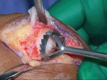
TECH FIG 3 • Alternative technique for joint preparation with specialized reamers. The Kirschner wire is placed in the center of the head to ensure concentric joint preparation.
Resect the ends of the metatarsal head and base of the phalanx, incorporating the chondral surfaces, with the cuts angled appropriately to produce the proper angles for subsequent positioning.3–5,7
Create multiple drill holes in the metatarsal head and phalangeal base with a Kirschner wire or small drill bit to augment bleeding and bony ingrowth.
Arthrodesis Positioning and Fixation
After preparing the joint surfaces, position the arthrodesis in 10 to 15 degrees of valgus, 15 degrees of dorsiflexion relative to the sole of the foot, and neutral pronation–supination.
Because it can be difficult to determine the plane of the sole with the patient on the table, a more predictable method of positioning the toe is to determine dorsiflexion relative to the first metatarsal axis. In most cases, the appropriate angle is about 25 to 30 degrees of dorsiflexion. 1,3,6,7,11,12,14,16–19
The hallux is held provisionally with Kirschner wires or partially threaded guidewires from a cannulated screw set.
 Confirm the positioning radiographically with a minifluoroscopy unit and clinically with use of a flat surface to simulate weight bearing (the cover of the screw set tray works nicely).
The hallux should be slightly off the surface with the heel on the cover (TECH FIG 4).
Placing a screwdriver handle under the heel simulates a shoe with a small heel; in this case, the pulp of the distal hallux should just barely engage the surface.

TECH FIG 4 • Positioning of the first metatarsophalangeal joint. A flat surface is used to position the toe properly. Note the positioning of the toe to allow for adequate clearance during gait.
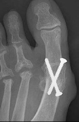
TECH FIG 5 • Postoperative radiograph shows the crossedscrew technique.

TECH FIG 6 • Alternative technique of fixation secondary to poor bone quality. Dorsal plating is used to augment the fixation.
Cannulated or solid screws can be used per the surgeon's preference. We use 4.0-mm cannulated screws in most patients; however, in some situations, such as for very large individuals, 4.5-mm screws can be used. Solid 3.5-mm cortical screws are an alternative (TECH FIG 5).
Insert one guidewire from the medial aspect of the phalangeal base just distal to the metaphyseal flare and advance it across the arthrodesis site through the dorsolateral cortex of the metatarsal neck.
Place the second wire from the medial aspect of the metatarsal neck, just proximal to the flare of the medial eminence; advance this wire distally and slightly plantarly across the arthrodesis site to engage the plantar–lateral cortex of the phalanx.
Check wire position and length with fluoroscopy.
Measure the wires percutaneously with the cannulated depth gauge and overdrill them with the cannulated drill bit. Then, countersink the cortex carefully to prevent subsequent cracking with screw placement.3
Place the partially threaded cannulated screws over the guidewires while compressing the hallux manually.
Alternatively, insert solid lag screws under fluoroscopic guidance.
In the event of suboptimal fixation or in patients with osteopenic bone (eg, secondary to rheumatoid arthritis or chronic oral corticosteroid usage), a dorsal plate can be used for augmented fixation.
This plate can be a precontoured, commercially available hallux MTP fusion plate or a standard minifragment plate that is cut and contoured to fit, and then affixed to the dorsal surface of the metatarsal and phalanx with small-diameter screws (eg, 2.7 mm) (TECH FIG 6).
Close the incision in layers with absorbable suture for the arthrotomy and subcutaneous layers and nonabsorbable monofilament for the skin.
ALTERNATIVE TECHNIQUE
A medial incision over the hallux MTP joint can be used in the presence of a previous surgical scar or at the surgeon's preference.
Carry out dissection at the level of the joint capsule, taking care to avoid the dorsomedial branch of the superficial peroneal nerve with elevation of the flap.
Perform a midline arthrotomy to expose the metatarsal head and base of the proximal phalanx.21
Prepare the joint surfaces with a saw blade.21
To allow for correct positioning, make the cut on the metatarsal head perpendicular to the sole of the foot, and avoid resecting excessive bone when making the cut on the proximal phalanx. Then, position the hallux, with attention to all three planes as described above.
Perform fixation with the crossed-lag-screw technique as described above, with supplemental dorsal plate fixation as needed.

POSTOPERATIVE CARE
In patients with an isolated arthrodesis that has good bone quality and solid fixation, weight bearing as tolerated on the heel and lateral border of the foot is allowed in a postoperative hard-soled shoe or fracture boot, restricting weight bearing on the forefoot.
If there are concerns about bone quality, suboptimal fixation, or potential noncompliance by the patient, strict non–weight-bearing in a below-the-knee cast is maintained for 6 to 8 weeks.
After 6 to 8 weeks, partial weight bearing is advanced, based on evidence of clinical and radiographic healing.
Full weight bearing usually is achieved by 10 to 12 weeks, at which time the patient transitions from the postoperative shoe or boot into sneakers or comfortable, low-heeled walking shoes.
At 14 to 16 weeks, with additional reduction in swelling, most patients can transition into unrestricted shoe wear; however, some individuals have permanent difficulty wearing fashion shoes or high heels.
Prolonged walking and athletic activities usually resumes at 4 to 5 months.
Custom-made orthotics with a build-up under the hallux to improve weight bearing of the first ray may dissipate forefoot stresses.
OUTCOMES
The clinical results after hallux MTP arthrodesis usually are excellent, with high rates of bony union, patient satisfaction, and pain relief.
Union rates for in situ arthrodesis range from 77% to 100%.4–8,11,13,16–18
Patient satisfaction rates also are high, regardless of the indications.5–8,10,11,13,16–18
MTP arthrodesis causes a rigid lever arm, resulting in an earlier toe-off in the gait cycle and decreasing the stress on the lesser metatarsals.6,8,14,16–18 This stiffness may result in increased stress across the hallux interphalangeal joint.14
After arthrodesis, the first ray shows improved weightbearing capacity, with the foot compensating for the relative stiffness during stance phase.10
COMPLICATIONS
Nonunion rates range from 5% to 10%.5,10,13,17,18 Nonunion may not be symptomatic and may not require revision surgery.13
Malunion after MTP arthrodesis can result in mild malalignment that is tolerated, but more severe malposition may be symptomatic.
Excessive dorsiflexion leads to unloading of the hallux and lesser toe transfer metatarsalgia.1
Positioning the hallux in relative plantarflexion may lead to interphalangeal joint irritation, callus formation, and later interphalangeal arthritis.1,22
Valgus positioning can lead to painful impingement on the second toe,11 whereas varus positioning causes impingement of the hallux against the toe box of the shoe.
Subsequent arthritis of the interphalangeal joint may occur in one third of cases.
Arthritis in the interphalangeal joint is more common than that of the first tarsometatarsal or other midfoot joints.
However, symptoms may be mild despite radiographic involvement and may take 10 years to develop.11,16
Severe symptoms may require secondary interphalangeal arthrodesis, which leads to extreme stiffness of the hallux.
Iatrogenic nerve injuries of the dorsomedial cutaneous nerve are more common than injuries to the plantar nerves.
These may result in neuroma formation, mild numbness, or persistent dysethesias that compromise an otherwise successful arthrodesis.
Prevention by proper incision placement and meticulous surgical dissection remains the best strategy.
REFERENCES
· Alexander IJ. Arthrodesis of the metatarsophalangeal and interphalangeal joints of the hallux. In Myerson M, ed. Current Therapy in Foot and Ankle Surgery. St. Louis: Mosby-Year Book, 1993:81–90.
· Castro MD, Klaue K. Technique tip: revisiting an alternative method of fixation for first MTP joint arthrodesis. Foot Ankle Int 2001;22: 687–688.
· Conti SF, Dhawan S. Arthrodesis of the first metatarsophalangeal and interphalangeal joints of the foot. Foot Ankle Clin 1996;1:33–53.
· Coughlin MJ. Arthrodesis of the first metatarsophalangeal joint. Orthop Rev 1990;19:177–186.
· Coughlin MJ. Arthrodesis of the first metatarsophalangeal joint with mini-fragment plate fixation. Orthopedics 1990;13:1037–1044.
· Coughlin MJ. Rheumatoid forefoot reconstruction: a long-term follow-up study. J Bone Joint Surg Am 2000;82:322–341.
· Coughlin MJ, Abdo RV. Arthrodesis of the first metatarsophalangeal joint with Vitallium plate fixation. Foot Ankle Int 1994;15: 18–28.
· Coughlin MJ, Mann RA. Arthrodesis of the first metatarsophalangeal joint as salvage for the failed Keller procedure. J Bone Joint Surg Am 1987;69:68–75.
· Curtis MJ, Myerson M, Jinnah RH, et al. Arthrodesis of the first metatarsophalangeal joint: a biomechanical study of internal fixation techniques. Foot Ankle 1993;14:395–399.
· DeFrino PF, Brodsky JW, Pollo FE, et al. First metatarsophalangeal arthrodesis: a clinical, pedobarographic and gait analysis study. Foot Ankle Int 2002;23:496–502.
· Fitzgerald JAW. A review of long-term results of arthrodesis of the first metatarso-phalangeal joint. J Bone Joint Surg Br 1969;51:488–493.
· Harper MC. Positioning of the hallux for first metatarsophalangeal joint arthrodesis. Foot Ankle Int 1997;18:827.
· Kitaoka HB, Patzer GL. Arthrodesis versus resection arthroplasty for failed hallux valgus operations. Clin Orthop Relat Res 1998;347: 208–214.
· Mann RA. Surgical implications of biomechanics of the foot and ankle. Clin Orthop Relat Res 1980;146:111–118.
· Mann RA, Katcherian DA. Relationship of metatarsophalangeal joint fusion on the intermetatarsal angle. Foot Ankle 1989;10:8–11.
· Mann RA, Oates JC. Arthrodesis of the first metatarsophalangeal joint. Foot Ankle 1980;1:159–166.
· Mann RA, Schakel ME II. Surgical correction of rheumatoid forefoot deformities. Foot Ankle Int 1995;16:1–6.
· Mann RA, Thompson FM. Arthrodesis of the first metatarsophalangeal joint for hallux valgus in rheumatoid arthritis. J Bone Joint Surg Am 1984;66:687–692.
· McKeever DC. Arthrodesis of the first metatarsophalangeal joint for hallux valgus, hallux rigidus, and metatarsus primus varus. J Bone Joint Surg Am 1952;34:129–134.
· Mizel MS, Alvarez RG, Fink BR, et al. Ipsilateral arthrodesis of the metatarsophalangeal and interphalangeal joints of the hallux. Foot Ankle Int 2006;27:804–807.
· Rydholm A, Rooser B. Surgical margins for soft-tissue sarcoma. J Bone Joint Surg Am 1987;69:1074–1078.
· Trnka HJ. Arthrodesis procedures for salvage of the hallux metatarsophalangeal joint. Foot Ankle Clin 2000;5:673–686.