
Hans-Joerg Trnka and Stefan G. Hofstaetter
DEFINITION
 First metatarsophalangeal arthrodesis is a reasonable alternative to a joint-sparing procedure in salvage of various great toe deformities.
These deformities comprise failed hallux valgus procedures, avascular necrosis of the metatarsal head, failed first metatarsophalangeal (MTP) joint arthroplasty, prior infection, rheumatoid arthritis, posttraumatic conditions, hallux rigidus, severe hallux valgus deformities, and neuromuscular disorders.
With minimal to moderate bone loss, we perform hallux MTP joint arthrodesis in situ, accepting slight shortening of the hallux. In our opinion, the slight shortening creates minimal cosmetic concerns and affords satisfactory functional improvement in a majority of cases.
With marked shortening of the hallux and associated lesser metatarsalgia, an in-situ hallux MTP joint arthrodesis may fail to restore satisfactory function.
We favor interposition structural bone graft to restore first ray length, which, in turn, should improve the weight bearing of the first metatarsal and hallux while alleviating lesser metatarsalgia.
Sources for structural interposition bone block arthrodesis include (a) structural allograft (usually contoured from a donor femoral head or iliac crest) or (b) structural autograft (typically obtained from the patient's iliac crest). In our hands, ipsilateral anterior iliac crest harvesting is ideal for foot and ankle surgery because this site is readily accessible in the patient positioned supine on the operating table.
Several configurations have been described for contouring the interpositional graft. We prefer the ball-and-socket technique, which affords three advantages over flat cuts or a conical preparation: (a) minimal resection of residual host bone, (b) optimal surface area at both ends of the graft for healing, and (c) relative ease of positioning the toe after preparing the arthrodesis without forfeiting contact area for fusion.
PATHOGENESIS
In our referral practice, we most commonly use the balland-socket interpositional bone block distraction technique for severe bone loss after:
Keller-Brandes procedure (FIG 1A), resection of the base of the proximal phalanx (this generally creates bone loss isolated to the hallux and not globally in the first ray)
Mayo procedure (FIG 1B), resection of the first metatarsal head (creates a more global bone loss in the first ray)
The Keller-Brandes and Mayo procedures have for the most part been abandoned because of their detrimental effects on forefoot function and the introduction of modern procedures that preserve anatomy.
Avascular necrosis of the first metatarsal head (FIG 1C), a relatively rare complication of a distal chevron osteotomy
Bone destruction after first MTP joint arthroplasty, particularly silicone implants (FIG 1D)
NATURAL HISTORY
The natural history after failure of the aforementioned procedures is one of functional imbalance of the forefoot. The first ray fails to provide physiologic support, creating an overload phenomenon, or transfer metatarsalgia, to the lesser metatarsal heads. While the lesser metatarsals may be shortened to compensate for the loss of first ray length, this is often undesirable because there is no pathology at the lesser toes.


FIG 1 • A. Bone loss after Keller-Brandes procedure. B. Bone loss after Mayo resectional arthroplasty. C. Bone loss after avascular necrosis following a chevron osteotomy. D. Bone loss after failed silicon implant.
PATIENT HISTORY AND PHYSICAL FINDINGS
The patient typically complains of pain and deformity in the hallux MTP joint and pressure and pain under the lateral forefoot.
Typical physical examination findings include:
 Short hallux or first ray (bone loss at the hallux only is typical with bone loss on the phalangeal or metatarsal head side; more global first ray bone loss occurs after the Mayo procedure, avascular necrosis of the metatarsal head, or a failed great toe implant)
Cock-up deformity of the hallux
Residual hallux valgus deformity and occasional hallux varus deformity
Pain and crepitance with range of motion of hallux
Pain and tenderness (and occasionally plantar callus formation) under the lesser metatarsal heads
The potential iliac crest harvest site should also be inspected for unanticipated soft tissue concerns or to confirm that no prior graft harvest has been performed.
IMAGING AND OTHER DIAGNOSTIC STUDIES
Weight-bearing foot radiographs, including AP, lateral, and oblique views
With avascular necrosis of the first metatarsal head, an MRI of the forefoot may prove useful in estimating the extent of necrotic bone and predicting the size of the interpositional graft.
NONOPERATIVE MANAGEMENT
In our experience, nonoperative treatment of the painful, shortened, and cocked-up hallux is generally unsuccessful. The shoe's toe box may be enlarged and an accommodative orthotic to post under the hallux may be considered, but this is generally of limited value.
Metatarsal support, however, may relieve the symptoms related to transverse metatarsalgia by unloading the lesser metatarsal heads. If metatarsal pads are effective, then a custom orthotic with metatarsal support may prove beneficial as well.
SURGICAL MANAGEMENT
Preoperative Planning
The decision to perform a structural femoral head or iliac crest allograft or an anterior iliac crest autograft must be made preoperatively. However, we recommend having the flexibility to use either method, with the patient's consent, if one graft proves ineffective based on intraoperative assessment. The patient should be aware of the risks of iliac crest graft harvest and the use of allograft bone.
Myerson et al5 in 2005 investigated the use of structural allografts in foot and ankle surgery and discussed the risks of using a structural allograft. One concern with the use of structural allograft is the possible transmission of disease and malignancy, but with the use of processed allografts the risk is practically zero. The risk of iliac crest bone harvest is donorsite morbidity, which includes local hematoma, local infection, and in rare cases local nerve irritation.
We routinely draw a preoperative plan for the structural graft, determining the approximate amount of bone resection required and the length of graft that needs to be acquired.
Positioning
The patient is positioned supine on the operating table, with a bump placed beneath the hip ipsilateral to the foot that will be operated on. This not only positions the foot in an ideal position (to allow improved assessment of proper hallux alignment) but also facilitates harvest of the iliac crest graft by making the anterior crest more accessible.
Approach
First MTP joint: A standard dorsal approach is recommended, starting about 4 cm proximal to the MTP joint and extending to the interphalangeal joint. When possible, previous incisions should be incorporated into the approach, to avoid a skin bridge that may be at risk, particularly when the toe will be distracted with the interpositional graft.
Iliac crest: An incision is made parallel and inferior to the anterior iliac crest, about 3 cm posterior to the anteriorsuperior iliac spine, to avoid injuring the lateral femoral cutaneous nerve.
TECHNIQUES
PREPARATION OF THE MTP JOINT
 Start the dorsal skin incision about 4 cm proximal to the first MTP joint and extend it to the interphalangeal joint (TECH FIG 1).
The extensor hallucis longus (EHL) tendon can be simply retracted if the first ray has minimal shortening. With moderate to severe shortening, particularly with associated cock-up toe deformity, the EHL tendon may need to be Z-lengthened to allow restoration of toe length and to avoid hyperextension at the hallux interphalangeal joint.

TECH FIG 1 • The joint capsule and the soft tissue coverage of the metatarsal and the phalanx are incised longitudinally straight down to the bone and then opened as an envelope. Subperiosteal preparation is mandatory to ensure sufficient release from the lateral soft tissue and scar adhesions.
Divide the first MTP joint capsule, scar tissue from prior surgery, and periosteum of the proximal phalanx and distal first metatarsal longitudinally and reflect them. While we subscribe to the principle of minimal soft tissue stripping, we believe that subperiosteal preparation is mandatory to afford sufficient mobilization of the hallux. However, we leave the plantar soft tissues to maintain the blood vessels supplying the residual metatarsal head and proximal phalanx (TECH FIG 2).
We routinely remove any osteophytes and additional soft tissue adhesions.
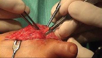
TECH FIG 2 • After the articular surfaces of the first metatarsophalangeal joint have been adequately freed, the big toe is brought into maximal plantarflexion.
REAMING OF THE METATARSAL HEAD AND BASE OF THE PROXIMAL PHALANX
After mobilizing the articular surfaces of the first MTP joint, maximally plantarflex the hallux (TECH FIG 3).
Insert a guidewire for the reamer set into the center of the metatarsal head. Place the appropriately sized “female” reamer over the guidewire (TECH FIG 4). Remove the sclerotic bone surface with the reamer down to cancellous bleeding bone.
Expose the base of the proximal phalanx (TECH FIG 5) and place a guidewire for the reamer set (TECH FIG 6).
In a similar manner, prepare the proximal phalanx with the “male” reamer counterpart (TECH FIG 7). Distract the toe to the desired length and measure the gap (TECH FIG 8).

TECH FIG 3 • After the articular surfaces of the first metatarsophalangeal joint have been mobilized, the hallux is maximally plantarflexed.

TECH FIG 4 • The adequately sized female reamer is then used to remove the sclerotic bone surface of the metatarsal head down to the cancellous bleeding bone.

TECH FIG 5 • Exposure of the base of the proximal phalanx.
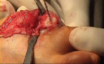
TECH FIG 6 • Placement of the guidewire of the reamer set.

TECH FIG 7 • The male reamer is now used to remove the sclerotic bone surface of the metatarsal head down to the cancellous bleeding bone.
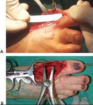
TECH FIG 8 • The toe can then be pulled into position of the desired length, and the exact extent of the gap in the joint is measured.
HARVEST OF THE ILIAC CREST BONE BLOCK
Make the incision for the tricortical iliac crest block 3 cm posterior to the anterior-superior iliac spine (TECH FIG 9). Carry dissection down to the intermuscular plane using electrocautery for hemostasis. Reflect the periosteum from the superior crest. Insert a Hohmann retractor on the inner and outer aspects of the iliac crest, deep to the periosteum (TECH FIG 10). Based on the desired length of the first ray and the gap created with first MTP joint preparation, mark the segment of iliac crest to be harvested. We use a microsagittal saw to cut the iliac crest and an osteotome to complete the separation of the structural graft (TECH FIG 11).

TECH FIG 9 • The incision for the tricortical iliac crest block is made 2 to 3 cm posterior to the anterior-superior iliac spine.
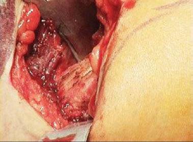
TECH FIG 10 • A Hohmann retractor is inserted on the inner and outer side of the iliac crest under the periosteum.
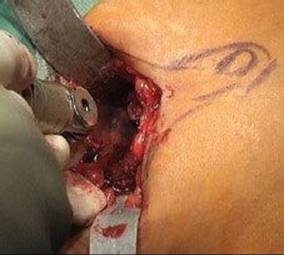
TECH FIG 11 • A saw is used to osteotomize the ends of the tricortical bone block.

TECH FIG 12 • The iliac crest bone block is retrieved.
Harvest the iliac crest structural graft (TECH FIG 12).
The defect in the iliac crest may be backfilled with allograft bone chips. Close the periosteum after placing a drain. Reapproximate the subcutaneous tissues and close the skin.
CONTOURING OF THE GRAFT
Secure the graft (either the harvested iliac crest graft or a femoral head graft) on the back table using a forceps, to be shaped into the desired length (TECH FIG 13).
Place a guidewire for the reamer set in the center of the graft's long axis. Use the same reamers that were used to prepare the first MTP joint to contour the ends of the graft. One end is prepared with the “female” reamer and the other using the “male” reamer to create optimal contact to the host bone (TECH FIG 14).

TECH FIG 13 • A. The graft is held on the table with a forceps and shaped into the desired length. B. The graft margins are marked on an allograft femoral head.
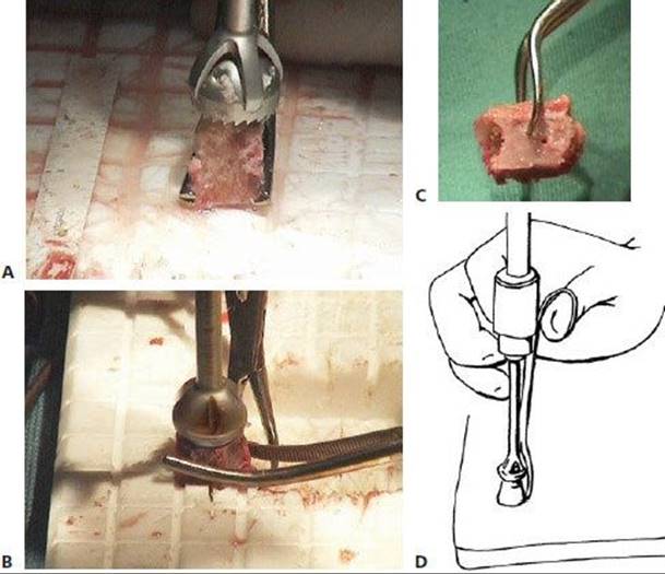
TECH FIG 14 • The same-sized female and male reamers are used to mold the two surfaces on the sides of the block, one as a ball and the other as a socket to fit to the ends of the proximal phalanx and the metatarsal head respectively.
INSERTION OF THE GRAFT AND FIXATION
Insert the contoured graft (TECH FIG 15) into the prepared gap between metatarsal and phalanx. With a ball-and-socket shape on either end of the prepared gap, the alignment of the lengthened hallux may be seamlessly adjusted in any direction. Place either a standard plate or a special revision plate dorsally (TECH FIG 16). Place temporary pins to maintain the reduction, and confirm proper arthrodesis and plate positions fluoroscopically.
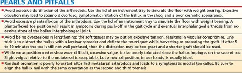
In our experience, optimal hallux position for arthrodesis is (a) neutral rotation (no pronation or supination), (b) about 15 degrees of dorsiflexion (relative to the plantar surface of the foot), and (c) 5 degrees of valgus with respect to the first metatarsal (TECH FIG 17).
To determine optimal sagittal plane position, a lid from an instrument tray may be used to simulate weight bearing. Ideally, the distal hallux tuft is 1 to 2 mm elevated from the plate when the ball of the foot and heel are contacting the instrument tray lid (TECH FIG 18).
Place a 3.0-mm or 3.5-mm screw from the medial aspect of the residual proximal phalanx across the graft to the lateral aspect of the residual metatarsal (TECH FIG 19).
Secure the plate to the construct, with screws in the proximal phalanx, graft, and metatarsal, while avoiding the initial screw (TECH FIG 20). Three or four absorbable deep sutures are generally adequate to cover the plate. We advocate the use of a small-diameter drain for 2 days postoperatively to reduce the risk of hematoma formation.
Reapproximate the subcutaneous layer and skin in a tension-free manner; perform the closure carefully since the soft tissues are already under some tension due to lengthening of the first ray.
After sterile dressings are placed on the wound, we routinely apply a well-padded short-leg cast that extends beyond the toes. We recommend univalving the cast.
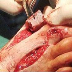
TECH FIG 15 • The molded graft is inserted into the gap between metatarsal and phalanx.

TECH FIG 16 • The special revision plate is placed dorsally.

TECH FIG 17 • The hallux is positioned in neutral rotation, with special attention paid to the position of the toenail.
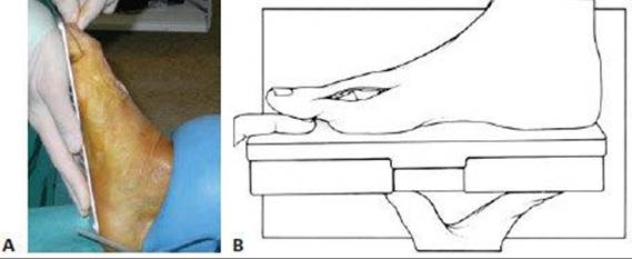
TECH FIG 18 • For the optimal extension, a lid of the instrument tray may be used to simulate floor contact.
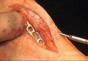
TECH FIG 19 • To add additional stability, a 3.0 AO screw is inserted from the medial aspect of the proximal phalanx of the great toe across the graft to the lateral side of the metatarsal.

TECH FIG 20 • The plate is fixed with adequate screws.
POSTOPERATIVE CARE
We recommend placing the patient in a short-leg cast that extends beyond the toes for a full 6 to 8 weeks. The patient should be touch-down weight bearing until suture removal and then weight bearing on the heel until 6 to 8 weeks.
OUTCOMES
Myerson et al4,5 treated 24 patients with hallux MTP joint arthrodesis using bone graft to restore first ray length (FIG 2).
This procedure was performed after bone loss subsequent to previous surgeries for the correction of hallux valgus and hallux rigidus with Silastic arthroplasty (n = 11), bunionectomy and distal metatarsal osteotomy (n = 6), Keller resection arthroplasty (n = 5), and total joint replacement (n = 2).
All patients were examined clinically and radiographically at a mean interval of 62.7 months after surgery (range 26 to 108 months).
Successful fusion was observed in 19 of the 24 patients (79.1%) at a mean of 13.3 weeks (range 11 to 16 weeks), and the first ray was lengthened by a mean of 13 mm (range 0 to 29 mm).

FIG 2 • A. A 45-year-old woman after Keller-Brandes arthroplasty. B. Postoperative photograph of a first metatarsophalangeal bone block fusion. C. 2-year follow-up after hardware removal.
Of the five nonunions noted radiographically, two were asymptomatic and three were managed successfully with further surgery.
Complications included one deep infection requiring intravenous antibiotics and irrigation and débridement of the graft repeat surgery for treatment of osteomyelitis and two minor superficial wound infections managed effectively with oral antibiotics and local wound care.
The mean AOFAS score improved from 39 points (range 22 to 60 points) to 79 points (range 64 to 90 points).
Brodsky et al1 reviewed 12 patients (12 feet) who underwent salvage first MTP arthrodesis with structural interposition autologous iliac crest bone graft.
Eight patients had a bony defect secondary to failed first MTP joint implant arthroplasties, two had avascular necrosis after failed bunion surgery, one had a nonunion of an attempted arthrodesis for failed bunion surgery, and one had been treated for osteomyelitis after cheilectomy.
Eleven cases had a single dorsal plate secured by screws and one case had two plates, one dorsal and one medial.
A plate, crossed screw(s), or Kirschner wire combinations were used in four cases.
Clinical arthrodesis was achieved after an average of 12 weeks (range 4 to 20).
 Radiographic arthrodesis was achieved in 11 of 12 feet at an average of 15 weeks (range 8 to 28), with one pseudarthrosis.
The AOFAS forefoot clinical rating score averaged 70 points (maximum 90 after first MTP arthrodesis) at an average follow-up of 22 months (range 5 to 70).
Sesamoiditis, prominent hardware, and scar sensitivity were complaints in four patients postoperatively. Two cases required flap coverage for skin necrosis. There was no symptomatic progression of interphalangeal degenerative change postoperatively.
COMPLICATIONS
Pseudarthrosis
Wound dehiscence or infection
Nerve irritation
Poor alignment
REFERENCES
· Brodsky JW, Ptaszek AJ, Morris SG. Salvage first MTP arthrodesis utilizing ICBG: clinical evaluation and outcome. Foot Ankle Int 2000;21:290–296.
· Machacek F Jr., Easley ME, Gruber F, et al. Salvage of a failed Keller resection arthroplasty. J Bone Joint Surg Am 2004;86A:1131–1138.
· Machacek F Jr., Easley ME, Gruber F, et al. Salvage of the failed Keller resection arthroplasty: surgical technique. J Bone Joint Surg Am 2005;87A(Suppl 1):86–94.
· Myerson MS, Schon LC, McGuigan FX, et al. Result of arthrodesis of the hallux metatarsophalangeal joint using bone graft for restoration of length. Foot Ankle Int 2000;21:297–306.
· Myerson MS, Neufeld SK, Uribe J. Fresh frozen structural allografts in the foot and ankle. J Bone Joint Surg Am 2005;87A:113–120.
· Trnka HJ. Arthrodesis procedures for salvage of the hallux metatarsophalangeal joint. Foot Ankle Clin 2000;5:673–686.