
Geert I. Pagenstert, Victor Valderrabano, and Beat Hintermann
DEFINITION
 Hallux sesamoid bone fracture is a break through the sesamoid bone or cartilage. Medial sesamoid bone fractures are more common than lateral sesamoid bone fractures.1,15
Fractures usually occur about perpendicular to the long axis of the elliptically shaped bone. Longitudinal and comminuted fractures are less common.5,17
In partite or bipartite sesamoid bones, the fracture always occurs in the fibrocartilaginous junctional zone (most often perpendicular to the long axis), which can disguise the fracture.15
ANATOMY
The hallux sesamoid bones usually are 13.5 ± 3 mm long. The sesamoid bones are larger in men than in women, and the medial sesamoid is more elliptically shaped and larger compared to the more circularly shaped lateral sesamoid.14
The hallux sesamoid bones are invested in the tendon sheath of the flexor hallucis brevis. They connect with the intersesamoid ligament to form a solid pedestal to elevate the first ray and absorb stress during gait2,3,14 (FIG 1A).
The sesamoid complex acts as a fulcrum to the flexor hallucis brevis and longus tendons, increasing their lever arms and big toe push-off power, e.g., the patella to the quadriceps tendon2,3 (FIG 1B).
Failure of the bone to ossify completely during childhood results in a multi-part sesamoid bone. Bipartite sesamoids are much more common than those with three or more parts. Despite incomplete ossification, the sesamoid parts are firmly connected with fibrocartilaginous tissue to act as one bone. Spontaneous fusion can occur later in life.10
Partite sesamoid bones are bilateral in only about 25% of cases; therefore, unilaterality cannot be relied upon as a criterion of fracture.10
The main blood supply is provided over the posterior tibial to the medial plantar artery to the sesamoids. Considerable variation exists, however, such as the main blood supply from the lateral plantar artery or even the dorsal arterial arch.7,14
In general, only one major artery pierces the cortex of the sesamoid bone at the plantar aspect of the proximal pole. Small vessels also enter from the plantar nonarticular side and over the capsular attachments as a second source of vascularity.7,14
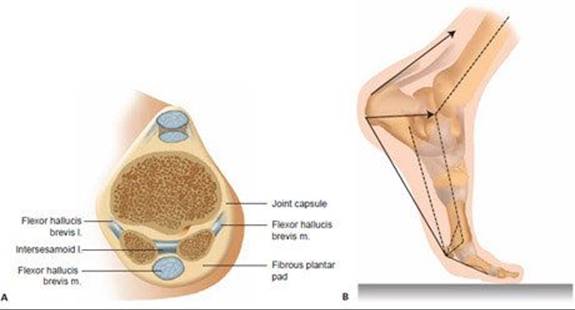
FIG 1 • Anatomy and biomechanics of the hallux sesamoid complex. A. The sesamoids elevate the first metatarsal bone. Fifty percent or more of body weight is transferred over the first ray. With sesamoid excision, preloading of the metatarsal bone is decreased, transferring the load to the lesser toes. B.Sesamoid bones increase the lever arm of the hallucis brevis and hallucis longus flexor tendons. Sesamoid excision reduces this lever and subsequently reduces push-off power of the big toe. (A: From Aper RL, Saltzman CL, Brown TD. The effect of hallux sesamoid resection on the effective moment of the flexor hallucis brevis. Foot Ankle Int 1994;15:462–470; B: From Aper RL, Saltzman CL, Brown TD. The effect of hallux sesamoid excision on the flexor hallucis longus moment arm. Clin Orthop Relat Res 1996;325:209–217.)
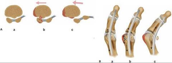
FIG 2 • Biomechanics of the sesamoid complex in hallux valgus deformity. A. Varus subluxation of the first metatarsal bone causes pressure concentration to the medial sesamoid bone. The intersesamoid crista enhances friction to the sesamoid joint surface. B. After stress fracture occurs, hallux deviation will cause constant fragment displacement. Therefore, immobilization may not suffice. Sesamoid excision will enhance hallux deviation if the deformity is not addressed. (A,B: From Pagenstert GI, Valderrabano V, Hintermann B. Medial sesamoid nonunion combined with hallux valgus in athletes. Foot Ankle Int 2006;27:135–140.)
PATHOGENESIS
Acute trauma or chronic overuse leads to acute or stress fractures, respectively, of the sesamoid bones.1,15
In the acute setting, the typical mechanism is excessive hyperextension of the big toe, also referred to as the “turf toe” injury seen in American football players. Disruption of the plantar joint capsule occurs as a trans-sesamoidal fracture–dislocation of the first metatarsophalangeal (MTP) joint.15
Typically, in the chronic setting, no trauma is remembered. Pain and swelling increase insidiously over weeks, months, or years. Diagnosis is significantly delayed. Endurance sports such as running and dancing have shown to be associated with chronic stress fractures of the hallux sesamoid bones.5,12
Foot deformities that concentrate pressure to the sesamoids increase the chance of suffering sesamoid stress fractures in both athletic and nonathletic persons. Cavus foot deformities with a steep plantar flexed first ray stress both sesamoid bones. Hallux valgus deformity with varus dislocation of the metatarsal head leads to pressure concentration at the medial sesamoid bone only12,13 (FIG 2).
NATURAL HISTORY
Acute fractures without mild dislocation heal normally with little or even no treatment.15
Chronic stress fractures usually do not heal without surgery, which is explained by the typical pathogenesis described earlier. During the prolonged time to diagnosis and the constant friction of fracture fragments, necrotic tissue accumulates at the fracture site and prevents healing. Brodsky et al,6 Van Hal et al,17 and Saxena and Krisdakumtorn16 independently reported on consecutive series of athletes with chronic sesamoid fractures. None of the sesamoid fractures in their series healed, even with prolonged nonsurgical regimens. Histologic examination after sesamoid excision revealed accumulation of necrotic tissue at the fracture site.6
Foot deformities can cause fragment separation and may prevent healing with immobilization.12
PATIENT HISTORY AND PHYSICAL FINDINGS
The patient history and physical examination must rule out the differential diagnoses.
 The typical patient history is discussed in the section Pathogenesis.
The physical examination includes examination of areas of localized pain and swelling, and hyperextension testing of the big toe.
Patients have localized pain and swelling around the first MTP joint (FIG 3).
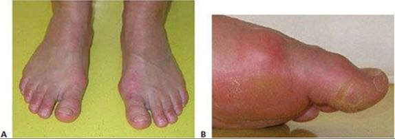
FIG 3 • Clinical appearance of sesamoid stress fracture. A. Swelling of the MTP joint with localized tenderness at the medial sesamoid bone. B. Evaluate the hallux valgus deformity on the left. Progression of the deformity was noted by the patient within the preceding 3 months.
A complete examination of sesamoid status includes examination of the whole foot and ankle, with special attention to cavus deformity with a flexed first ray or hallux valgus deformity12 (see Fig 2B).
IMAGING AND OTHER DIAGNOSTIC STUDIES
Sesamoid oblique and tangential (“skyline”) views are useful to evaluate sesamoid fracture displacement (FIG 4A).
Partite sesamoid bones are bilateral in only about 25% of cases.10 Therefore, radiographs of the contralateral foot do not rule out fracture. In addition, fractures of bipartite sesamoid bones occur at the fibrocartilaginous junctional zone.15
A longitudinal CT scan of the foot has been shown to be very effective in demonstrating sesamoid stress fracture in difficult settings4 (FIG 4B).
MRI11 and bone scans8 are nonspecific for diagnosing stress fracture or distinguishing between a traumatized bipartite sesamoid and a stress fracture. Bone edema is seen on MRI in bone contusion, inflammatory disease, avascular necrosis, and infection.11 Localized sesamoid scintigraphic activity has been demonstrated in about 26% to 29% of cases in asymptomatic active and sedentary populations.8
On full weight-bearing radiographs of the lateral whole foot, the angle between the talus and the first metatarsal is evaluated. In a normal foot, it is straight or in up to 10 degrees of flexion. A greater amount of flexion demonstrates a flexed first ray, whereas flexion of less than 0 degrees demonstrates medial arch insufficiency, which is connected with hallux valgus formation.
On full weight-bearing dorsoplantar radiographs of the foot, the hallux valgus, sesamoid position, metatarsus primus varus, and talo–first metatarsal angle are evaluated for stress concentration to the medial sesamoid bone. The talonavicular joint congruence is examined to identify excessive forefoot abduction with pes plano valgus or excessive adduction with neurogenic pes cavo varus.
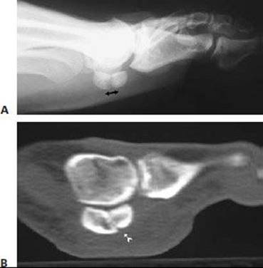
FIG 4 • Radiologic examination of sesamoid fractures. A. Conventional radiographs demonstrating horizontal sesamoid fracture dislocation. B. CT scan shows fracture line of chronic painful sesamoid, which was not visible on conventional radiographs.
DIFFERENTIAL DIAGNOSIS
Hallux rigidus or sesamoid–first metatarsal bone osteoarthritis
Hallux valgus
First MTP joint capsuloligamentous disruptions (turf toe)
Osteomyelitis and septic arthritis
Podagra of gout and pseudogout
Inflammatory arthritis
Avascular necrosis of sesamoid or metatarsal head
NONOPERATIVE MANAGEMENT
Acute fractures with up to 5 mm dislocation are treated with a forefoot immobilization shoe (stiff and convex sole) for 6 to 8 weeks.15
Treatment of chronic fractures is controversial. Despite frequent failure after nonsurgical treatment attempts5,6,12,13,16,17 and the already long time it takes to establish diagnosis, many physicians try immobilization with a shoe or cast, sometimes with the patient non–weight-bearing on crutches. Recommendations for the duration of this approach before surgery is advocated range from 6 to 12 weeks.16,17
If the diagnosis of stress fracture was established soon after the symptoms began, activity modification and use of a stiffsoled shoe for 6 weeks may be successful. Modification in athletic training and eating habits, with running on soft ground only, a change of sole stiffness in the athletic shoe, and increased intake of calcium and vitamin D 3 may be reasonable adjuncts in the future.
SURGICAL MANAGEMENT
Severe (>5 mm) acute trans-sesamoidal fracture dislocations of the first MTP joint require open repair of the capsule and flexor muscles.15 The sesamoid bone fixation can be done with a compression screw or heavy no. 1 suture.
Indications for percutaneous compression screw fixation include a transverse sesamoid stress fracture, transverse nonunion, or transverse symptomatic bipartite sesamoid. Fragments must be at least 3 mm to allow screw fixation.13
Contraindications include infection, longitudinal sesamoid fractures, and comminuted fractures with multiple fragments that are too small for screw fixation. In these cases, partial or total sesamoid resection is indicated.
Combined medial sesamoid fracture and hallux valgus deformity are best treated with conventional open correction of the hallux and open reduction and fixation of the sesamoid fracture by heavy no. 1 suture or compression screw.12 Débridement of the necrotic fracture zone and grafting can be done to enhance healing.1 In cases with less than 2 mm dislocation, the fracture zone can be stabilized by grafting only. The flexor brevis tendon sheath acts as tension band fixation.1
In patients who are likely to be noncompliant, a temporary 2.5-mm K-wire can be placed through the first MTP joint to prevent hallux dorsiflexion and stress to the fragments.
Combined hindfoot and first ray deformities with chronic sesamoid fractures must be addressed in the same surgery.12
Preoperative Planning
Acute transsesamoidal fracture dislocations of the first MTP joint require open stabilization sometimes with an extended medioplantar L-shaped incision to reach the lateral aspect of the joint. Sesamoid fracture fixation is part of the plantar capsule or plate repair.15
In chronic sesamoid fracture, preoperative planning should incorporate treatment of any underlying foot deformities.
A metatarsus primus flexus is treated with a dorsal extension osteotomy or arthrodesis.
A metatarsus primus varus and hallux valgus are addressed with appropriate osseous or soft tissue procedures.
Reduction of mechanical stress to the sesamoid bones is thought to be the main factor contributing to fracture healing. Surgical stress reduction alone may result in fracture healing even without sesamoid osteosynthesis in marked foot deformities.
In the combined setting, medial sesamoid stress fractures are treated open because deformity correction is done at the same time as arthrotomy of the first MTP joint. Lateral sesamoid stress fractures are treated percutaneously, because deformity correction does not include arthrotomy of the first MTP joint.
The least invasive approach can be used in the absence of foot deformities.
Chronic sesamoid fractures can be addressed by percutaneous compression screw fixation alone.
Surgery can be performed under local anesthesia, and the stab incision of the skin can be closed with Steri-Strips.
Healing is thought to occur because of reaming (vitalizing) of the fracture zone and fracture stabilization. Ossification of the bipartite sesamoids occurs.13
Grafting of sesamoid nonunions (bipartite sesamoids) is inherently stabilized by the flexor brevis tendon sheath.1 In cases of persistent instability after grafting, additional suture or screw fixation is advisable.
Positioning
The patient is placed in the supine position for isolated sesamoid bone fixation or combined deformity corrections. A tourniquet is needed, except in percutaneous fixation.
Approach
A medial internervous or medioplantar L-shaped approach to the lateral aspect of the first MTP joint is used for acute turf toe repair, including sesamoid fracture fixation or partial removal.15
A standard medial internervous approach is used for grafting of medial sesamoid nonunions and combined hallux correction.12
In the case of percutaneous fixation, a stab incision is made distal to the pole of the fractured sesamoid bone and distal to the weight-bearing area of the first MTP joint. Lateral sesamoid fractures usually are treated with percutaneous screw fixation.13
TECHNIQUES
ANDERSON-MCBRYDE TECHNIQUE OF GRAFTING SESAMOID NONUNIONS 1
 A medial internervous skin incision is made over the first MTP joint (TECH FIG 1A).
Longitudinal capsulotomy and subperiosteal limited exposure of the medial sesamoid wall are done.
Débridement of the necrotic tissue at the fracture site is performed with a small curette from an extra-articular medial approach (TECH FIG 1B).
Fenestration of the MT head is performed to enable autologous bone harvesting (TECH FIG 1C).
The sesamoid fracture zone is grafted and stuffed, with care not to disrupt the fracture line in the joint surface.
If stability is in doubt, fixation with no. 1 resorbable suture is performed to leave the least amount of foreign material in situ. Cannulated compression screws are used as well and may provide higher compression. (Screw placement is described in the next section.)
The suture needle is introduced from the proximal lateral pole along the internal lateral cortex to the distal lateral pole. Backstitching is done outside the bone under the medial sesamoid suspensory (capsule) ligament, back to the proximal medial pole, and knotted tight to stabilize the sesamoid joint line (TECH FIG 1D).
The capsule and skin are closed as usual.
A compressive dressing is applied with the foot in the neutral hallux position.
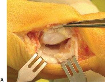

TECH FIG 1 • Anderson-McBryde technique. A. Medial internervous approach. B. Débridement of the fracture with a small curette using an extra-articular approach to the necrotic tissue. C. Harvesting of autologous bone from the first metatarsal head. D. Suture cerclage of the fractured sesamoid.
PREFERRED TECHNIQUE OF PERCUTANEOUS SESAMOID SCREW FIXATION 13
The hallux is held in dorsiflexion, and the sesamoid bone is pressed against the MT head to level the fracture fragments against the joint line of the MT head (TECH FIG 2A).
One 3-mm stab incision is done distal to the fractured sesamoid bone and distal to the weight-bearing area of the first MTP joint (TECH FIG 2B).
The guidewire (1.5-mm wire for 2.4-mm self-tapping Bold screws [Newdeal, Lyon, France]) is introduced under fluoroscopic control from the distal pole, perpendicular to the fracture line and subchondral to the sesamoid joint line (TECH FIG 2C).
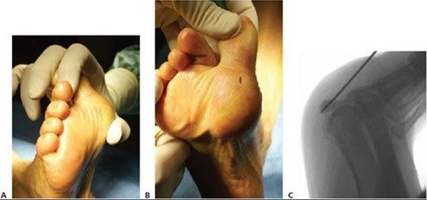
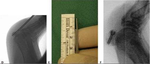
TECH FIG 2 • Preferred technique for percutaneous sesamoid screw fixation. A. Fixation of the hallux in hyperextension. Compress the sesamoid against the metatarsal head to level the fracture fragments against the joint line. B. Place the stab incision distal to the sesamoid outside the weight-bearing area of the MTP joint. C. Place the guidewire perpendicular to the fracture line, subchondral from proximal to distal. D. The guidewire should just pierce the proximal cortex. The second guidewire is advanced to the distal cortex for exact measurement. E. Measurement using two K-wires. F. The definitive screw should incorporate both cortices for optimal compression. The usual length of the screw ranges between 12 and 16 mm.
The length of the headless cannulated compression screw is measured as the difference to a second guidewire that is held next to the first and is advanced to the sesamoid cortex. The usual range is between 12 and 16 mm. The shortest screw available is 10 mm (Bold screws; TECH FIG 2D and E).
The screw should pierce the proximal cortex to enhance stability (TECH FIG 2F).
The stab incision is closed with sterile strips.
Apply compression dressing in neutral hallux position.

POSTOPERATIVE CARE
Full weight bearing over the heel is allowed immediately after surgery.
A shoe with a stiff and convex sole is used to prevent dorsiflexion of the first MTP joint for 6 weeks after surgery, after which time conventional shoes are allowed.
Return to full athletic activity is not recommended before 12 weeks after surgery.
Anderson and McBryde1 treated their patients with 4 weeks non–weight-bearing and another 4 weeks with a weightbearing cast. In our experience with the Anderson-McBryde procedure, hallux correction or turf toe repair requires no adaptations to the postoperative program outlined earlier.
No suture removal or wound care is needed with percutaneous sesamoid fixation, because the stab incision has been closed by a sterile strip.
With combined deformity correction, the type of correction performed dictates postoperative management.
OUTCOMES
Blundell and colleagues5 repaired nine sesamoid fractures in athletes with percutaneous cannulated screws and achieved excellent results. All of the athletes returned to their previous level of activity, with no complications reported. Blundell et al concluded that percutaneous screw fixation is a safe and fast procedure. They also questioned the importance of diagnosing the etiology of painful sesamoid fragments, because treatment is the same regardless of the cause.
Anderson and McBryde1 performed autogenous bone grafting of medial sesamoid nonunions in 21 athletic and nonathletic patients. Of these, 19 grafts healed, whereas 2 grafts failed because the initial fracture dislocation was greater than 2 mm. These two sesamoids were excised. All patients returned to their preinjury activity levels. No hallux deviations have been reported.
At our institution, we performed screw fixation in eight athletes and suture fixation with grafting in two nonathletic women and had excellent results with full recovery.
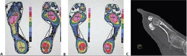
FIG 5 • Postoperative clinical and radiographic results. A. Preoperative pedobarogram shows functional amputation of the first MTP joint as a result of painful sesamoid nonunion in the left foot. B. Pedobarogram 8 weeks postoperatively shows normalization of pressure distribution of the left foot after sesamoid screw fixation. C. CT scan 8 weeks postoperatively shows the healed sesamoid fracture with a screw in place.
The “athletic group” included six women and two men, all of whom were endurance athletes (eg, running, dancing).
We treated two lateral and eight medial sesamoid bone nonunions.
 In one patient, an accompanying forefoot-driven pes cavovarus was corrected with extension osteotomy of the first metatarsus.
In four patients, concomitant hallux valgus deformity was corrected in combined open surgery. In two of these patients, screws were used, and in two other patients sutures were used to stabilize the sesamoid bone during the open approach.
The rest of the patients were treated percutaneously. Local anesthesia was sufficient in one of these cases.
All of the patients returned to their preinjury athletic or occupational activity level within 12 weeks after surgery.
Clinical healing was documented with pedobarography (FIG 5A,B), and osseous healing of the fractures was proved by CT scan in three cases (FIG 5C). One screw had to be removed because of intermittent pain with exercise 1 year after surgery.
Since then, we have used suture cerclage in open approaches, but we also continue to use percutaneous screw fixation.
No sesamoid has had to be excised, and no hallux deformity has occurred.
COMPLICATIONS
Persistent sesamoid pain may be caused by:
Unrecognized foot deformity and continuous stress to the hallux sesamoids
Development of arthritis or avascular necrosis
Screw irritation
Focused therapy (eg, deformity correction, screw removal) may prevent total excision as a definitive treatment of persistent sesamoid pain.
Hallux varus after lateral sesamoid excision, hallux valgus after medial sesamoid excision, and cock-up deformity after both sesamoids were excised have been consistently described in 10% to 20% of cases in the current literature.6,16,17 No hallux deviation has been described after fixation of sesamoid bone fractures.1,5,12,13
A lever arm for flexor tendons and consecutive hallux pushoff can be reconstructed with sesamoid fixation and may be important for the running athlete.2,3
This biomechanical advantage has been proven in vitro2,3 but has an uncertain use in praxis, given the excellent functional results if only one sesamoid bone is excised.6,15,16,17
REFERENCES
1. Anderson RB, McBryde AM. Autogenous bone grafting of hallux sesamoid nonunions. Foot Ankle Int 1997;18:293–296.
2. Aper RL, Saltzman CL, Brown TD. The effect of hallux sesamoid resection on the effective moment of the flexor hallucis brevis. Foot Ankle Int 1994;15:462–470.
3. Aper RL, Saltzman CL, Brown TD. The effect of hallux sesamoid excision on the flexor hallucis longus moment arm. Clin Orthop Relat Res 1996;325:209–217.
4. Biedert R. Which investigations are required in stress fracture of the great toe sesamoids? Arch Orthop Trauma Surg 1993;112: 94–95.
5. Blundell CM, Nicholson P, Blackney MW. Percutaneous screw fixation for fractures of the sesamoid bones of the hallux. J Bone Joint Surg Br 2002;84B:1138–1141.
6. Brodsky JW, Robinson AHN, Krause JO, et al. Excision and flexor hallucis brevis reconstruction for the painful sesamoid fractures and non-unions: Surgical technique, clinical results and histo-pathological findings. J Bone Joint Surg Br 2000;82B:217.
7. Chamberland PDC, Smith JW, Fleming LL. The blood supply to the great toe sesamoids. Foot Ankle Int 1993;14:435–442.
8. Chisin R, Peyser A, Milgrom C. Bone scintigraphy in the assessment of the hallucal sesamoids. Foot Ankle Int 1995;16:291–294.
9. Coleman SS, Chestnut WJ. A simple test for hindfoot flexibility in cavovarus foot. Clin Orthop Relat Res 1977;123:60–62.
10. Inge GAL, Ferguson AB. Surgery of sesamoid bones of the great toe. Arch Surg 1933;27:466–489.
11. Karasick D, Schweitzer ME. Disorders of the hallux sesamoid complex: MR features. Skeletal Radiol 1998;27:411–418.
12. Pagenstert GI, Valderrabano V, Hintermann B. Medial sesamoid nonunion combined with hallux valgus in athletes. Foot Ankle Int 2006;27:135–140.
13. Pagenstert GI, Valderrabano V, Hintermann B. Percutaneous screw fixation of hallux sesamoid fractures. In: Scuderi GR, Tria AJ, eds. Minimally Invasive Orthopaedic Surgery. In press.
14. Prettenklieber ML. Dimensions and arterial vascular supply of the sesamoid bones of the human hallux. Acta Anat 1990;139:86–90.
15. Rodeo SA, Warren RF, O’Brien SJ, et al. Diastasis of bipartite sesamoids of the first metatarsophalangeal joint. Foot Ankle Int 1993;14:425–434.
16. Saxena A, Krisdakumtorn T. Return to activity after sesamoidectomy in athletically active individuals. Foot Ankle Int 2003;24: 415–419.
17. Van Hal ME, Keene JS, Lange TA, et al. Stress fractures of the great toe sesamoids. Am J Sports Med 1982;10:122–128.