
Javier Maquirriain
DEFINITION
 Posterior ankle impingement syndrome (PAIS) is a clinical disorder characterized by posterior ankle pain that occurs during forced plantarflexion.13,14
ANATOMY
The posterior ankle region comprises the soft tissues structures situated behind the tibiotalar joint and the dorsal aspect of the calcaneus.
This region extends superiorly to a horizontal line 4 cm above the tip of the lateral malleolus and inferiorly to a curved line 4 cm below the lateral malleolus.3
The Achilles tendon constitutes the central axis of this region. Neurovascular and musculoskeletal structures in the medial and lateral retromalleolar sulcus surround the calcaneal tendon.
The posterior talar process protrudes posterior to the articular surface of the ankle joint. The body of the posterior process extends both posteriorly and medially from the talus and has two projections designated as the posteromedial process and posterolateral process. These processes are divided by a groove containing the flexor hallucis longus (FHL) tendon (FIG 1).
The posterolateral process, injuries of which are the most common cause of posterior ankle impingement syndrome, is also called the trigonal process.
When the posterolateral process remains separated from the talus, it is called os trigonum.
PATHOGENESIS
The etiology of the posterior ankle pain in forced plantarflexion is varied and may involve any part of the posterior ankle anatomy. PAIS compression pathogenesis has been likened to a “nut in a nutcracker”9(FIG 2).

FIG 1 • Posterior talar process anatomy. Superior view of the talus shows the close relationship of the flexor hallucis longus tendon and the trigonal process.
Trigonal process pathology includes fractures, disrupting of a pre-exisitng synchondrosis, or compression/impingement phenomena.
FHL tendon pathology, including stenosing tenosynovitis and impingement from a prominent posterior talar process, is another cause of PAIS.
PAIS may be produced by intrinsic tibiotalar pathology resulting from ankle trauma. Articular chondral damage or bony injury may cause pain in extreme plantarflexion.
Posttraumatic thickened, inflamed, and sometimes calcified soft tissues in the posterior ankle, including the capsule, synovium, and ligaments, may contribute to chronic impingement with ankle plantarflexion.
A prominence of the posterior calcaneal process also can impinge on the hindfoot.
In posterior ankle impingement, combined pathologies are common. For example, ballet dancers frequently present with associated trigonal process injury with FHL tenosynovitis.8
NATURAL HISTORY
Once injured, patients typically compensate for the loss of plantarflexion by placing the foot in an antalgic position. For example, dancers may begin to assume a more inverted en pointe position to decrease impingement of the posterior structures; doing so, however, may place increased loads on the anterior tibiofibular ligament, which thus predisposes the dancers to frequent ankle sprains.


FIG 2 • The “nutcracker” phenomenon involving the os trigonum.
Calf strain and contractures, plantar foot pain, and toe curling are also typical compensatory problems in dancers that result from efforts to force the foot into a better en pointe position.7
Subtalar joint pathology, especially in posterior articular facets, can produce posterior ankle pain in forced plantarflexion. Furthermore, in chronic PAIS, with limited ankle motion, the subtalar joint may show degenerative changes as a result of higher compensatory loads imposed to maintain ankle range of motion (ROM) during closed kinetic athletic activities.
PATIENT HISTORY AND PHYSICAL FINDINGS
The patient usually reports chronic or recurrent posterior ankle pain caused or exacerbated by forced plantarflexion or push-off activities, such as dancing, kicking, downhill running, and walking on high heels. Biomechanical analysis showed that ankle plantarflexion is required during the swing limb phase and foot contact with the ball of kicking.2 Pain is usually deep and mechanical.
There may be a recent or remote history of ankle trauma, but overuse should be considered.
Tibiotalar, subtalar, and hallux ROM should be measured and recorded.
The diagnostic approach should be based on cause-related conditions.
 The forced plantarflexion test tries to reproduce the typical painful motion of PAIS. It also allows one to estimate the passive ROM limitation.
The Maquirriain test tries to reproduce the typical painful motion of PAIS in a closed position. It also allows one to estimate the passive ROM limitation.
The one-leg hop test provides valuable functional information to rule out Achilles tendon pathology.
IMAGING AND OTHER DIAGNOSTIC STUDIES
A complete history and physical examination is often sufficient to diagnose PAIS. However, ancillary imaging studies should be done to establish the cause, thus allowing proper and timely treatment.
Ankle radiographs should be obtained routinely with lateral view clearly defining trigonal process anatomy. This projection also is used to measure ankle ROM.14
Demonstration of the presence of an os trigonum by conventional radiographs is not necessarily indicative of current clinical relevance.13 Lateral views may show fracture lines, but they cannot differentiate chronic or acute pathology.
Bone scintigraphy is a helpful diagnostic tool. Increased activity is present in all patients with an acute fracture of the trigonal process and synchondrosis disruption.15 A normal bone scan virtually rules out trigonal process pathology.11
MRI is considered the technique of choice to investigate patients with PAIS5,20 because it enables determination of the nature of the osseous and soft tissue lesions and excludes other causes of posterior ankle pain (FIG 3A,B). Bone contusions of the trigonal process are prevalent in individuals with PAIS5 (FIG 3C).
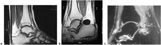
FIG 3 • A. Parasagittal T1-weighted image showing a posterior tibia fracture, which was occult in the initial radiographs. B. Parasagittal T1-weighted image showing a synovial cyst from the tibiotalar joint causing PAIS. C. Parasagittal STIR image showing bone marrow edema as a result of contusion in trigonal process injury.
DIFFERENTIAL DIAGNOSIS
Misdiagnosis is common among patients with posterior ankle pain. Frequently patients had been previously treated for a Achilles tendinopathy.
When a patient has pain in the posterolateral aspect of the ankle, the differential diagnosis includes Achilles tendinopathy, peroneal tendinopathy or tear, retrocalcaneal bursitis, Sever’s disease, and sural neuralgia.
When the patient has pain in the posteromedial aspect of the ankle, the differential diagnosis includes a posterior deltoid sprain, osteochondral lesion of the talus, soleus syndrome, posterior tibial tendinopathy, tarsal tunnel syndrome, and posteromedial tarsal coalition.
NONOPERATIVE MANAGEMENT
Initial treatment for PAIS due to overuse includes rest, nonsteroidal anti-inflammatory drugs, cryotherapy, and avoidance of activities that require forced plantarflexion.
Casting is rarely indicated, but acute articular or bony injuries may benefit from a brief period of immobilization and limited weight bearing.
Physical therapy is indicated to improve ankle and subtalar ROM, as well as strength and flexibility of regional muscles.
Successful nonoperative treatment has been reported in about 60% of patients with PAIS.8
Corticosteroid injection for trigonal process pathology and other chronic causes of PAIS can effectively provide pain relief and should be done at least once before surgery is undertaken.14,15
SURGICAL MANAGEMENT
Indications for surgical intervention include failure of nonsurgical treatment and rehabilitation exercises and a positive response to a diagnostic posterior ankle injection.
Simultaneous bilateral posterior ankle surgery is not recommended.8 While bilateral mechanical posterior ankle impingement is possible, this presentation should prompt a careful workup for systemic causes of posterior ankle pain.
Preoperative Planning
All imaging studies are reviewed.
The ankle–hindfoot score from the American Orthopaedic Foot and Ankle Society (AOFAS) scale is determined.
Ankle and subtalar ROM are tested under anesthesia.
Positioning
The patient is placed in a prone position and a tourniquet is applied on the thigh. Both feet are suspended off of the end of the bed, and a small triangular support is placed under the lower leg, making it possible to move the ankle freely and allow fluoroscopic examination. A support is placed at the ipsilateral side of the pelvis to allow slight rotation of the operating table in a safe manner when needed.19
The surgeon must be aware of the potential complications of this position, such as damage to the genitalia, brachial plexus, and ulnar nerves, among other structures.
Approach
Posterior ankle disorders can be approached by either an open or endoscopic technique.
Open medial and lateral approaches have been described, with both approaches carrying an inherent risk of damaging neurovascular structures. A posterolateral approach should be used to treat isolated bony impingement.8 A medial approach should be used when both FHL tendinopathy and bony impingement are being treated.8
In our experience, hindfoot endoscopy is an advanced procedure with a learning curve that needs to be overcome by practicing on cadavers. The difficulty with endoscopic procedure is the initial orientation in the posterior ankle to achieve a safe access to the pathologic structures.18
TECHNIQUES
POSTEROMEDIAL APPROACH
 A 4-cm curvilinear incision is made posterior to the medial malleolus at the level of the superior border of the calcaneus, following the underlying course of the neurovascular bundle.
The bundle and the FHL are retracted posteriorly with a blunt retractor.
The bony disorder of the posterior talar process (ie, symptomatic os trigonum) is removed.
The area is rasped smooth, and hypertrophic capsulitis and inflamed tissue are débrided.
Finally, the FHL excursion is checked and the tunnel is released as needed from proximal to distal to the level of the sustentaculum tali.
POSTEROLATERAL APPROACH
A curvilinear incision is begun at the posterior ankle mortise in line with the posterior border of the peroneal tendons so that it lies anterior to the sural nerve.12
A capsulotomy is performed with the ankle in slight dorsiflexion, and the lateral talar process or os trigonum is identified lateral to the tunnel. According to Hamilton et al,8 the fibro-osseous tunnel of the FHL tendon cannot be released safely from the lateral side.
Adequate osseous decompression is assessed by plantarflexing the foot and palpating for any bone-on-bone impingement.
POSTERIOR ANKLE ENDOSCOPY
According to van Dijk’s technique, 18 the posterolateral portal is made first at the level or slightly above the tip of the lateral malleolus, just lateral to the Achilles tendon; a clamp is directed anteriorly, pointing in the direction of the first interdigital space (TECH FIG 1A).
When the tip of the clamp touches bone, it is exchanged for a 4.5-mm arthroscope shaft with blunt trocar pointing in the same direction. The blunt trocar is situated extra-articularly at the level of the ankle joint, but it is not necessary to enter the joint capsule.
The posteromedial portal is made just medial to the Achilles tendon at the same level as the posterolateral portal in the horizontal plane.
A clamp is introduced and directed toward the arthroscope shaft to guide the anterior travel of the clamp.
The blunt trocar is exchanged for a 30-degree 4.0-mm scope using a lateral view direction to prevent lens damage. The scope in pulled backward until the tip of the clamp comes into view.
The fatty tissue and adhesions overlying the joint capsule are partially removed using a 3.5-mm full-radius shaver. The posterior compartment of the subtalar joint can be visualized, including the posterior talar process and the FHL tendon. The FHL is an important landmark to prevent damage to the more medially located neurovascular bundle.
By applying manual distraction to the os calcis, the posterior compartment of the ankle joint opens and allows better visualization. The talar dome can be inspected over almost its entire surface as well as the complete tibial plafond. An osteochondral defect or subchondral cystic lesion can be identified, débrided, and drilled.
Removal of a symptomatic os trigonum or a nonunion of a fracture of the posterior talar process involves partial detachment of the posterior talofibular ligament and release of the flexor retinaculum, both of which attach to the posterior talar prominence (TECH FIG 1B,C).
Releasing the FHL involves detachment of the flexor retinaculum from the posterior talar process.
Bleeding is controlled, the new ankle range of motion is checked and recorded, and portals are sutured closed.

TECH FIG 1 • A. Cross-section of the ankle joint at level of the arthroscope. (1) The arthroscope is placed through the posterolateral portal, pointing in the direction of the webspace between the first and second toe. (2) The full-radius resector is introduced through the posteromedial until it touches the arthroscope shaft. (3) It then glides into an anterior direction until it touches bone. B. Posterior left ankle endoscopic view. The os trigonum synchondrosis was released, and the ossicle is ready for excision. The FHL tendon is marked. C. Posterior ankle endoscopic view after trigonal process resection.
PEARLS AND PITFALLS
 During endoscopy, motion of the hallux intraoperatively is a good marker for identification of the posterior anatomy because of the neurovascular bundle’s being medial and the trigonal process’s being lateral to the FHL tendon.
In the hindfoot, the crural fascia can be quite thick. This local thickening is called the Rouvière ligament. During the endoscopic procedure, it needs to be partially excised or sectioned to approach the posterior ankle joint.
POSTOPERATIVE CARE
An elastic bandage is applied, and the ankle is placed in a walker boot.
Weight bearing is allowed after 2 or 3 days. Chondral lesion débridement requires longer non–weight-bearing period.
Early motion (especially plantarflexion) is emphasized.
OUTCOMES
Most experts agree that results of surgery for PAIS are highly satisfactory.1,4,6,8–10,14,19,21
Outcomes after endoscopic treatment of PAIS reported in the literature compare favorably with results of open surgery.19 Advantages include decreased morbidity, less scarring, and the potential for faster recovery.19,21
In a series 55 patients treated endoscopically, van Dijk et al19 reported an average improvement of the AFOS score from 75 points preoperatively to 90 points after surgery.
Results may vary according to the cause of symptoms. Patients treated for PAIS caused by overuse have better results than those treated following trauma.19 Furthermore, patients with osseous impingement do better postoperatively than do patients with soft tissue impingement.
Involvement in a workers’ compensation claim has been shown to influence patient outcome.1
COMPLICATIONS
Sural nerve damage (after posterolateral approach)
Peroneal tendon fibrosis (after posterolateral approach)
Tibial nerve injury (after posteromedial approach)
FHL injury through lateral approach; also during endoscopic technique
Reflex sympathetic dystrophy
Infection
Wound healing problems
Ankle stiffness
Deep vein thrombosis
REFERENCES
1. Abramowitz Y, Wollstein R, Barzilay Y, et al. Outcome of resection of a symptomatic os trigonum. J Bone Joint Surg Am 2003; 85:1051–1057.
2. Barfield WR. The biomechanics of kicking in soccer. Clin Sports Med 1998;17:711–728.
3. Bouchet A, Cuilleret J. Región posterior de la garganta del pie. In: Cuilleret BA, ed. Anatomía Descriptiva, Topográfica y Funcional. Buenos Aires: Editorial Médica Panamericana, 1995:223–234.
4. Brodsky AE, Khalil MA. Talar compression syndrome. Am J Sports Med 1986;14:472–476.
5. Bureau NJ, Cardinal E, Hobden R, et al. Posterior ankle impingement syndrome: MR imaging findings in seven patients. Radiology 2000; 215:497–503.
6. Calder JD, Sexton SA, Pearce CJ. Return to training and playing after posterior ankle arthroscopy impingement in elite professional soccer. Am J Sports Med 2010;38:120–124.
7. Frey C. Injuries of the subtalar joint. In Pffeffer GB, ed. Chronic Ankle Pain in the Athlete. Rosemont, IL: American Academy of Orthopedic Surgeons, 2000.
8. Hamilton WG, Geppert MJ, Thompson FM. Pain in the posterior aspect of the ankle in dancers: differential diagnosis and operative treatment. J Bone Joint Surg Am 1996;78A:1491–1500.
9. Hedrick MR, McBryde AM. Posterior ankle impingement. Foot Ankle 1994;15:2–8.
10. Jaivin JS, Ferkel RD. Arthroscopy of the foot and ankle. Clin Sports Med 1994;13.
11. Karasick D, Schweitzer ME. The os trigonum syndrome: imaging features. Am J Radiol 1996;166:125–129.
12. Lawrence SJ, Botte MJ. The sural nerve in the foot and ankle: an anatomic study with clinical and surgical implications. Foot Ankle Int 1994;15:490–494.
13. Lawson JP. Symptomatic radiographic variants in the extremities. Radiology 1985;157:625–631.
14. Maquirriain J. Posterior ankle impingement syndrome. J Am Acad Orthop Surg 2005;13:365–371.
15. Mouhsine E, Crevoisier X, Leyvraz PF, et al. Post-traumatic overload or acute syndrome of the os trigonum: a possible cause of posterior ankle impingement. Knee Surg Sports Traumatol Arthrosc 2004; 12:250–253.
16. Paulos LE, Johnson CL, Noyes FR. Posterior compartment fractures of the ankle. Am J Sports Med 1983;11:439–443.
17. Scholten PE, Sierevelt IN, van Dijk CN. Hindfoot endoscopy for posterior ankle impingement. J Bone Joint Surg Am 2008;90A: 2665–2672.
18. van Dijk CN, Scholten PE, Krips R. A 2-portal endoscopic approach for diagnosis and treatment of posterior ankle pathology. Arthroscopy 2000;16:871–876.
19. van Dijk CN, de Leeuw PA, Scholten PE. Hindfoot endoscopy for posterior ankle impingement. J Bone Joint Surg Am 2009;91A (Suppl 2):287–298.
20. Wakeley CJ, Johnson DP, Watt I. The value of MR imaging in the diagnosis of the os trigonum syndrome. Skel Radiol 1996;25:133–136.
21. Willitis K, Sonneveld H, Amendola A, et al. Outcome of posterior ankle arthroscopy for hindfoot impingement. Arthroscopy 2008; 24:196–202.