
Beat Hintermann and Victor Valderrabano
DEFINITION
 Pronation injuries of the ankle joint complex may result in a partial or complete disruption of the superficial anterior bundles of the deltoid ligament.
Chronic medial ankle instability may cause a secondary posterior tibial dysfunction over time, as the tendon may become elongated, ruptured, or both.
Medial ankle instability may also be the result of a posterior tibial dysfunction with chronic overload of the deltoid ligaments and consecutive step-by-step disruption.
Medial ankle instability must be suspected if the patient complains of “giving way,” especially medially, when walking on even ground, downhill, or downstairs, pain at the anteromedial aspect of the ankle, and sometimes pain on the lateral ankle, especially during dorsiflexion of the foot.
ANATOMY
The deltoid ligament is a multibanded complex with superficial and deep components.
It may be wise to differentiate the superficial and deep portions of the deltoid complex with respect to the joints they are spanning. The superficial ligaments cross two (the ankle and the subtalar joints) and the deep ligaments cross one joint (only the ankle joint), although differentiation is not always absolutely clear.10
The three superficial and more anterior bands are the tibionavicular, tibiospring, and tibiocalcaneal ligaments; the three deep bands are the anterior, intermediate, and posterior tibiotalar ligaments (FIG 1).1
As the tibioligamentous portion of the superficial deltoid has a broad insertion on the “spring ligament,” this ligament complex may interplay with the deltoid ligament in the stabilization of the medial ankle joint, and thus functionally not be separated from it (Fig 1).3
PATHOGENESIS
Acute injuries to the medial ankle ligaments can occur during running downstairs, landing on an uneven surface, and dancing while the body is simultaneously rotated in the opposite direction. A key feature of the history is whether the patient has sustained a pronation (eversion) trauma—for instance, an outward rotation of the foot during simultaneous inward rotation of the tibia.
Complete deltoid ligament ruptures are sometimes seen in association with lateral malleolar fractures, or in specific bimalleolar fractures.
Chronic deltoid ligament insufficiency can be seen in a number of conditions, including posterior tibial tendon disorder, traumatic and sports-related deltoid disruptions, as well as valgus talar tilting in patients with previous triple arthrodesis or total ankle arthroplasty.
NATURAL HISTORY
There is evidence that the medial ankle ligaments are more often injured than generally believed.4,5,7,8
Several structures contribute to the stabilization of the medial ankle, and in the case of injury they are not involved in a uniform way. Medial ankle instability is thus not a single entity, and this has most important consequences for treatment.
The findings of an exploratory, prospective study on 51 patients (53 ankles) have supported our belief that medial ankle instability without posterior tibial tendon dysfunction does exist as an entity.7 It is, however, not clear yet whether, or to what extent, such a medial ankle instability may cause a secondary posterior tibial dysfunction over time, as the tendon may become elongated, ruptured, or both.
What is clear from the literature is that a coexisting pronation deformity of the foot will lead to further deterioration over time, as the medial ankle ligaments are chronically overstretched.
PATIENT HISTORY AND PHYSICAL FINDINGS
The diagnosis of medial ankle instability is made on the basis of the history and the results of physical examination, including special maneuvers, and plain roentgenography.
Medial instability is suspected if the patient complains of “giving way,” especially medially, when walking on even ground, downhill, or downstairs, pain at the anteromedial aspect of the ankle, and sometimes pain on the lateral ankle, especially during dorsiflexion of the foot.
A history of chronic instability, manifested by recurrent injuries with pain, tenderness, and sometimes bruising over the medial and lateral ligaments, is considered to indicate combined medial and lateral instability that is thought to result in rotational instability of the talus in the ankle mortise.
Acute injuries may present with tenderness and hematoma at the side of the deltoid ligament.

FIG 1 • Anatomic situs of medial ankle. The superficial and deep deltoid consists of three distinct bundles each.
Physical examination methods for chronic medial ankle instability should include:
 Standing test. Inspect for malalignment, deformity, asymmetry, and swelling. Asymmetric planus and pronation deformity of the affected foot may indicate medial ankle instability: distinct, moderate, important.
Palpation of anteromedial ankle. Pain in the medial gutter is typically provoked by palpation of the anterior border of the medial malleolus. It is the result of underlying synovitis due to chronic shifting of the talus within the ankle mortise.
Anterior drawer test is a highly sensitive test for medial ankle instability.
A complete examination of the hindfoot should also include evaluating associated injuries and ruling out other possible causes. These include, among others:
Fracture of medial malleolus: After an acute injury, radiographic analysis must be performed routinely to exclude a fracture of the medial malleolus (eg, bony avulsion of the deltoid ligament) or fibula fracture with or without syndesmotic disruption.
Loss of posterior tibial function after partial or complete rupture: The patient cannot correct the deformity while standing or create supination power to the foot.
Talonavicular coalition: The subtalar joint is not mobile; so there is no varusization of the heel while going into the tiptoe position.
Neurologic disorder: There is partial or complete palsy of one or more muscles due to deficient neurologic control.
IMAGING AND OTHER DIAGNOSTIC STUDIES
Acute injury: Plain radiographs, including AP and lateral views, should be obtained to rule out bony avulsion fractures or associated injuries.
Chronic injury: Plain weight-bearing radiographs, including AP views of the foot and ankle and a lateral view of the foot, should be obtained to rule out old bony avulsion fractures, secondary deformity of the foot (eg, valgus malalignment of the heel, dislocation at the talonavicular joint), and tibiotalar alignment (eg, medial gapping of the joint due to incompetence of the deltoid ligament) (FIG 2).

FIG 2 • Incompetence of deltoid ligament. AP weight-bearing radiograph shows a gapping of the medial tibiotalar joint.

FIG 3 • Proximal avulsion of deltoid ligament. AP MR imaging reveals a complete avulsion of the deltoid ligament to the medial malleolus, resulting in a medial gapping of the tibiotalar joint.
Stress radiographs may be helpful to identify incompetence of the deltoid ligament in the treatment of acute ankle fractures,13 but they are not helpful in chronic conditions.10
A CT scan may be obtained to detect a talocalcaneal coalition or bony fragmentation that involves the articular surfaces.
MR imaging may show an injury to the deltoid ligament (FIG 3), particularly in acute conditions, and it may also reveal pathologic conditions of the posterior tibial tendon.
DIFFERENTIAL DIAGNOSIS
Bony avulsion fracture of the medial malleolus (with or without fracture of the fibula or syndesmotic disruption)
Fixed flatfoot deformity (eg, acquired flatfoot deformity in adults after posterior tibial dysfunction)
Osteochondral injury
Talocalcaneal coalition
NONOPERATIVE MANAGEMENT
Although nonoperative management is controversial, patients with less instability, particularly whose who have less of a “giving-way” feeling, and those who are less involved with highlevel pronation sports activities, may be treated nonoperatively.
Nonoperative treatment consists of three components:
Medial foot arch supports
Physiotherapy for strengthening the invertor muscles
A neuromuscular rehabilitation program
SURGICAL MANAGEMENT
Preoperative Planning
All imaging studies are reviewed.
Plain films should be reviewed for fractures, cartilage lesions, hindfoot and midfoot malalignment, and the presence of any hardware (from previous procedures) or foreign bodies.
Associated fractures, cartilage lesions, foot malalignment, and tendon disruption should be addressed concurrently.
Examination under anesthesia should be performed to compare with the contralateral ankle.
Positioning
The patient is in the supine position with the feet at the edge of the table.
A commercially available knee holder is used to support the distal femur and to place the foot into a hanging position (FIG 4).
This allows the surgeon to move the foot freely while arthroscopy is done before open reconstruction.
After the arthroscopy, the knee holder is removed, leaving the foot on the table.

FIG 4 • Positioning for arthroscopy and medial ligament reconstruction. A knee holder is used to support the distal femur so that the foot is hanging on the table. A. View from the bottom of the table. B.Medial view.
Approach
 An anteromedial approach is used for ankle arthroscopy.4
A gently curved incision of 3 to 5 cm is made, starting 1 cm cranially of the tip of the medial malleolus and running toward the medial aspect of the navicular bone.
If there is additional instability of the lateral ankle ligaments, as found on the clinical examination and confirmed by arthroscopy, a lateral approach to the ankle is also performed to explore the anterior talofibular and calcaneofibular ligaments.
TECHNIQUES
ANKLE ARTHROSCOPY
Arthroscopy is done to visualize the internal structures and to assess medial and lateral ankle stability.4
After visual evaluation of the ligaments, test lateral and medial ligament stability by applying gentle varus, valgus, and anterior pull stress to the ankle joint under arthroscopic control.
Ligament lesions are graded as distended if the ligament is thinned or elongated, and as ruptured if continuity is lost.7 Because most ligament tears are located on the proximal insertion, this is usually best seen by a completely free insertion area of the ligament on the malleoli (TECH FIG 1).
As the foot is everted and pronated, the deltoid ligament is considered incompetent when it is tensioned, but obviously no strong medial buttress is created with this maneuver (TECH FIG 2). An excessive lifting away of the talus from the medial malleolus by pulling the foot anteriorly is also considered an indicator of stretching of this ligament.

TECH FIG 1 • Avulsion of anterior superficial layers from medial malleolus. Arthroscopy typically reveals a completely free insertion area of the ligament on the medial malleolus.
Lateral instability is considered to be present when talar tilting occurs by supination stress of the foot.
As evaluated for both the medial and lateral side, the ankle joint is graded as stable when there is some translocation of the talus, but not enough to open the tibiotalar joint by more than 2 mm (as measured by the 2-mm hook) and not enough to introduce the 5-mm arthroscope into the tibiotalar space; as moderately unstable when the talus moves to some extent out of the ankle mortise, allowing introduction of the 5-mm arthroscope into the tibiotalar space, but not enough to open the tibiotalar joint by more than 5 mm; and as severely unstable when the talus moves easily out of the ankle mortise, typically allowing free insight into the posterior aspect of the ankle joint without significant pulling stress on the heel.7
TECH FIG 2 • Incompetent deltoid ligament. A. As the foot is everted and pronated, the deltoid ligament is considered incompetent when it is tensioned, but obviously no strong medial buttress is created with this maneuver. B. An excessive lifting away of the talus from the medial malleolus by pulling the foot anteriorly is also considered an indicator of stretching of this ligament.
MEDIAL ANKLE LIGAMENT RECONSTRUCTION
Complete acute rupture: Because the rupture is mostly situated proximally of the deltoid ligament (TECH FIG 3), reattachment to the medial malleolus is achieved by interosseous sutures; a bony anchor can also be used for refixation to the bone.8
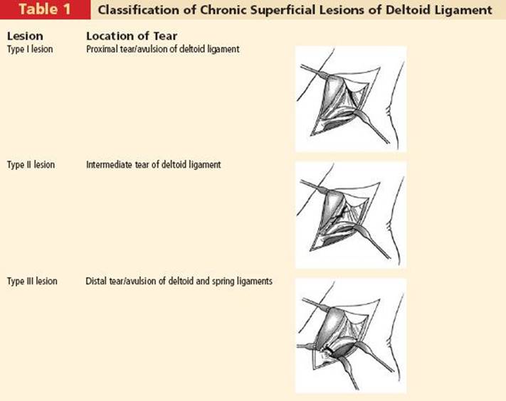
Chronic ruptures of the superficial deltoid ligament are classified as shown in Table 1.7,8
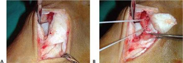
Chronic rupture of the superficial deltoid ligament (type I lesion): Expose the anterior border of the medial malleolus by making a short longitudinal incision between the tibionavicular and tibiospring ligaments, where there is usually a small fibrous septum without adherent connective fibers between the two ligaments (TECH FIG 4A). After roughening the medial aspect of the medial malleolus, place an anchor (Panalock®) 6 mm above the tip of the malleolus (TECH FIG 4B); this serves for refixation of the tibionavicular and tibiospring ligaments to the medial malleolus, and to shorten both the tibionavicular and tibiospring ligaments (TECH FIG 4C–E). Use additional no. 0 resorbable sutures to refix the tibionavicular and tibiospring ligaments.
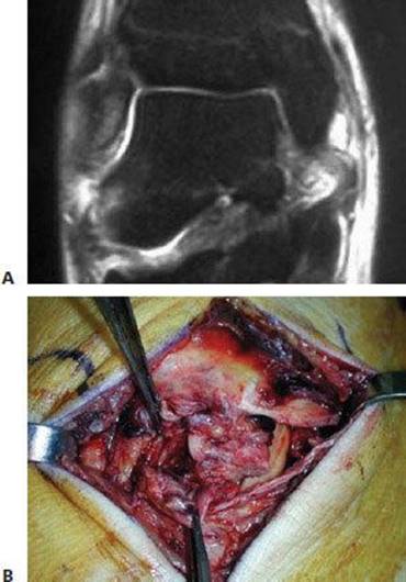
TECH FIG 3 • Acute deltoid rupture. This 28-year-old soccer player sustained a valgus trauma, causing an acute “giving way” of the foot. A. MR imaging reveals complete disruption of the ligament close to its proximal insertion to the medial malleolus. B. Surgical exploration confirms complete disruption of the deltoid ligament, although the posterior tibial tendon remained intact.

TECH FIG 4 • Chronic rupture of the superficial deltoid ligament (type I lesion). A. The rupture is located between the tibionavicular and tibiospring ligaments, where a small fibrous septum without adherent connective fibers between the two ligaments is usually present. B. After roughening the medial aspect of the medial malleolus, an anchor (Panalock®) is placed 6 mm above the tip of the malleolus. C. It serves for refixation of the tibionavicular and tibiospring ligaments to the medial malleolus, and to shorten both ligaments. D. Final reconstruction after some additional no. 0 resorbable sutures. E. Principle of reconstruction.
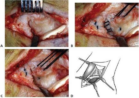
Chronic rupture of the superficial deltoid ligament (type II lesion): Divide the scarred insufficient ligament (TECH FIG 5A) into two flaps: the deep flap remains reattached distally; the superficial flap remains reattached to the medial malleolus. Place two anchors (Panalock®) 6 mm above the tip of the malleolus (TECH FIG 5B), and place one anchor (Panalock®) at the superior edge of the navicular tuberosity (TECH FIG 5C). Two anchors serve for refixation of the deep flap to the medial malleolus (TECH FIG 5D), and the superficial flap to the navicular tuberosity (TECH FIG 5E), thereby creating a strong and well-tightened ligament reconstruction (TECH FIG 5F). The second superior anchor on the medial malleolus serves for reattachment of the tibionavicular ligament (TECH FIG 5G). Use additional no. 0 resorbable sutures to further stabilize the reconstructed tibionavicular and tibiospring ligaments (TECH FIG 5H,I).
Chronic rupture of the superficial deltoid ligament (type III lesion): If necessary, débride the tear (TECH FIG 6A). Then place two nonresorbable sutures in the spring ligament (TECH FIG 6B). If the tibionavicular ligament is completely detached from its insertion, place an anchor (Panalock®) at the superior edge of the navicular tuberosity. After tightening the sutures (TECH FIG 6C,D), use additional no. 0 resorbable sutures to further stabilize the reconstructed tibionavicular and spring ligaments.
Chronic rupture of the deep deltoid ligament: Because this condition usually includes an extended tear of the superficial anterior bundles of the deltoid ligament, any

TECH FIG 5 • Chronic rupture of the superficial deltoid ligament (type II lesion). A. The superficial deltoid ligament is scarred and incompetent. B. Two anchors (Panalock®) are placed 6 and 9 mm above the tip of the medial malleolus. C. Another anchor (Panalock®) is placed into the tuberosity of the navicular bone. D. The deep flap is reattached to the medial malleolus using the distal anchor suture. E. The superficial flap is reattached to the tuberosity of the navicular bone using the anchor suture. F. A strong and well-tightened ligament reconstruction is thus obtained. G. The second superior anchor on the medial malleolus serves for reattachment of the tibionavicular ligament. H. Additional no. 0 resorbable sutures are used to further stabilize the reconstructed tibionavicular and tibiospring ligaments. I.Principle of reconstruction.
TECH FIG 6 • Chronic rupture of the superficial deltoid ligament (type III lesion). A. The distal tear in the spring ligament is exposed and débrided. B. Two nonresorbable sutures are placed in the spring ligament. C. The sutures are tightened. D. Principles of reconstruction.
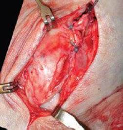
TECH FIG 7 • Chronic rupture of the deep deltoid ligament. After the posterior tibial tendon has been split into two bundles, both bundles are inserted into a drill hole at the tip of the medial malleolus (arrow). One bundle is conducted through the anterior tunnel at the anterior aspect of the medial malleolus, and the posterior bundle is conducted through the posterior tunnel at the posterior aspect of the medial malleolus.
reconstructive surgery should attempt to address the whole deltoid ligament. The posterior tibial tendon can be used as a graft for augmentation of the reconstructed deltoid ligament by passing it through a drill hole from the tip of the medial malleolus to the medial aspect of the distal tibia (TECH FIG 7). This technique was found to be disappointing, however, as it does not sufficiently reinforce the deep tibiotalar ligaments (Hintermann, unpublished data). Most recently, the use of a bone–tendon–bone transplant has been proposed for reconstruction of the deltoid ligament (TECH FIG 8).2 In this in vitro study, two limbs were created on a distal transplant; one was fixed to the medial aspect of the talus and the other to the sustentaculum tali. The proximal end was fixed to the distal tibia, the medial malleolus, or the lateral tibia. Less than 2.0 degrees of angulation was found while applying valgus stress of 5 daN for all fixation methods. However, the authors advised against fixation of the proximal limb in the medial malleolus.
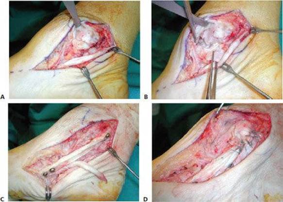
TECH FIG 8 • Chronic rupture of the deep deltoid ligament. A. Exposure of the posterior tibial tendon reveals a tear. B. Exposure of the deltoid ligament reveals an extended disruption and incompetence of the superficial and deep layers. C. A bone–tendon–bone transplant is fixed by screws distally into the navicular bone and, after tightening, proximally to the posterior aspect of the medial malleolus. D.Multiple nonabsorbable and absorbable sutures are used for further reconstruction of the ligament.
LATERAL ANKLE LIGAMENT RECONSTRUCTION
About 75% of patients with chronic medial ankle instability were found to have an associated avulsion of the anterior talofibular ligament that resulted in a complex rotational instability of the talus within the ankle mortise.7
If the condition of the anterior talofibular ligament and the calcaneofibular ligament allows an adequate primary repair, these ligaments can be reconstructed by shortening and reinsertion (TECH FIG 9).
When no substantial ligamentous material is present, augmentation with a free plantaris tendon graft is performed (TECH FIG 10).12
Lateral Lengthening Calcaneal Osteotomy
Inspect the posterior tibial tendon meticulously during surgery, especially in the case of a type II or type III lesion of the anterior deltoid ligament.
If there is degeneration of the tendon, débride the tendon.
If there is elongation of the tendon, consider shortening the tendon.
If there is an accessory bone (os tibiale externum), consider reattaching the bone with the tendon insertion; the posterior tibial tendon can also be tightened if the bone is reattached more distally to the navicular bone (TECH FIG 11).9
A transfer of the flexor digitorum tendon might be considered in the case of a diseased or ruptured tendon, but this is seldom the case.
Posterior Tibial Débridement and Reconstruction
This procedure is considered in the case of a pre-existing valgus and pronation deformity of the foot (eg, when a valgus and pronation deformity is also present on the contralateral, asymptomatic foot) or in the case of a severe attenuation or defect of the tibionavicular, tibiospring, or spring ligaments.
A calcaneal osteotomy is performed along and parallel to the posterior facet of the subtalar joint, from lateral to medial, preserving the medial cortex intact (TECH FIG 12A–D).6
As the osteotomy is widened, the pronation deformity of the foot is seen to disappear (TECH FIG 12E).
Fashion a tricortical graft from the iliac crest to the length required and place it into the osteotomy site (TECH FIG 12F–H).
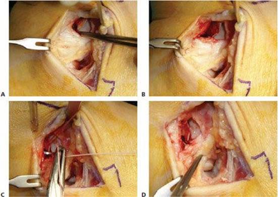
TECH FIG 9 • Primary anatomic repair of lateral ankle ligaments. A. Exposure of lateral ligaments and arthrotomy of ankle and subtalar joints that are débrided. The scarred anterior portion of the lateral ligaments is widely disconnected from the anterior border of the fibula. B. The anterior border of the fibula is roughened. C. An anchor or transosseous sutures are used to reattach the avulsed lateral ligaments (eg, the anterior tibiofibular and calcaneofibular ligaments at their common insertion 8 to 10 mm above the tip of lateral malleolus). D. A strong and well-tightened ligament reconstruction is thus obtained.
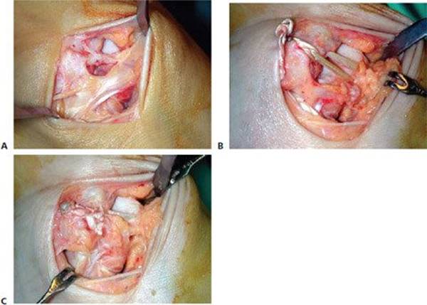
TECH FIG 10 • Reconstruction of lateral ankle ligaments with a free plantaris tendon graft. A. The remaining scarred ligaments do not allow primary repair of lateral ankle ligaments. B. A free plantaris tendon graft is used for reconstruction of the anterior talofibular and the calcaneofibular ligaments. C. A strong and well-tightened ligament reconstruction is thus obtained.
Double Arthrodesis
This procedure is considered when the medial ankle instability is so excessive that a valgus tilt of the talus within the mortise is seen on a standard AP view of the ankle while the foot is loaded (Fig 2).11
Be sure to fully correct the whole deformity (eg, valgus malalignment of the heel, and the peritalar dislocation of talus).
Expose the talonavicular joint from medially through the same incision (TECH FIG 13A,B).
Use a distraction spreader (Hintermann spreader) to open the joint; this allows for cartilage removal and débridement (TECH FIG 13C,TD).
Expose the subtalar joint from medially through the same incision.

TECH FIG 11 • Unstable os tibiale externum. A. An unstable accessory bone (os tibiale externum) is found to weaken the pull of posterior tibial tendon. B. The accessory bone is mobilized and 3 to 5 mm of bone is removed on both sides of the pseudarthrosis. C. This allows for reattachment of the accessory bone more distally to the navicular bone, using screws and nonabsorbable sutures.
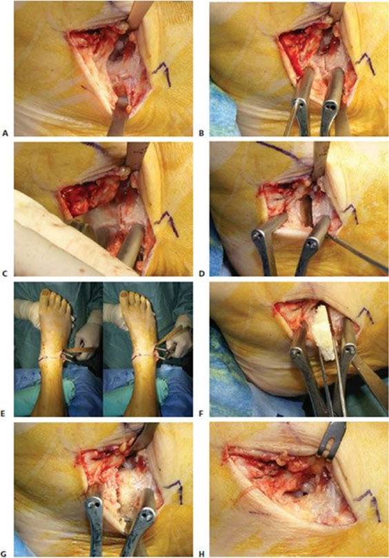
TECH FIG 12 • Calcaneal lengthening osteotomy. A. The neck of the calcaneus is exposed using a lateral incision. B. The osteotomy is marked by a cisel to be directed through the sinus tarsi along the anterior border of the posterior facet of the subtalar joint. Two Kirschner wires for the Hintermann retractor are inserted. C. Osteotomy is performed using a saw. D. The osteotomy is opened using the retractor. E. As the osteotomy is widened, the pronation deformity of the foot is seen to disappear. F. A tricortical graft from the iliac crest or an allograft is fashioned to the length required and placed into the osteotomy site. G. The border of the inserted graft is smoothed. H. A regular bony contour on the bottom of the sinus tarsi is thus obtained.
Use the distraction spreader to open the joint; this allows for cartilage removal and débridement (TECH FIG 13E–G).
Correct the deformity first by reducing the former talonavicular joint, making sure to correct the frontal plane position of the navicular (eg, to achieve full correction of any forefoot supination deformity) (TECH FIG 13H–L).
Stable fixation is achieved by triple screw fixation at the talonavicular and double screw fixation at the subtalar joint (TECH FIG 13M–O).
WOUND CLOSURE
Close the wounds in layers.
Close the subcutaneous tissue and skin in standard fashion.

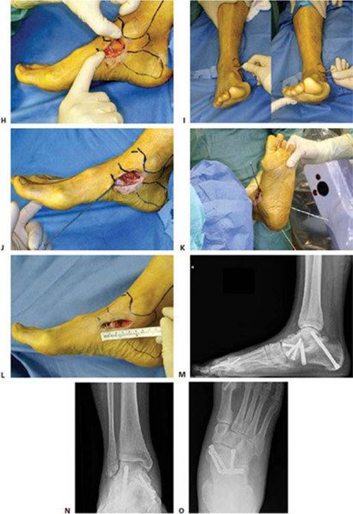
TECH FIG 13 • Double arthrodesis. A. Skin incision just above the posterior tibial tendon; the surgeon should stop proximally at a perpendicular line through the medial malleolus (eg, so as not to damage the deep bundles of the deltoid ligament). B. Incision of skin and dissection of the medial ankle ligaments by sharp incision along the spring ligament. C. The talonavicular joint is exposed first. The Hintermann retractor serves to expose the joint. D. Cartilage is removed and the joint is cleaned to subchondral bone. E. A third Kirschner wire is inserted into the sustentaculum tali of the calcaneus. This allows the surgeon to open the subtalar joint using the Hintermann distractor. F. The cartilage is removed. G. Final inspection shows complete débridement of the subtalar joint, including the sinus tarsi. H. The Kirschner wires in the navicular and talar bones are kept in place and serve to reduce the talonavicular joint properly. I. Frontal view showing the frontal realignment at the talonavicular joint using both Kirschner wires as joysticks. J. A first guiding Kirschner wire is inserted through the tuberosity of the navicular into the talus. Afterwards, two other guidewires will be used to properly stabilize the talonavicular joint in the frontal plane. K. After inserting two additional guidewires from the bottom through the subtalar joint, fluoroscopy is used to insert the cannulated screws (QUIX, Newdeal/Integra). L. The deltoid ligament is reattached to the spring ligament using nonabsorbable sutures. The foot looks properly positioned at the end of surgery. Note the short incision that is used for this procedure. At 2 months, weight-bearing radiographs are obtained. M. Lateral view. N. AP view of the ankle. O. AP view of the foot.

POSTOPERATIVE CARE
The foot is protected by a plaster cast for 6 weeks, and full weight bearing is allowed as soon as pain-free loading is possible. In the case of double arthrodesis, initial plaster immobilization for 8 weeks is recommended.
The rehabilitation program starts after cast removal. It includes passive and active mobilization of the ankle joint, training of the muscular strength, and protection with a walker or stabilizing shoe when walking.
A walker or stabilizing shoe can be used for 4 to 6 weeks after cast removal, depending on regained muscular balance of the hindfoot.
We recommend continued use for walks on uneven ground, for high-risk sports activities, and for professional work outside.
OUTCOMES
With appropriate surgical technique, success rates for ligament reconstruction of the medial ankle are on the order of 85% to 90% in terms of return to former sports and professional activities.7
As associated malalignment has been addressed more aggressively in the last years, the success rate has further increased.
The most troubling problem remains a chronic incompetence of the deep deltoid ligament, which results in valgus tilt of the talus while loading the foot. Despite the use of tendon augmentation, most attempts at isolated ligament reconstruction have failed; the main step is probably a double arthrodesis in getting a stable and well-aligned hindfoot. An alternative may be a tibiocalcaneal arthrodesis.
COMPLICATIONS
Deficient stability because of inappropriate ligament reconstruction
Recurrent instability because valgus deformity was not addressed
Suture granuloma at the anterior margin of the medial malleolus when using nonresorbable sutures and placing the suture knot onto a bony surface
Deep venous thrombosis
Infection
Scarring in the anteromedial ankle causing soft tissue impingement
REFERENCES
1. Boss AP, Hintermann B. Anatomical study of the medial ankle ligament complex. Foot Ankle Int 2002;23:547–553.
2. Buman EM, Khazen G, Haraguchi N, et al. Minimally invasive deltoid ligament reconstruction: a comparison of three techniques. Proceedings of the 36th Annual Winter Meeting, Specialty Day, AOFAS, Chicago, March 25, 2006, p. 25.
3. Harper MC. Deltoid ligament: an anatomical evaluation of function. Foot Ankle 1987;8:19–22.
4. Hintermann B, Boss A, Schäfer D. Arthroscopic findings in patients with chronic ankle instability. Am J Sports Med 2002;30:402–409.
5. Hintermann B. Medial ankle instability. Foot Ankle Clin 2003;8: 723–738.
6. Hintermann B, Valderrabano V. Lateral column lengthening by calcaneal osteotomy. Techn Foot Ankle Surg 2003;2:84–90.
7. Hintermann B, Valderrabano V, Boss AP, et al. Medial ankle instability: an exploratory, prospective study of 52 cases. Am J Sports Med 2004;32:183–190.
8. Hintermann B, Knupp M, Pagenstert GI. Deltoid ligament injuries: diagnosis and management. Foot Ankle Clin 2006;11:625–637.
9. Knupp M, Hintermann B. Reconstruction in posttraumatic combined avulsion of an accessory navicular and the posterior tibial tendon. Techn Foot Ankle Surg 2005;4:113–118.
10. Milner CE, Soames RW. The medial collateral ligaments of the human ankle joint: anatomical variations. Foot Ankle Int 1998; 19:289–292.
11. Nelson DR, Younger A. Acute posttraumatic planovalgus foot deformity involving hindfoot ligamentous pathology. Foot Ankle Clin 2003;8:521–537.
12. Pagenstert GI, Hintermann B, Knupp M. Operative management of chronic ankle instability; plantaris graft. Foot Ankle Clin 2006;11: 567–583.
13. Tornetta P, III. Competence of the deltoid ligament in bimalleolar ankle fractures after medial malleolar fixation. J Bone Joint Surg Am 2000;82A:843–848.