
Nicholas A. Ferran, Ansar Mahmood, and Nicola Maffulli
DEFINITION
 Rupture of the Achilles tendon is common.
More than 20% of acute injuries are misdiagnosed, leading to chronic or neglected ruptures.4
ANATOMY
The two heads of the gastrocnemius arise from the condyles of the femur, the fleshy part of the muscle extending to about the midcalf. As the muscle fibers descend they insert into a broad aponeurosis that contracts and receives the tendon of the soleus on its deep surface to form the Achilles tendon.9
The Achilles tendon is the thickest and strongest tendon in the body. About 15 cm long, it originates in the midcalf and extends distally to insert into the posterior surface of the calcaneus. It receives muscle fibers from the soleus on its anterior surface throughout its length.9
PATHOGENESIS
The most common mechanism of injury is pushing off with the weight-bearing forefoot while extending the knee. Sudden unexpected dorsiflexion of the ankle or violent dorsiflexion of a plantarflexed foot may also result in ruptures.5
Corticosteroids, fluoroquinolone use, tendon pathology, and poor vascularity of the Achilles tendon have been associated with rupture.5
NATURAL HISTORY
A delay in treatment of Achilles tendon rupture results in the formation of a discrete gap. The gap between ruptured tendon ends may fill with fibrous nonfunctional scar. Patients find walking and ascending stairs difficult, and standing on tiptoes on the affected limb impossible.
PATIENT HISTORY AND PHYSICAL FINDINGS
Patients often give a history of feeling a blow to the posterior aspect of the leg and may describe an audible snap followed by pain and inability to bear weight.
In acute tendon ruptures, a gap in the Achilles tendon is usually palpable. In delayed presentation, edema may fill this gap, making palpation unreliable.
Active plantarflexion of the foot is usually preserved due to the action of the tibialis posterior and the long toe flexors.
The calf squeeze test, first described by Simmonds in 19577 but often credited to Thompson, is performed with the patient prone and the ankles clear of the table. The examiner squeezes the fleshy part of the calf, causing deformation of the soleus, and resulting in plantarflexion of the foot if the Achilles tendon is intact. The affected leg should be compared to the contralateral leg.
The knee flexion test is performed with the patient prone and the ankles clear of the table. The patient is asked to actively flex the knee to 90 degrees. During this movement the foot on the affected side falls into neutral or dorsiflexion and a rupture of the Achilles tendon can be diagnosed.6
IMAGING AND OTHER DIAGNOSTIC STUDIES
The diagnosis of acute ruptures is usually a clinical one.
Plain lateral radiographs may reveal an irregular configuration of the fat-filled triangular space anterior to the Achilles tendon and between the posterior aspect of the tibia and the superior aspect of the calcaneus.
DIFFERENTIAL DIAGNOSIS
Ankle sprain
NONOPERATIVE MANAGEMENT
Acute ruptures may be managed conservatively in an equinus cast for 6 to 8 weeks before being converted to a functional brace.
Conservative management may result in tendon lengthening, thus altering function.1
SURGICAL MANAGEMENT
Percutaneous repair3 was originally described as a compromise between open surgery and conservative management. A percutaneous repair aims to provide the optimal functional outcome of open repair while decreasing the problems associated with it in terms of wound healing and skin breakdown.
Preoperative Planning
Once the diagnosis is made, an assessment of general health and comorbidities should be performed.
The preoperative functional status should be noted.
The skin quality and neurovascular status of the affected limb should be examined.
The status of the sural nerve should be documented.
We recommend that the patient be maintained on deep venous thrombosis prophylaxis.
The procedure can be performed under general anesthesia or a local anesthetic, with a 50:50 mixture of 10 mL of 2% lignocaine hydrochloride (Antigen Pharmaceuticals Ltd, Roscrea, Ireland) and 10 mL of 0.25% bupivacaine hydrochloride (Astra Pharmaceuticals Ltd, Kings Langley, England) instilled into an area of between 8 and 10 cm around the ruptured Achilles tendon.
Positioning
The patient is placed prone, and a pillow is placed beneath the anterior aspect of the ankles to allow the feet to hang free.
The operating table is angled down 20 degrees cranially to reduce venous pooling in the feet and ankles.
A tourniquet is not necessary for this procedure.
Approach
Previous approaches such as those described by Ma and Griffith3 using three medial and three lateral stab incisions have been abandoned in light of the relatively increased incidence of sural nerve entrapment.
We will present two techniques that we employ. The first is an approach similar to that described by Webb and Bannister.8 Three 3-cm transverse skin incisions are made. The middle one is made over the palpable gap, and the proximal and distal incisions are placed 4 cm proximal and distal to the middle incision respectively (FIG 1). The second is currently our favored technique: the results for acute ruptures are as good as the first technique, but it is even less invasive.
In the first technique the proximal incision is made more medial to the others to avoid the sural nerve. The stab incisions used in the five-incision latter procedure do not usually give rise to sural nerve problems.
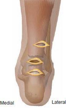
FIG 1 • Incisions. The proximal incision is placed medially.
TECHNIQUES
MINIMALLY INVASIVE REPAIR OF ACUTE ACHILLES TENDON RUPTURE WITH MODIFIED KESSLER SUTURE PATTERN
 Use a small hemostat to free the tendon sheath from the overlying subcutaneous tissue.
Pass a 1 PDS II (Ethicon, Johnson & Johnson Intl, Brussels, Belgium) double-strand suture on a long curved needle transversely through the distal incision, passing through the substance of the tendon and out through the same incision (TECH FIG 1A).
Reintroduce the needle medially into the distal incision through a different entry point in the tendon, and pass it longitudinally through the tendon to lock the tendon. Direct the needle toward the middle incision and out through the ruptured tendon end (TECH FIG 1B).
Rethread the suture still protruding from the distal incision onto the needle and reintroduce it laterally into the distal incision and into the tendon. Pass it proximally through the tendon to exit from the middle incision (TECH FIG 1C).

TECH FIG 1 • A. The suture is passed transversely through the distal stump of the tendon. B. The suture is passed medially into the distal incision through a different entry point in the tendon and passed longitudinally and brought out through the middle incision. C. The suture still protruding from the distal incision is rethreaded onto the needle and reintroduced laterally into the tendon and brought out through the medial incision. D. The procedure is repeated in the proximal stump. E. The suture ends are then tied with the ankle in physiologic plantarflexion.
Apply traction to the suture to ensure satisfactory grip of the tendon.
Carry out the same procedure for the proximal stump of the ruptured tendon (TECH FIG 1D).
A further 1 PDS II (Ethicon) double-stranded suture can be placed in the tendon ends as described above to produce an eight-strand repair.
Tie the sutures with the ankle in physiologic plantarflexion (TECH FIG 1E).
Assess the tension by observing the contralateral limb as the sutures are tied.
Close the skin wounds with undyed subcuticular 3-0 Vicryl (Ethicon, Edinburgh, UK) suture and apply nonadherent dressings.
Apply a full plaster-of-Paris cast in the operating room with the ankle in physiologic equinus.
PERCUTANEOUS REPAIR OF ACUTE ACHILLES TENDON RUPTURE USING FIVE STAB INCISIONS
Local anesthetic infiltration is used. Instill a 50:50 mixture of 10 mL of 2% lignocaine hydrochloride (Antigen Pharmaceuticals) and 10 mL of 0.25% bupivacaine hydrochloride (Astra Pharmaceuticals) into an area 8 to 10 cm around the ruptured Achilles tendon.
The patient is placed prone, and a pillow is placed beneath the anterior aspect of the ankles to allow the feet to hang free.
Angle the operating table down about 20 degrees cranially to reduce venous pooling in the feet and ankles.
The affected leg is prepared with antiseptic and sterile draped. We do not use a tourniquet.
Make five stab incisions over the Achilles tendon (TECH FIG 2A). The first is directly over the palpable defect and measures about 2 cm in a transverse direction.
The other incisions are about 4 cm proximal and 4 cm distal to the first incision and are vertical 1-cm stab incisions on the medial and lateral aspect of the Achilles tendon.
We advocate blunt dissection with a small hemostat directly onto the Achilles tendon. This avoids damaging the sural nerve, which crosses the lateral border of the Achilles tendon about 10 cm proximal to its insertion into the calcaneus.
Use a small hemostat to free the tendon sheath from the overlying subcutaneous tissue (Tech Fig 2A).
Pass a 1 PDS II (Ethicon) double-stranded suture on a long curved needle transversely through the lateral proximal stab incision, passing it through the substance of the tendon and out through the medial proximal stab incision (TECH FIG 2B).
Reintroduce the needle into the medial proximal stab incision through a different entry point in the tendon and pass it longitudinally and distally through the tendon to lock into the tendon. Direct the needle toward the middle incision and out through the ruptured tendon end (TECH FIG 2C).
Rethread the suture that is still protruding from the lateral proximal stab incision onto the needle and reintroduce it via the lateral proximal stab incision into the tendon substance. Also pass it longitudinally and distally through the tendon to exit from the middle incision. Apply traction to the suture to ensure a satisfactory grip within the tendon. If the suture pulls through, repeat the procedure. We sometimes use an eight-stranded method by doubling the sutures used for the Kessler-type technique we are describing.
Carry out the same procedure for the distal half of the ruptured tendon.
Tie the sutures with the ankle in physiologic plantarflexion and bury them into the tissues using a hemostat (TECH FIG 2E).
Close the skin wounds with undyed subcuticular 3-0 Vicryl (Ethicon) suture and apply nonadherent dressings.
Apply a full plaster-of-Paris cast in the operating room with the ankle in physiologic equinus. Split the cast on both medial and lateral sides to allow for swelling (TECH FIG 2F).
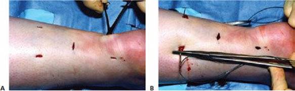

TECH FIG 2 • A. The five stab incisions around the Achilles tendon rupture. A hemostat is used to free the Achilles from any subcutaneous and peritendinous adhesions. B. The needle is introduced into the lateral proximal stab incision through the substance of the tendon. C. The needle is reintroduced into the medial proximal stab incision through a different entry point in the tendon and passed longitudinally and proximally through the tendon, directed toward the middle incision and out through the ruptured tendon end. The same is done with the suture protruding from the lateral proximal stab incision once it is rethreaded onto a needle. Traction is applied to the suture to ensure a satisfactory grip within the tendon. The same procedure is then repeated for the distal segment. D. The sutures are then tied with the ankle in physiologic plantarflexion. E. A hemostat is used to bury the suture into the tissues. F. A full plaster-of-Paris cast is applied in the operating room with the ankle in physiologic equinus. The cast is split on both medial and lateral sides to allow for swelling.

POSTOPERATIVE CARE
The postoperative care regimen and rehabilitation are similar for both techniques.
Patients are discharged on the same day of the operation.
The neurovascular status of the limb is assessed.
After assessment by a physiotherapist, making sure that the patient is safe and comfortable in the cast, the patient can be discharged.
The full cast is retained for 2 weeks, and patients are allowed to bear weight as comfort allows. During the period in the cast, patients are advised to perform gentle isometric contractions of the gastroc–soleus complex.
At 2 weeks, patients are reviewed as outpatients, the cast is split, and the wounds are inspected. An anterior splint is worn with the foot in plantarflexion for a further 4 weeks.
Patients are advised to mobilize with partial weight bearing initially, increasing to weight bearing as able by 4 weeks.
The splint is then removed, and physiotherapy follow-up for gentle mobilization is arranged. Light weight-bearing exercise can be started 2 weeks after cast removal, and the patient should be fully weight bearing by 10 weeks.
OUTCOMES
Lim et al,2 in a randomized controlled trial, advocated percutaneous repair over open surgical techniques after finding no significant differences in functional results, a lower infection rate with the percutaneous repair, and a subjectively more acceptable cosmetic appearance of the percutaneous operative site.
We reviewed 31 patients who underwent percutaneous repair in our tertiary referral center between 2001 and 2003.10 Eleven patients (35.5%) received general anesthesia and 20 (64.5%) had local anesthesia. The average length of cast time was 5.97 weeks. One (3.2%) patient sustained a major complication, a small pulmonary embolism, which was managed successfully with warfarin. There were no reruptures, and six (19.4%) patients had minor wound complications.
COMPLICATIONS
Early complications include sural nerve damage and hematoma.
Intermediate superficial and deep wound infections may occur.
The most important late complication is rerupture.
REFERENCES
1. Bohnsack M, Ruhmann O, Kirsch L, et al. Surgical shortening of the Achilles tendon for correction of elongation following healed conservatively treated Achilles tendon rupture. Z Orthop Ihre Grenzgeb 2000;138:501–505.
2. Lim J, Dalal R, Waseem M. Percutaneous vs. open repair of the ruptured Achilles tendon: a prospective randomized controlled study. Foot Ankle Int 2001;22:559–568.
3. Ma GWC, Griffith TG. Percutaneous repair of acute closed ruptured Achilles tendon: a new technique. Clin Orthop Relat Res 1977;128:247–255.
4. Maffulli N. Clinical tests in sports medicine: more on Achilles tendon. Br J Sports Med 1996;30:250.
5. Maffulli N. Rupture of the Achilles tendon. J Bone Joint Surg Am 1999;81A:1019–1036.
6. Matles AL. Rupture of the tendo Achilles: another diagnostic sign. Bull Hosp Joint Dis 1975;36:48–51.
7. Simmonds FA. The diagnosis of the ruptured Achilles tendon. Practitioner 1957;179:56–58.
8. Webb JM, Bannister GC. Percutaneous repair of the ruptured tendo Achillis. J Bone Joint Surg Br 1999;81B:877–880.
9. Williams PL. Gray's Anatomy, 38th ed. Edinburgh: Churchill Livingstone, 1995.
10. Young J, Sayana MK, McClelland D, et al. Percutaneous repair of acute rupture of Achilles tendon. Tech Foot Ankle Surg 2006;5:9–14.