
William C. McGarvey and Thomas O. Clanton
DEFINITION
 The term insertional Achilles tendinitis (IAT) is actually a misnomer. The condition is more typically a degenerative process, and the nomenclature should reflect this condition, more appropriately, as a tendinosis or tendinopathy.5,7,8,9,14
As the name suggests, insertional Achilles tendinopathy is identified by a painful condition at the musculotendinous insertion of the tendo Achilles on the posterior calcaneus.
 It represents about 10% to 20% of all Achilles pathology.2
It is most commonly seen as an overuse injury in athletes, eg., runners and “push-off” athletes such as basketball or volleyball players, or in more sedentary patients as a degenerative process.
ANATOMY
The Achilles tendon (TA) is the largest tendon in the body. Its chief function is plantarflexion of the foot and ankle.
It is viscoelastic and strong, elongating up to 15% under loads and bearing up to 10 times body weight in single-legged stance during running.5,9
The Achilles insertion is a broad expanse that envelops the entire tuberosity of the os calcis and sends Sharpey's fibers to the medial, lateral, and plantar borders of the bone.1
Immediately anterior to the Achilles tendon lie the retrocalcaneal bursa and a variably sized posterolateral prominence of calcaneus, often known as Haglund's deformity.
More anteriorly lies the deep posterior compartment musculature, which includes the flexor hallucis longus (FHL), the neurovascular bundle, including the tibial nerve, and vessels.
The FHL originates from the fibula and interosseous membrane, travelling obliquely and distally to pass under the sustentaculum tali, through a fibro-osseous tunnel, and on to the master knot of Henry to attach to the hallux.
PATHOGENESIS
Repetitive stress can lead to a combination of inflammatory and degenerative changes.
Degeneration or tendinosis occurs as the already compromised vascularity of the tendon is further reduced by age and injury.8,9
Microscopic and macroscopic changes occur, leading to scarring and slow regeneration or repair.
Tenocytes are reduced in number and quality, contributing to poor repair potential.
Inflammatory changes are manifested as paratenonitis involving the investing layer surrounding the TA, but not the tendon itself. This leads to thickening and adherence of the paratenon to the TA.
Additionally, the continuum of injury and inadequate repair capacity create a cycle of collagen and calcium deposition in an effort to stabilize the musculotendinous enthesis, leading to enlargement of the insertion site; generation of abundant, poor quality tissue; and irritation of the surrounding tisssues, causing a painful thickening of the insertion of the TA.
NATURAL HISTORY
Untreated IAT has not been studied extensively. However, surgical findings and histologic analyses have provided some information.
Persistent IAT leads to continued pain and swelling of the retrocalcaneal heel.
A vicious cycle occurs in which further injury induces more attempts at repair and scar formation, leading to more irritation of surrounding tissues, decreased vascularity, and further microscopic injury.
The posterior heel is more difficult to accommodate in a shoe.
Range of motion is reduced, leading to increased susceptibilty to injury with any activity that places the TA under strain.
Calcific debris is generated, both as reactive tissue reponse to injury and intratendinous hematoma formation as a result of injury. This compromises the viscoelasticity and, therefore, the integrity of the tendon, making it more apt to tear, either partially or completely.4,14
A less pliable, less resilient TA is the end product. Insertional avulsion or rupture may occur, presenting a difficult treatment dilemma.
PATIENT HISTORY AND PHYSICAL FINDINGS
Patients provide fairly accurate descriptions and complain of pain at the bone–tendon interface on the retrocalcaneal heel.
Pain may be worse after activity, but gradually becomes more pervasive.
Athletes may note an increase in symptoms with increases in training intensity or duration or changes in surface or shoes.
Examination demonstrates tenderness directly posteriorly on the heel or, often, posterolaterally.
In advanced cases, thickening, nodularity, or hardening may be palpated.
Dorsiflexion of the ankle may be reduced compared with the contralateral leg.
Methods for examining the Achilles tendon and its insertion include:
Direct palpation
 The posterior heel and TA are inspected for visual or palpable swelling, tenderness, nodularity, or gapping, all of which are suggestive of diseased tendon.
Thompson test
With the patient prone, squeeze the calf at the gastrocnemius–soleus junction to elicit plantarflexion of the foot. Compare with the contralateral side. A positive test is identified when the excursion of the injured side is far less than its uninjured counterpart. This is evidence for complete rupture.
IMAGING AND OTHER DIAGNOSTIC STUDIES
Radiographs
Radiographs often are not necessary for diagnosis, but they are helpful in determining the presence of calcific debris, which is a poor prognostic sign14 (FIG 1).
Plain lateral and axial radiographic views usually are sufficient.
Ultrasonography
Ultrasonography is a relatively inexpensive and accurate way to determine tendon quality, integrity, and function.
It has the advantage of being used dynamically to watch active tendon excursion, if so desired. It also may be used to follow the course of healing.
It is a highly user-dependent tool.
MRI
MRI probably is the most comprehensive study available for investigation and evaluation of a damaged Achilles tendon (FIG 2A,B).
This study gives the most accurate information regarding degree of TA involvement, quality of surrounding tissue, presence or absence of rupture, and other concomitant pathology.
DIFFERENTIAL DIAGNOSIS
Retrocalcaneal bursitis
Haglund syndrome
Inflammatory arthritides
Seronegative spondyloarthropathies
Gout
Familial hyperlipidemia
Sarcoidosis
Diffuse idiopathic skeletal hyperostosis
Pharmacologically induced pathology
Fluoroquinolone use
Chronic corticosteroid use

FIG 1 • Plain radiographs demonstrate intratendinous calcification at the Achilles insertion.
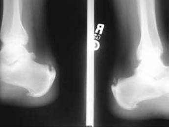
FIG 2 • MRI scan of insertional Achilles tendinosis. In this T2 sequence, the degenerative areas of tendon are demonstrated by increased uptake within the substance of the tendon in the sagittal (A) and axial (B) planes.
NONOPERATIVE MANAGEMENT
Nonoperative treatment is successful in over 90% of patients with this process.5,8
Success rates decline in the face of greater age at time of presentation, longstanding symptoms, and evidence of calcific tendinosis.8
Initial phases of treatment include NSAID use, heel lifts, eccentric stretching, and shoe modifications to widen and soften the heel counter.
More advanced situations may call for formal orthsoses to correct any biomechanical abnormallity, night splinting to apply continual stretch to the TA, and therapy modalities such as ice, contrast baths, and iontophoresis.
Severe cases may require immobilzation in a cast or boot, followed by gradual reintroduction to cross-training before return to regular sports or activities.
SURGICAL MANAGEMENT
Surgical decision making should reflect failed conservative efforts and continued symptoms and functional impairments.
Younger, more athletic patients often respond to a simple débridement of damaged TA, which usually accounts for less than 50% of the total tendon. A midline tendon-splitting approach to this débridement is our preferred procedure for these patients.
Preoperative Planning
Tendon integrity becomes more questionable with involvement of more than 50%, so augmentation is entertained at this point. This extensive involvement may become evident on preoperative testing or, alternatively, on intraoperative evaluation.
Ideally, the surgeon will already have a good idea before beginning the procedure as to whether an augmentation is necessary.
This is readily apparent on imaging.
One must be ready to add this procedure if it is found to be necessary intraoperatively.
Positioning
The patient is placed in the prone position (FIG 3).
Both feet are prepped into the surgical field, up to the level of the knees.
Approach
A midline incision currently is preferred (FIG 4), beginning about 2 to 3 cm proximal to the insertion and extending distally to expose the entire insertion of the TA.

FIG 3 • Positioning for flexor hallucis longus (FHL) transfer. The patient is in the prone position with both feet prepped into the surgical field. Note the evidence of acute Achilles avulsion as resting tension on the foot is lost.

FIG 4 • Surgical approach. The planned incision for Achilles insertional débridement and FHL transfer is marked on the skin.
TECHNIQUES
RETROCALCANEAL DÉBRIDEMENT
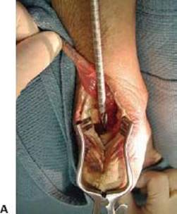
 Full-thickness flaps should be developed medially and laterally, including the substance of the Achilles tendon (TECH FIG 1A).
All nonviable or suspect-appearing tissue should be removed (TECH FIG 1B).
Once adequate débridement takes place, the retrocalcaneal bursa also can be excised.
Any enlarged or impinging posterolateral prominence of calcaneus should be aggressively removed with a ronguer, oscillating saw, or osteotome.

TECH FIG 1 • A. A midline tendon-splitting incision is made with full-thickness flaps through the TA. B. The retrocalcaneal bursa is visualized through the tendon.
FLEXOR HALLUCIS LONGUS HARVEST
The FHL muscle belly usually is easily visible after the débridement is complete (TECH FIG 2A).
Trace the FHL into its fibro-osseous tunnel (TECH FIG 2B,C).
Plantarflex the ankle and hallux maximally.
With a no. 15 blade, transect the tendon as distally as possible while an assistant pulls posteriorly on the proximal portion of the tendon (TECH FIG 2D).
An interlocking suture of 2-0 braided nonabsorbable material is sewn into the tendon stump.

TECH FIG 2 • A, B. The flexor hallucis longus (FHL) tendon and the tibial nerve (medial or to the right) lie parallel and in close proximity to one another separated by a fibro-osseous sheath. C. The FHL is harvested throught the posterior incision, with care taken to avoid injuring the nerve, which lies just outside or medial to the fibro-osseous tunnel. D. A Krackow interlocking suture technique is used to secure the FHL to be brought into the tunnel.
ATTACHING THE GRAFT
A 6.5-mm vertical tunnel is created in the posterior calcaneus about 1 cm anterior to the previous Achilles insertion (TECH FIG 3A).
The FHL suture is passed through the tunnel and through the plantar skin with a Beath pin, bringing the FHL tendon into the tunnel (TECH FIG 3B).
Tensioning is ensured by using the other foot as a reference at about 15 to 20 degrees of resting equinus. If the most medial and lateral remnants of native Achilles are maintained, they will also serve as reference for proper tensioning.
An absorbable interference screw is introduced for graft fixation (TECH FIG 3C).
Tension and range of motion are assessed and should be roughly equal to that of the unijnjured limb (TECH FIG 3D).
The FHL muscle belly is then sewn in a side-to-side tenodesis fashion to the remaining TA to promote vascularity and help restore power to push-off (TECH FIG 3E).

TECH FIG 3 • A. A bony tunnel is reamed to match the size of the interference screw to be used. B. FHL pulled through the bony tunnel with tension equal to that of the other leg. C. Interference screw placement. D.Completed FHL transfer. E. A side-to-side tenodesis connecting the FHL muscle belly to the Achilles remnant is performed.
WOUND CLOSURE
Layered closure is performed with a 2-0 absorbable monofilament in the paratenon, if any is left.
2-0 monocryl suture is used for subcutaneous fat.
Skin is closed with 3-0 nylon suture.

POSTOPERATIVE CARE
Immediately postoperatively, the foot is splinted in 15 to 20 degrees of equinus for 2 weeks.
The patient is then placed in a walking boot with a 2-inch heel lift for another 2 weeks and allowed to gently touch down to the floor. (Noncompliant patients receive a walking cast.)
At 1 month, patients are instructed on gentle active-assisted range of motion and permitted to progress to full weight bearing as tolerated.
Over the next 2 months, the heel lift is gradually reduced in height until a painless plantigrade foot is achieved.
Physical therapy is begun at 6 to 8 weeeks after surgery.
OUTCOMES
Treatment of IAT by simple débridement is useful in younger patients, but more unpredictable as the extent of disease or patient age increases.8
Several studies have shown substantial healing times after débridement alone, and even further compromise and poor predictability with evidence of intratendinous calcific debris.14
The success of the procedure depends on how thoroughly débridement of involved tissue is performed; however, beyond 50% tendon compromise, the stability of the insertion comes into question.
Augmentation with the FHL tendon has been shown to be technically reproducible and statistically successful.6,12,13,15
In one series, 20 patients undergoing this procedure for chronic Achilles tendon insufficiency revealed no postoperative reruptures, tendinopathy recurrences, or wound complications.15
Despite presumed and reported differences in calf circumference and push-off strength, these differences seem well tolerated and acceptable to patients when compared to the substantial amount of pain relief and restoration of function they receive.15
The technique, as described, modifies classic descriptions in several ways:
Deviation from the classic two-incision technique,6,12,13 thus reducing morbidity of another surgical site
Maintaining more native FHL bulk and function by preserving distal vincular tendon11 attachments at the master knot of Henry. Theoretically, this will reduce the push-off strength morbidity associated with this procedure.
Decision regarding augmentation can be made after Achilles débridement determines insertional integrity, because the FHL harvest may be performed through the same incision as the débridement of the TA.
Less tendon is needed because tendon transfer fixation is equal to or better than the side-to-side single-looped method because of interference screw fixation of the tendon directly to bone.3,10
COMPLICATIONS
Wound complications
Inadequate tendon débridement
Inadequate bone resection
Tibial nerve injury
Fracture through bone tunnel
Overor under-tensioning the tendon transfer
REFERENCES
1. Chao W, Deland JT, Bates JE, Kenneally SM: Achilles tendon insertion: An anatomic in vitro study. Foot Ankle Intl 1997;18:81–84.
2. Clain MR, Baxter DE. Achilles tendonitis. Foot Ankle Intl 1992; 13:482–487.
3. Cohn JM, Sabonghy EP, Godlewski CA, et al. Tendon fixation in flexor hallucis longus transfer: A biomechanical study comparing a traditional technique versus bioabsorbable interference screw fixation. Tech Foot Ankle Surg 2005;4:4214–4221.
4. Fiamengo SA, Warren RF, Marshall JL, et al. Posterior heel pain associated with a calcaneal step and Achilles tendon calcification. Clin Orthop Relat Res 1982;167:203–211.
5. Gerken AP, McGarvey WC, Baxter DE. Insertional Achilles tendinitis. Foot Ankle Clin 1996;1:237–248.
6. Kann JN, Myerson MS. Surgical management of chronic ruptures of the Achilles tendon. Foot Ankle Clin 1997;2:535–545.
7. Marks RM. Achilles tendinopathy, peritendinitis, pantendinitis, and insertional disorders. Foot Ankle Clin 1999;4:789–810.
8. McGarvey WC, Palumbo RC, Baxter DE, et al. Insertional Achilles tendinosis: Surgical treatment through a central tendon splitting approach. Foot Ankle Intl 2002;23:19–25.
9. Myerson MS, McGarvey WC. Disorders of the Achilles tendon insertion and Achilles tendinitis. Instr Course Lect 1999;48:211–218.
10. Sabonghy EP, Wood RM, Ambrose, et al. Tendon transfer fixation: Comparing a tendon to tendon technique versus bioabsorbable interference-fit screw fixation. Foot Ankle Intl 2003;24:260–262.
11. Wapner KL, Hecht PJ, Shea JR, et al. Anatomy of second muscular layer of the foot: Consideration for tendon selection in transfer for Achilles and posterior tibial tendon reconstruction. Foot Ankle Intl 1994;15:420–423.
12. Wapner KL, Hecht PJ. Repair of chronic Achilles tendon rupture with flexor hallucis longus tendon transfer. Oper Tech Orthop 1994;4:132–137.
13. Wapner KL, Pavlock GS, Hecht PJ, et al. Repair of chronic Achilles tendon rupture with flexor hallucis longus tendon transfer. Foot Ankle 1993;14:443–449.
14. Watson AD, Anderson RB, Davis WH. Comparison of retrocalcaneal decompression for retrocalcaneal bursitis and insertional Achilles tendinosis with calcific spur. Foot Ankle Intl 2000; 21:638–642.
15. Wilcox DK, Bohay DR, Anderson JG. Treatment of chronic Achilles tendon disorders with FHL tendon transfer augmentation. Foot Ankle Intl 2000;21:1004–1010.